Expansion microscopy (ExM) is fast gaining traction as a powerful technique that greatly improves upon the diffraction limit of conventional optical microscopes by making samples physically larger rather than attempting to optically magnify further. The technique relies on embedding fixed or preserved tissues in a sodium polyacrylate polymer hydrogel, the same hyper-absorbent material found in diapers. Swelling results in an isotropic expansion of the tissue sample ~4-5X in linear dimension, enabling imaging of cellular structures at the nanoscale level. Although electron microscopy and super-resolution microscopy (SRM) techniques also offer nanoscale resolution, their application for human clinical samples has been limited due to their high complexity, cost, and requirements for specialized equipment and training.
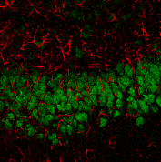
In a recent Nature Protocols article, Bucur et al. provide a detailed procedure for the application of ExM on human clinical pathology samples. Referred to as expansion pathology (ExPath), the authors demonstrate its broad applicability for a variety of human tissues and clinical specimens. A rapid ExPath procedure for use on thin (<5 um) tissue sections for time-sensitive applications is also described. The workflow for both conventional and rapid ExPath is straightforward and only requires a conventional fluorescence microscope and commercially available reagents. The process is optimized for uniform expansion and low distortion across various samples and tissue types. To withstand polymerization conditions and subsequent dilution of the label upon expansion, the use of bright, oxidation-resistant, and photostable fluorescent dyes such as CF®405M and CF®633 are recommended. In addition to finer resolution of complex structures, ExPath also offers significantly reduced autofluorescence. This is due to a water-based expansion that generates a transparent sample and the removal of unanchored biomolecules by protease treatment during mechanical tissue softening.
The main limitation of ExPath is that can only be used on fixed samples. In addition, as high labeling density is critical for the quality of ExPath images, labeling reagents and immunostaining conditions need to be thoroughly validated. Due to the aggressive proteinase K digestion conditions used, the method is also not suitable for downstream immunostaining. ExPath is also limited to the analysis of 3-5 targets based on the colors that can be spectrally distinguished on a conventional fluorescence microscope.
Learn more about our bright and photostable CF® dyes, especially our rhodamine-based CF® dyes that are well-suited for expansion microscopy due to their superior signal, chemical stability, and photostability over spectrally similar Alexa Fluor® dyes. We also offer a full selection of CF® dye labeled antibodies and bioconjugates.
Full Citation
Bucur O, Fu F, Calderon M, et al. 2020 Nat Protoc. 15(5):1649-1672. Nanoscale imaging of clinical specimens using conventional and rapid-expansion pathology. doi:10.1038/s41596-020-0300-1