 New Products
 Earth-Friendly Products
 Biotium Choice Antibodies
 Special Offers


Content #1
Content #1
Content #1
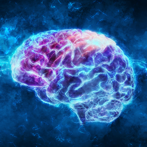
Comprehensive cellular analysis of intact organs provides crucial information for biological research and pathological diagnosis. Current methods for whole-organ cell profiling involve time intensive tissue-clearing and high-resolution volumetric imaging methods. Clear, unobstructed brain imaging cocktails and computational analysis (CUBIC) procedures are recent advancements that provide workflow pipelines aimed at improving preparation and analysis of isolated organ tissue.
In a recent publication in Nature Protocols, Matsumoto et al. describe an advanced CUBIC pipeline for tissue clearing, high-speed volumetric imaging, and high-throughput computational analysis by a cell-nucleus-detection algorithm. The pipeline provides two protocols for tissue-clearing: a rapid and high-quality organ clearing protocol (CUBIC-L and CUBIC-R+), and a tissue expansion-based protocol for high-resolution imaging (CUBIC-X). The study also describes methods for significantly enhancing the image acquisition rate using a novel system MOVIE (moving observation and efficient real-time autofocus for volumetric imaging). The system integrates continuous acquisition (MOVIE-scan), real-time autofocusing (MOVIE-focus) and the skipping of blank images (MOVIE-skip). The authors apply these advanced tissue-clearing and imaging methods to demonstrate simultaneous dual-color imaging of whole mouse brain with H2B-mCherry and RedDot™2, a far-red nuclear stain. Lastly, the CUBIC method implements a cell nucleus detection algorithm based on 3D Hessian-based difference of Gaussian (3D-HDoG). This algorithm allows for highly accurate image acquisition as well as the ability to leverage the computing power of multiple CPUs and graphics processing units (GPUs). Overall, the CUBIC method described provides significant improvements for preparation and analysis of whole organ tissues. Limitations that may be addressed in the future involve the inability to clear certain pigments, such as melanin and lipofuscin, as well as the limited resolution of the MOVIE system.
Learn more about RedDot™2 far-red nuclear stain for dead or fixed cells, and RedDot™1 far-red nuclear stain for live cells. Also see our full selection of nuclear and other cellular stains on our comprehensive and interactive Cellular Stains page.
