 New Products
 Earth-Friendly Products
 Biotium Choice Antibodies
 Special Offers


Content #1
Content #1
Content #1
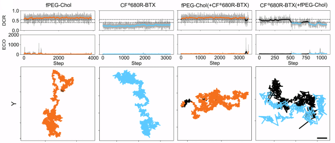
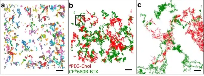
In membrane biology, the diffusion and interaction of proteins and lipids are fundamental to cellular signaling and function. Neurotransmitter receptors such as the nicotinic acetylcholine receptor (nAChR) are known to display cholesterol-sensitive diffusion patterns in the plasma membrane, but conventional microscopy has lacked the spatiotemporal resolution to resolve these dynamics. Capturing rapid, heterogeneous motions requires Minimal Photon Flux (MINFLUX), a super-resolution microscopy method that merges Stimulated Emission Depletion (STED) and single-molecule localization (SMLM) to deliver nanometer precision with sub-millisecond resolution.
In a 2025 Nature Communications publication, Reina et al. applied two-color MINFLUX single-molecule tracking to visualize the joint motion of nAChRs and fluorescent cholesterol in live mammalian cells. To enable this approach, the authors used Biotium’s CF®640- and CF®680R-labeled α-bungarotoxin conjugates to selectively tag nAChRs. CF®680R Dye provided the critical spectral separation needed for multiplex detection of a cholesterol analogue labeled with STAR Red dye. This allowed the researchers to reliably co-track receptor and lipid molecules under single-wavelength excitation, revealing heterogeneous diffusion modes and direct instances of receptor–cholesterol co-diffusion.
The authors also showed that cholesterol availability modulates receptor motion, with distinct regions of confined and joint diffusion observed. These findings, made possible by Biotium's wide selection of CF® Bungarotoxin conjugates, underscore cholesterol’s role in shaping receptor dynamics and highlight how high-resolution, multiplexed MINFLUX microscopy can uncover lipid–protein interactions previously imperceptible with conventional methods.
Biotium offers a wide range of bright and photostable CF® Dyes validated for advanced imaging modalities such as MINFLUX, STED, and STORM. Explore our full selection of labeling reagents for super-resolution microscopy and live-cell imaging applications.
Full Citation:

Content #1
Content #1
Content #1
Content #2
Content #3