 New Products
 Earth-Friendly Products
 Biotium Choice Antibodies
 Special Offers


Content #1
Content #1
Content #1
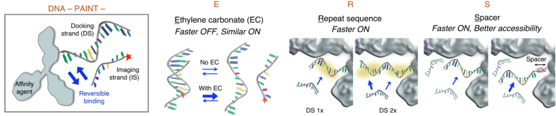
DNA-PAINT (points accumulation for imaging in nanoscale topography) is a single-molecule localization microscopy (SMLM) technique that takes advantage of the high specificity of DNA hybridization for super-resolution imaging. The docking strand, linked to an affinity agent such as an antibody, localizes to a target in the cell. The localizations arise from the reversible hybridization between a docking strand and the freely diffusing fluorescently labeled imaging strand. DNA-PAINT thus increases the number of observations possible compared to other super-resolution techniques such as STORM or PALM, which are constrained by the photon budget of the fluorophore. However, DNA-PAINT suffers from slow imaging speed inhibiting its potential for observing biological systems.
In a recent publication in Nature Communications, Civitci et al. developed a method that improves the imaging speed of DNA-PAINT by increasing the localization kinetics of the imaging strand labeled with the photostable CF®660R. The goal was to devise a strategy compatible with a large panel of oligonucleotide pairs validated for DNA-PAINT and can be readily adopted into the existing workflows.
The authors hypothesized that increasing the imaging strand’s off-rate without slowing down the binding would improve the localization kinetics and improve the imaging speed. The authors demonstrate that the addition of ethylene carbonate (EC), a water-soluble aprotic solvent shown to speed up hybridization between target DNA and oligonucleotides, in the imaging buffer increases the off-rate without significantly affecting the on-rate. The authors further optimized DNA hybridization kinetics and efficiency by implementing tandem sequence repeats (R) in the docking strands and a PEG spacer (S) between the docking oligonucleotide and antibody. Cumulatively, these modifications (ERS) to DNA-PAINT increased both super-resolution imaging speed and quality.
Learn more about our bright and photostable CF® dyes for super-resolution applications, including single-label secondary antibodies for STORM. Also, learn more about our rapid and versatile Mix-N-Stain™ Antibody Labeling Kits for labeling antibodies with CF® dyes and other labels.
Full Citation
Civitci, F., Shangguan, J., Zheng, T., Tao, K., Rames, M., Kenison, J., Zhang, Y., Wu, L., Phelps, C., Esener, S., Nan, X. (2020). In Vivo Single-Molecule Detection of Nanoparticles for Multiphoton Fluorescence Correlation Spectroscopy to Quantify Cerebral Blood Flow. Nature Communications. https://doi.org/10.1038/s41467-020-18181-6
