 New Products
 Earth-Friendly Products
 Biotium Choice Antibodies
 Special Offers


Content #1
Content #1
Content #1
Fluorescence bleed-through and cross-talk are major sources of error in multicolor fluorescence microscopy, particularly as experiments increase in multiplexing complexity.
This app note explains the physical basis of these phenomena, outlines experimental and instrumentation factors that exacerbate signal interference, and provides practical strategies to ensure accurate, reproducible fluorescence imaging results.
Fluorescence microscopy is a fundamental technique in modern cell biology and clinical diagnostics, enabling researchers to visualize specific molecular components within complex biological specimens. It is routine to stain a sample with multiple fluorescent conjugates simultaneously to detect multiple targets; however, the increasing sophistication of multicolor fluorescence experiments brings significant technical challenges that one must carefully consider. Among these, fluorescence signal interference represents a critical limitation that can lead to misinterpretation of data and false conclusions if not properly addressed.
Bleed-through and cross-talk refer to two phenomena that result in the unwanted detection of fluorescence from one fluorophore in a channel intended for another. Bleed-through is when emission of a fluorophore extends into the detection range set for another fluorophore, while cross-talk refers more broadly to any unwanted signal interference between fluorophores and includes excitation and emission cross-talk. Both events can create artifacts that can be mistaken for genuine colocalization or expression patterns and, therefore, must be minimized. With the appropriate controls, experimental design, instrumentation settings, and post-acquisition processing, researchers can maximize signal specificity while minimizing confounding spectral interference.
Bleed-through and cross-talk can significantly compromise the accuracy and reliability of fluorescence-based experiments. Understanding and mitigating sources that contribute to cross-talk is essential for producing reliable, reproducible fluorescence data.
The fundamental challenge of fluorescence cross-talk arises from the physical properties of fluorophores. While we typically focus on peak excitation and peak emission for a given fluorophore, the spectra are usually much broader than the peak. For example, while FITC has peak excitation at 495 nm and peak emission at 519 nm, its actual excitation and emission curves span dozens of nanometers on each side of these peaks. This characteristic creates inevitable overlap zones when multiple fluorophores are used simultaneously, which can lead to cross-talk (Figure 1). In addition to spectral overlap, the relative brightness of each fluorophore plays a critical role as bright fluorophores are more likely to bleed into channels intended for dimmer fluorophores.
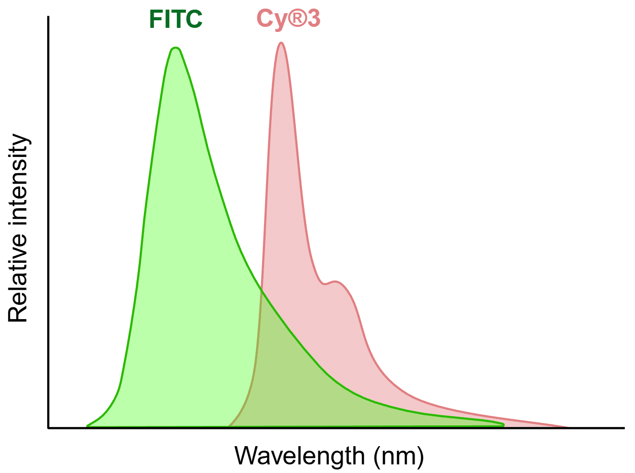
Figure 1. Example of spectral overlap that can lead to cross-talk. The FITC and Cy®3 emission peaks are well separated, but there is still an overlapping area that presents a potential for cross-talk. Created in https://BioRender.com
Signal intensity imbalance across multiplexed fluorescent probes is another key contributor to cross-talk. For example, the signal from fluorescent conjugates targeting highly expressed proteins can bleed into adjacent channels for fluorescent conjugates targeting less abundant proteins, particularly when imaging parameters are optimized to detect low-abundance targets. (Figure 2). If the signal intensities from all probes used are not carefully balanced, there is an increased likelihood of cross-talk.
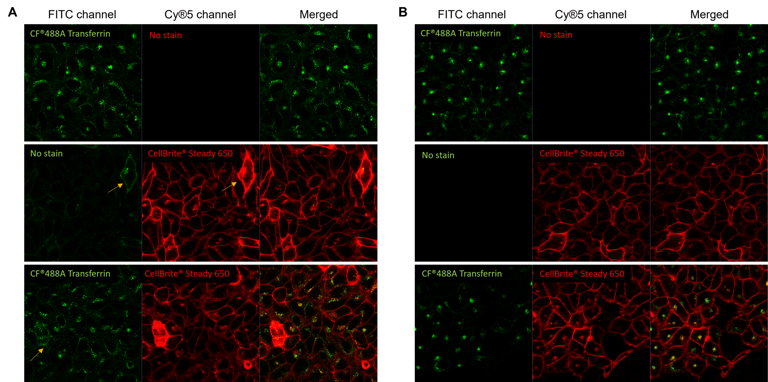
Figure 2. Unbalanced fluorophore signal intensities lead to cross-talk during simultaneous excitement. Live HeLa cells were labeled with CF®488A-transferrin receptor (TfR; green) and CellBrite® Steady 650 (red), then imaged with the 488 nm and 639 nm lasers simultaneously. (A) CF®488A-TfR produces a dim signal, requiring increased detector sensitivity in the FITC channel. Under these conditions, the much brighter CellBrite® Steady 650 signal bleeds into the FITC channel. Areas of colocalization (yellow in merged image) may be artifacts of bleed-through. (B) After titration to balance signal intensities, CF®488A-TfR and CellBrite® Steady 650 are cleanly detected in their respective channels with minimal cross-talk, enabling more confident assessment of colocalization.
Hardware configuration dictates how the signal is captured and instrument configuration is another important consideration for signal separation.
Bandpass filters play an important role in spectral separation. These filters act as gates that selectively pass a narrow range of wavelengths while blocking others, which allows only specific excitation or emission light to be isolated (Figure 3A). The bandpass filter’s width, or bandwidth, dictates the range of wavelengths it transmits, where narrower bandwidths provide better contrast and broader bandwidths capture more wavelengths. Longpass filters and shortpass filters are similar to bandpass filters, but they only transmit wavelengths longer than or shorter than a specific cutoff wavelength, respectively (Figure 3B). Users should always ensure that they choose fluorophores that are compatible with their instrument’s filters.
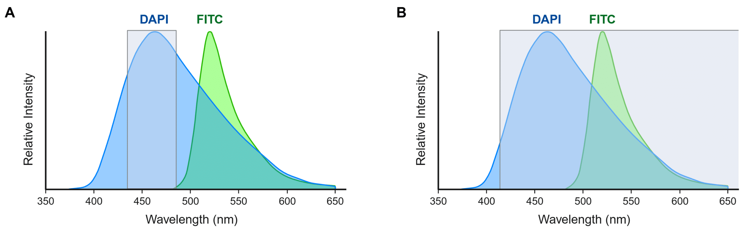
Figure 3. Effect of emission filter selection on spectral overlap in multiplex fluorescence imaging. Emission spectra of DAPI (blue) and FITC (green) are shown with wavelengths that will be detected with a 460/50 bandpass filter (A) or a 415 longpass filter (B) overlaid (gray). (A) A 460/50 bandpass filter will restrict detection to DAPI emission wavelengths and avoid cross-talk. (B) A 415 longpass filter will capture emission wavelengths from both DAPI and FITC. Created in https://BioRender.com
Confocal systems generally provide better spectral separation compared to widefield epifluorescence microscopes due to narrowband laser excitation and more customizable detection settings. While simpler epifluorescence systems and older confocal systems have fixed wavelength filters to isolate specific portions of emission spectra and prevent bleed-through from fluorophores in the adjacent channels, newer confocal systems use tunable dichroic mirrors that can be optimized for the emission wavelengths of the fluorophores used. However, because tunable systems offer more flexibility for the user to set a wider or narrower emission window, the user must take care to use settings appropriate for their experiment. A wider emission window can result in brighter signal for a single fluorophore, but may lead to cross-talk in a multicolor experiment.
Unrecognized cross-talk can have severe consequences, including false colocalization, inaccurate signal quantification, and misidentified cellular features. To minimize these risks, researchers should implement experimental design strategies and instrument-based considerations.
First and foremost, it is critical to use strategic fluorophore selection when designing a multiplex experiment to ensure fluorophores have maximal spectral separation. Biotium’s Spectra Viewer enables simultaneous visualization of multiple fluorophore spectra alongside instrument-specific excitation sources and emission filters, facilitating informed fluorophore selection and instrument compatibility. Additionally, consider the relative brightness of each fluorophore–conjugates with brighter fluorophores should be used for less abundant targets, and probes should be titrated to balance signal intensities (see Antibody Optimization and Signal Balancing below).
Even when best practices are followed, dyes with broader excitation/emission peaks are more likely to show cross-talk than those with narrower peaks. For example, some environmentally sensitive cellular stains, such as membrane dyes, nerve terminal dyes, or organelle stains, have broader emission spectra than small molecule dyes for antibody labeling, like CF® Dyes. Users should always visualize the spectra of dyes when designing experiments with a tool like our Spectra Viewer to minimize cross-talk risk.
Optimized sample preparation is essential for high-quality fluorescence imaging. There are numerous fixation and permeabilization methods commonly used during immunofluorescence, and each method should be optimized to preserve the fluorescent signal while minimizing autofluorescence. If autofluorescence is an issue, consider using autofluorescence quenching treatments such as TrueBlack® Lipofuscin Autofluorescence Quenchers, and perform unstained controls to determine the level of autofluorescence to help correct for it. Also, be sure to include appropriate blocking steps to reduce nonspecific antibody binding because nonoptimal blocking methods can lead to an increase in background and exacerbate the effects of cross-talk.
Each antibody chosen for an immunofluorescence experiment should be carefully optimized individually. Titrate each antibody to determine the minimum concentration needed for specific detection of each target with high signal-to-noise and validate antibody specificity with the appropriate positive and negative controls. This is especially important for colocalization studies which rely on balanced signals from each probe to prevent one signal overwhelming the other (see Figure 2). In addition, antibodies should be selected and validated to avoid cross-reactivity artifacts. Highly cross-adsorbed secondary antibodies should be used for multiplexing and to avoid direct binding of secondary antibodies to endogenous IgG in tissues. The appropriate controls with secondary antibody alone and different combinations of primary and secondary antibodies should be done to rule out antibody cross-reactivity as a source of cross-talk.
Sequential imaging should always be performed for adjacent channels when imaging a multi-stained sample, meaning you should excite with one laser and image one fluorophore at a time, then computationally combine images. It generally is possible to image spectrally distinct dyes in well-separated channels simultaneously (with more than one laser on), but the appropriate controls should always be included. It is recommended to use the lowest laser power possible to image each fluorophore to limit cross-excitation of fluorophores intended for other channels. Using excessively high laser power leads to excited-state saturation, where the fluorophore selected to be excited does not increase in signal intensity, and unintended fluorophores are also excited. Furthermore, when using DAPI or Hoechst nuclear stains, be aware that UV light from mercury arc lamp illumination can cause artifacts from DAPI/Hoechst photoconversion. For more information, read our Tech Tip: Avoiding Artifacts from UV Photoconversion of DAPI and Hoechst.
Filters and dichroic mirrors must be carefully selected to minimize bleed-through between channels because fluorophores in multiplex panels often have partially overlapping spectra, Optimizing these parameters ensures that each probe is detected within a well-defined, non-overlapping range of emission wavelengths.
Single-stain are samples prepared identically to the multicolor experimental samples but stained with only one fluorophore at a time to provide direct measurement of actual bleed-through for each fluorophore under the same experimental conditions. Single-stain controls allow one to account for sample-specific optical properties and instrument settings, unlike using theoretical spectral data or previous experimental data. These controls are the foundation of proper cross-talk management.
The preparation of single-stain controls should match that of the experimental samples, meaning the user utilizes the same tissue or cell type as well as identical fixation, permeabilization, labeling, and mounting methods. Ideally, the controls are processed alongside the experimental samples to reduce variability. Similarly, the controls should be imaged during the same session as the experimental samples, using the same exposure times, laser powers, and detector gains for each channel to be imaged in the multiplexed sample. It is important to remember that bleed-through is very likely to occur at high laser power and gain/exposure settings, even for well-separated fluorophores. Therefore, users should disable settings such as auto-exposure and auto-gain that will maximize signal in each channel, to ensure that the single-stain controls replicate the settings for the multiplexed sample, and thus reflect the true intensity relationships between fluorophores.
Many newer imaging systems have spectral unmixing capabilities that can be used to separate overlapping signals after imaging is complete. These instruments can measure a spectral signature for discrete regions of interest and then use computational algorithms to distinguish overlapping fluorophores. Spectral unmixing is increasingly used to expand the options for multicolor panels beyond what’s possible using conventional imaging. However, generally it is advisable to minimize cross-talk/bleed-through between fluorophores as much as possible using the methods discussed here before attempting to separate overlapping signals using spectral unmixing software.
Fluorescence cross-talk and bleed-through represent fundamental challenges in immunofluorescence microscopy that can significantly impact data interpretation if not properly addressed. Effective cross-talk management starts with the implementation of comprehensive single-stain controls, imaged under identical conditions as experimental samples. As imaging technology continues to advance toward higher multiplexing capabilities, the importance of rigorous cross-talk control only increases. By incorporating the best practices outlined in this guide, researchers can ensure their immunofluorescence data accurately represents biological reality rather than technical artifacts.
Below is a sample workflow for an immunofluorescence experiment to demonstrate how to effectively incorporate single-stain controls.
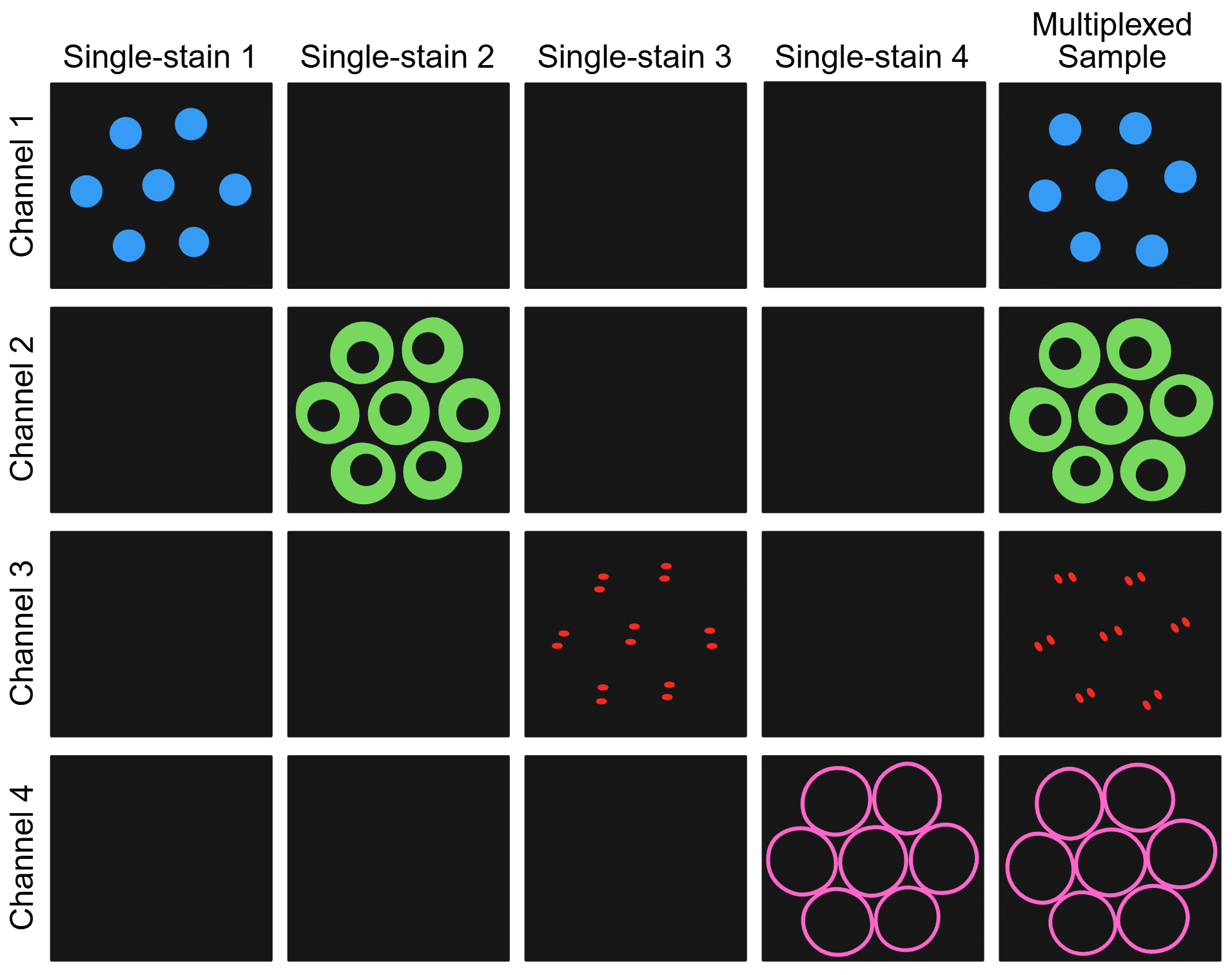
Figure 4. Sample and channel matrix illustrating how single-stain controls should only show signal in the on-target channel while multiplexed samples should only show specific signal in each on-target channel. Created in https://BioRender.com
CY DYE is a registered trademark of Cytiva.

Content #1
Content #1
Content #1
Content #2
Content #3