 New Products
 Earth-Friendly Products
 Biotium Choice Antibodies
 Special Offers


Content #1
Content #1
Content #1
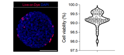
The increasing rise of liver diseases such as cancer and cirrhosis necessitates advanced research methods that overcome the temporal and visual limitations of in vitro models based on liver spheroids. In vivo liver spheroid models also exist and show promise towards understanding liver diseases, however low anatomical fidelity and time constraints represent major challenges, along with poor imaging accessibility from their commonly intraperitoneal and subcutaneous placements. In contrast, the anterior chamber of the eye (ACE) is a transplantation site that allows the growth of transplanted liver spheroids to be imaged through the transparent cornea by non-invasive confocal microscopy. In addition, the transplanted tissue can acquire innervation and vascularization from nerves and blood vessels in the iris, providing an ideal site for spheroid engraftment and growth.
In a 2024 publication in Nature Communications, Lazzeri-Barcelo et al. performed imaging studies of liver spheroids transplanted into mouse cornea, utilizing our Live-or-Dye NucFix™ Red dye to assess viability of the liver spheroids in vitro before being placed into the ACE. The authors discovered that liver spheroids transplanted into the ACE became vascularized and received sympathetic innervation, resembling endogenous liver tissue. Blood flow dynamics, bile acid transport, LDL uptake, and lipid droplet accumulation within the transplanted liver spheroids were also observed. This groundbreaking method not only overcame previous imaging limitations but also provided insights into the vascular and neural interactions crucial for liver function. Future applications of this particular technique may be used as a unique tool to study liver physiology and disease progression. In addition, this study confirmed the application of Live-or-Dye NucFix Red™ for spheroids, thus validating another tool for researchers assessing viability of 3D cultures.
Learn more about Biotium’s high-performance Live-or-Dye™ Fixable Viability Staining Kits, featuring a wide variety of dye options for standard flow, spectral flow, and fluorescence microscopy. Also learn more about our NucSpot® Live Cell Nuclear Stains and our wide selection of Cell & Organelle Stains.
Full Citation:
Lazzeri-Barcelo, F., Oliva-Vilarnau, N., Baniol, M. et al. Intraocular liver spheroids for non-invasive high-resolution in vivo monitoring of liver cell function. Nat Commun 15, 767 (2024). https://doi.org/10.1038/s41467-024-45122-4
