 New Products
 Earth-Friendly Products
 Biotium Choice Antibodies
 Special Offers


Content #1
Content #1
Content #1
Kinesin motor-dependent anterograde transport is critical in cells, particularly neurons, to transport cargo and organelles from the cell body to distal sites. To address the multi-kinesin anterograde transport mechanism, the authors of a recent paper in Molecular Biology of the Cell studied the functional relationships of axonal kinesins-1 and 3 in the movement of dense core vesicles filled with a GFP-tagged neuropeptide in the Drosophila nervous system.
Visualization and co-localization of kinesins-1 and 3 in primary neuronal cultures was performed using a combination of immunofluorescence and super-resolution microscopy. As both kinesin antibodies were generated in the same host species, a combination of direct and indirect immunofluorescence was performed (see our Tech Tip for more information). Kinesin-1 (anti-KHC) antibody was directly labeled with Biotium's next-generation CF® 568 dye, using the Mix-N-Stain™ CF® dye antibody labeling kit for direct immunofluorescence, followed by co-localization studies performed by super-resolution, 3-D structured illumination microscopy (SIM), a novel application for CF® 568 dye.
To read the original article, click here.
Lim A, Rechtsteiner A, Saxton WM. Two kinesins drive anterograde neuropeptide transport. Mol Biol Cell. 2017 Sep 13. pii: mbc.E16-12-0820. doi:10.1091/mbc.E16-12-0820.
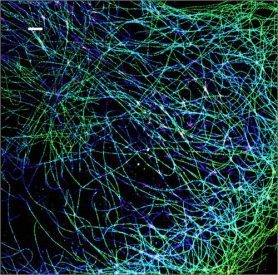
Tubulin staining (left) by direct immunofluorescence in MCF-7 cells using CF™568 dye-conjugated monoclonal anti-Ep-CAM (red) and (right) by 3-D STORM microscopy in fixed cells using CF™568 dye-conjugated anti-mouse secondary antibody.
Learn more about Biotium's Mix-n-Stain™ CF™ dye antibody labeling kits and CF™ dyes for super-resolution microscopy.
Also see our Tech Tip: Combined Direct and Indirect Immunofluorescence Using Primary Antibodies from the Same Host.
