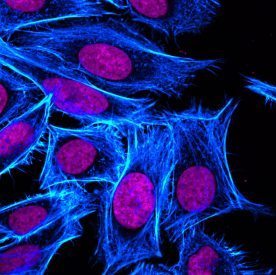
Histological examination of tissue pathology specimens has traditionally been performed on 5 um thin sections. Imaging of 3-dimensional tissue architecture using thin sections is challenging, requiring sequential imaging of many sequential sections and digital reconstruction of the tissue. Fluorescence imaging of thick tissue sections has been limited by the diffusion of light through opaque tissues. Now, a recent publication from van Royen and colleagues from Erasmus Medical Center in Rotterdam reported a novel optical clearing method that allowed them to perform 3-dimensional fluorescence imaging of both fresh and archival FFPE prostate clinical specimens. The authors performed 3-D reconstruction of prostate by staining thick tissue sections with anti-CK5 (basal cell marker), anti-CK8-18 (luminal epithelial cell marker), and the far-red nuclear counterstain RedDot™2. Optical clearing allowed fluorescence imaging to a depth of 800 um, more than 10 times the thickness that could be imaged on uncleared tissue, providing unprecedented opportunities for the study of normal™ and pathological tissue architecture using confocal microscopy.
To read the original article, click here.
van Royen ME, Verhoef EI, Kweldam CF, van Cappellen WA, Kremers GJ, Houtsmuller AB, van Leenders GJ. Three-dimensional microscopic analysis of clinical prostate specimens. Histopathology. 2016 Jun 29. doi: 10.1111/his.13022.
To learn more about RedDot2 and the other cell and organelle stains that Biotium has to offer, click here.