 New Products
 Earth-Friendly Products
 Biotium Choice Antibodies
 Special Offers


Content #1
Content #1
Content #1
Matching the refractive index of mounting medium and coverglass in microscopy allows optimal imaging, by minimizing the bending of light as it travels through the coverglass and the specimen. Antifade mounting media commonly used with fixed cells and tissue sections usually have refractive indices close to glass. However, these media cannot be used with live cells or organisms due to toxicity. Now, Boothe and colleagues report in an eLife publication that a non-toxic media additive can be used to optimize the refractive index of culture media for imaging of live cells, organoids, and organisms like zebrafish and worms. By optimizing the refractive index while imaging live planarian flatworms, they were able to image through several cell layers to detect nuclear staining using Biotium's far-red live nuclear stain RedDot™1 with greater sensitivity and signal:noise ratio.
To read the original article, click here.
Boothe T, Hilbert L, Heide M, Berninger L, Huttner WB, Zaburdaev V, Vastenhouw NL, Myers EW, Drechsel DN, Rink JC. (2017). A tunable refractive index matching medium for live imaging cells, tissues and model organisms. Elife. 14;6. DOI: 10.7554/eLife.27240
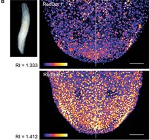
Refractive index matching improves imaging of RedDot™1 nuclear staining in a live planarian flatworm. From Figure 4b in Booth et al. Elife 14;6 (2017).
Learn more about RedDot™1 far-red nuclear stain for live cells, RedDot™2 far-red nuclear stains for dead or fixed cells, plus our full selection of Cellular Stains.
