 New Products
 Earth-Friendly Products
 Biotium Choice Antibodies
 Special Offers


 Powered by Bioz


Content #1
Content #1
Content #1
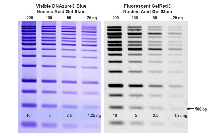
A blue nucleic acid gel stain to visualize dsDNA in agarose or polyacrylamide gels by the unaided eye.
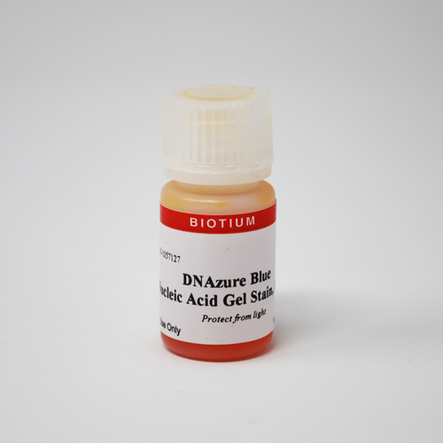

Check out DNAzure® 2.0 Visible Blue DNA Gel Stain Kit, a new and improved formulation of the original DNAzure® Blue Nucleic Acid Gel Stain that delivers enhanced sensitivity for visible staining of dsDNA in both agarose and polyacrylamide gels.
Visualize DNA bands in gels by unaided eye and without UV light sources. Detection is highly sensitive and rivals most fluorescence-based gel stains.
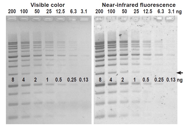
DNAzure® Blue Nucleic Acid Gel Stain is a DNA-binding dye that turns from colorless to deep blue upon exposure to bright light. After color development, the stain also has broad emission near-infrared fluorescence that can be imaged using the LI-COR®, Odyssey®, or similar near-IR imaging systems. The sensitivity of detection is similar for visible color and near-IR imaging. DNAzure® is compatible with agarose gels or polyacrylamide gels as well as downstream applications such as sequencing and cloning. The dye efficiently removed from DNA by common gel extraction kits that utilize silica-based DNA purification columns.

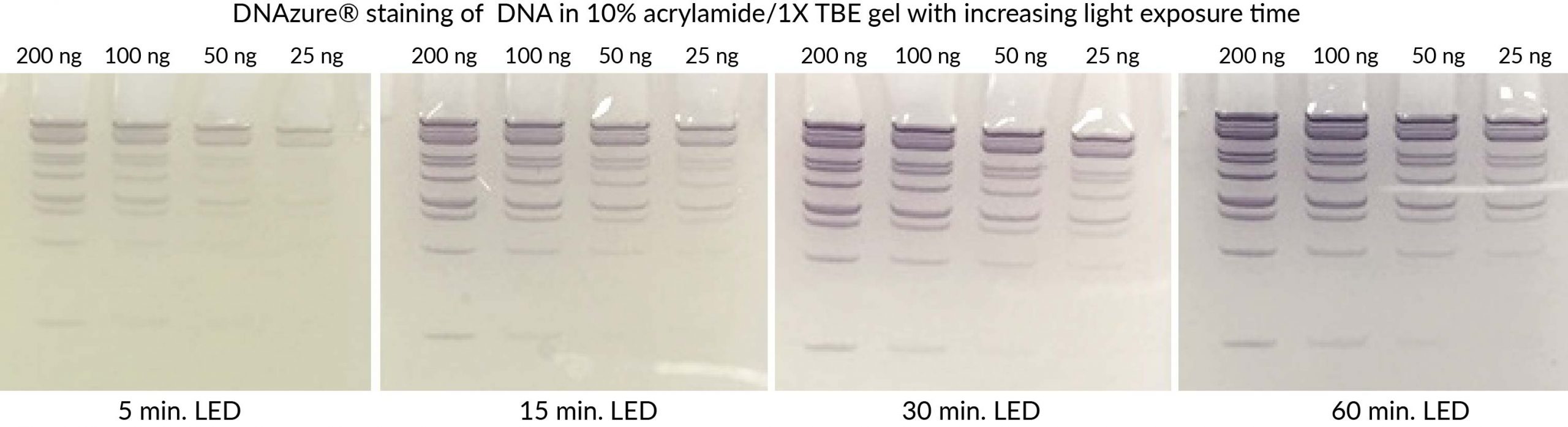
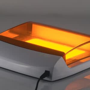
Light exposure can be performed with a variety of white and blue light sources. For best results, we recommend performing the light exposure with the Glo-Plate™ White Photoactivation Device or Glo-Plate™ 2.0 Blue LED Illuminator.
We recommend DNAzure® 2.0 Visible Blue DNA Gel Stain Kit as an improved alternative to the original DNAzure® Blue Nucleic Acid Gel Stain. The improved formulation delivers enhanced sensitivity for visible staining of dsDNA in both agarose and polyacrylamide gels. Also learn about the Original GelRed® Nucleic Acid Gel Stain or GelGreen® Nucleic Acid Gel Stain. GelRed® 3X in water is ready-to-use for post-electrophoresis gel staining, and is supplied in a 4L Cubitainer®. Higher concentrations of Original GelRed® are available as 10,000X in water or DMSO. We also offer GelRed® Agarose and GelRed® Prestain Plus 6X Loading Dye. GelGreen® Nucleic Acid Gel Stain is a safer replacement for SYBR® gel stains and is compatible with visible light excitation. Our Go-Go™ Fast DNA Gel Running Buffer allows running gels 3X faster than with TAE or TBE buffer.
| Product / Method | Procedure | Advantages | Disadvantages | Recommended for |
|---|---|---|---|---|
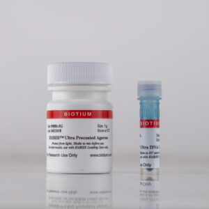
| DNA staining with EMBER™ Ultra DNA Gel Kit | Agarose is supplied pre-coated with EMBER™ Ultra Dye, just dissolve, heat, and pour. | • Safer and more convenient, no need to handle concentrated dye • Superior sensitivity, detect as little as ≤1 ng DNA • No need for post-electrophoresis staining • Optimal for blue LED gel imagers | • Not suitable for PAGE, DGGE, EMSA, or PFGE gels • Dye may cause band migration issues when loading larger amounts of DNA (more than ~200 ng/band), or for some restriction digests | • Routine agarose gels |
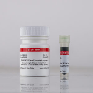
| RNA staining with EMBER™ Ultra RNA Gel Kit | Agarose is supplied pre-coated with EMBER™ Ultra Dye, just dissolve, heat, and pour. | • Safer and more convenient stain for RNA, no need to handle concentrated dye • Superior sensitivity, detect as little as ≤5 ng RNA • No need for post-electrophoresis staining • Included loading dye contains formamide for denaturing • Optimal for blue LED gel imagers | • Will stain DNA as well as RNA • Dye may cause band migration issues when loading larger amounts of RNA (more than ~200 ng/band) | • Routine RNA gel electrophoresis • Evaluate total RNA integrity and DNA contamination |
| DNA prestaining with GelRed® Prestain Plus 6X DNA Loading Dye | GelRed® loading buffer is added directly to the DNA sample before loading | • Fast & simple: one-step sample loading & DNA staining • Less concentrated dye for safer handling • Can re-run a gel to use empty lanes | • Not recommended for PAGE, DGGE, EMSA, or PFGE gels • Dye may cause band migration issues when loading larger amounts of DNA (more than ~100 ng/band), or for some restriction digests | • Routine agarose gels • Recommended loading 50-200 ng ladder or 2-5 uL PCR product ( ~100 ng/band or less) |
| Precast staining with GelRed® 10,000X in water or GelGreen® 10,000X in water | GelRed® or GelGreen® is mixed with molten agarose before gel casting | Familiar protocol, rapid results | ||
| Precast staining with GelRed® Agarose LE or GelGreen® Agarose LE | Agarose is supplied pre-coated with GelRed® or GelGreen®, just dissolve, heat, and pour | Safer & more convenient, no need to handle concentrated dye | ||
| Post-electrophoresis staining with GelRed® 10,000X in water or GelGreen® 10,000X in water - or - GelRed® 3X in water | No fluorescent dye is added to the gel, it is stained in 3X GelRed® or 3X GelGreen® solution after electrophoresis | • Most accurate sizing/sharpest bands • Staining solution can be re-used • Enhance sensitivity by adding NaCl | Extra staining step (up to 30 minutes) after electrophoresis (some customers report good results after only 5 minutes if dye is not reused) | • Highly accurate band sizing • Gels with more than ~100 ng DNA per band • Analyzing restriction digests |
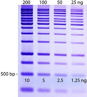
| Post-electrophoresis staining with DNAzure® 2.0 Visible Blue DNA Gel Stain Kit | No fluorescent dye is added to the gel, it is stained in DNazure® 2.0 solution and then exposed to a bright light source to generate visible blue DNA bands. We recommend the Glo-Plate™ White Photoactivation Device as a light source for developing DNAzure® 2.0-stained gels | • Allows visualization of DNA bands by the naked eye, no need for a UV light source • Detect as little as ~1 ng DNA • Stained bands are stable in gel for weeks • Also emits near-IR fluorescence (~700 nm) for detection on near-IR imaging systems | Extra staining step (up to 30 minutes) followed by a light exposure step (up to 30 minutes) to generate visible blue DNA bands | • Routine DNA agarose gels • Visualizing gels without the UV light or expensive imaging systems • Recommended loading 50-200 ng DNA per lane |
| Post-electrophoresis staining of PAGE gels using PAGE GelRed® 10,000X or 1X in water | No fluorescent dye is added to the gel, it is stained in 1X PAGE GelRed® solution after electrophoresis | • Formulated for efficient penetration and staining of polyacrylamide gels • Like the classic GelRed®, it is safe and environmentally friendly | Extra staining step of approx. 30 minutes after electrophoresis | Staining of nucleic acids in PAGE gels |
We have tested a variety of different light sources for the development of bands using the original DNAzure® and DNAzure® 2.0 in an agarose gel. We have found that most light sources will work, but how long the development takes is dependent on the brightness of the light. The fastest band development is seen with our Glo-Plate™ White Photoactivation Device. We have also seen good results with other bright, white LED lights. Other light sources that work but take more time include an LED desk lamp and a cell phone light. We found that a 600W halogen lamp did not work well- it was too hot, which melted the gel.
We recommend storing DNAzure® Blue Nucleic Acid Gel Stain and DNAzure® 2.0 Visible Blue DNA Gel Stain Kit at 4°C. After storage at room temperature, we have seen some loss of dye stability.
The 1X DNAzure® staining solution from the original DNAzure® Blue Nucleic Acid Gel Stain can be re-used for multiple gels, under certain conditions. Importantly, the staining solution must be removed from the gel before the gel is exposed to light to develop the bands. If the staining solution undergoes the light exposure, it will not be able to stain another gel. The used staining solution should be stored in the refrigerator, protected from light, between uses. We have successfully re-used the staining solution stored for up to 2 weeks, and used up to 4 times with little loss of signal (after 5 or 6 uses, the sensitivity was noticeably lower).
We do not recommend reusing or storing the 1X staining solution from the DNAzure® 2.0 Visible Blue DNA Gel Stain Kit. The 1X staining solution should be prepared on the day of use.
