 New Products
 Earth-Friendly Products
 Biotium Choice Antibodies
 Special Offers


 Powered by Bioz


Content #1
Content #1
Content #1
Buffer system optimized for fluorescence-based western provides superior specificity, sensitivity, and background suppression.
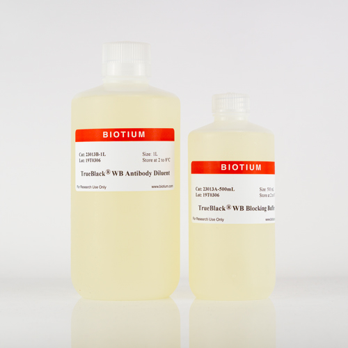
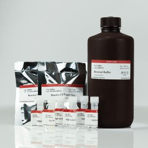
The TrueBlack® WB Blocking Buffer Kit is a ready-to-use buffer system for fluorescence-based western blotting (WB). The buffers are designed to achieve optimal specificity and sensitivity by blocking background from the non-specific interaction between the dye-labeled antibodies and blotting membranes.
Note: The TrueBlack® WB Blocking Buffer (Cat. No. 23013A-500ML) and TrueBlack® WB Antibody Diluent (23013B-1L) are available as standalone products. The standalone TrueBlack® WB Antibody Diluent is particularly useful for users who have evaluated the kit and prefer to use the antibody diluent but not the blocking buffer.
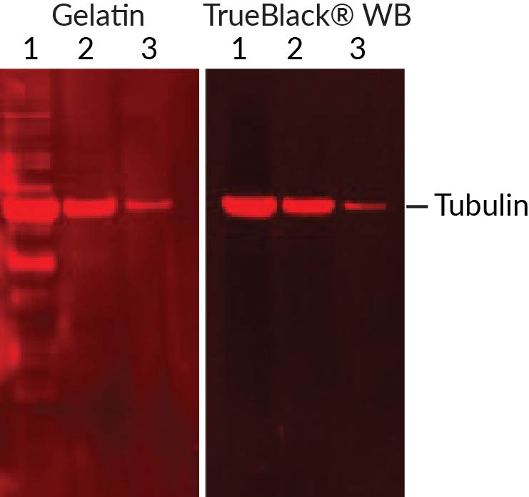
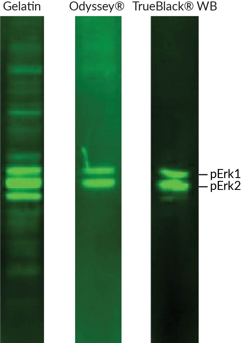
Non-specific background in fluorescence-based western blotting can arise from multiple sources, including antibody cross-reactivity with off-target proteins, non-specific antibody adsorption to the membrane, and membrane autofluorescence. Another potential cause of background that is not well-known is the effect of fluorescent dyes themselves on the specificity of labeled antibodies. Next-generation fluorescent dyes like Alexa Fluor® or CF® dyes often carry multiple negative charges to improve dye solubility and brightness of conjugates. However, the extra charge carried by the dye can result in non-specific antibody binding and background fluorescence. The TrueBlack® WB Blocking Buffer Kit blocks background from multiple sources including charged dye conjugates. It is especially advantageous for phosphoprotein detection, significantly improving specificity compared to conventional blocking buffers.
The TrueBlack® WB Blocking Buffer Kit includes blocking buffer and antibody diluent for primary and secondary antibody incubation steps. The number of membranes that can be processed per kit is based on 10 mL per incubation (blocking, primary antibody, and secondary antibody); the actual number of membranes may vary depending on protocol used and membrane size. When compared to LI-COR's Odyssey® Blocking Buffer, the TrueBlack® WB Blocking Buffer works as well or better, and is priced lower on a per membrane basis. See below for a size comparison.
Comparison between TrueBlack® WB Blocking Buffer Kit and Odyssey® Blocking Buffer
| Product | TrueBlack® WB Blocking Buffer Kit | Odyssey® Blocking Buffer |
|---|---|---|
| Trial Size | For 10 membranes | 125 mL for 4 membranes |
| Full Size | For 50 membranes | 500 mL for 16 membranes |
The TrueBlack® WB Blocking Buffer Kit belongs to our TrueBlack® line of background reducing agents for fluorescence applications, which includes TrueBlack® Lipofuscin Autofluorescence Quencher for tissue staining, and TrueBlack® IF Background Suppressor System (Permeabilizing) for blocking non-specific immunofluorescence staining.
Download curated list of TrueBlack® References
TrueBlack® Lipofuscin Autofluorescence Quencher, 20X in DMF (Cat. No. 23007) is the original formulation. TrueBlack® Lipofuscin Autofluorescence Quencher, 30X in DMSO (Cat. No. 23011) is a newer, more concentrated formulation in DMSO, which is a less toxic solvent than DMF. The quencher stock solution formulation does not affect the performance in staining.
TrueBlack® Lipofuscin Autofluorescence Quencher and TrueBlack® Plus Lipofuscin Autofluorescence Quencher were not designed for colorimetric detection, but they will stain lipofuscin black and be visible in light microscopy.
TrueBlack® Lipofuscin Autofluorescence Quencher (Cat. No. 23007, 23011) and TrueBlack® Plus Lipofuscin Autofluorescence Quencher (Cat. No. 23014) are designed to reduce autofluorescence from lipofuscin in tissue samples such as mouse and human brains and retina. While TrueBlack® Lipofuscin Quenchers have been reported to reduce autofluorescence from other sources, such as collagen, elastin, red blood cells, and general background fluorescence, they are not as effective at quenching these types of autofluorescence as for lipofuscin autofluorescence. However, they may improve background from a variety of sources in different experimental systems.
TrueBlack® IF Background Suppressor System (Cat. No. 230120) is a buffer system designed for optimal blocking of non-specific antibody binding as well as direct interaction of fluorescent dyes on antibodies with cells or tissue sections to eliminate non-specific staining for immunofluorescence (IF).
The TrueBlack® WB Blocking Buffer Kit (Cat. No. 23013) is a ready-to-use buffer system for blocking non-specific interactions of dye-labeled antibodies with proteins and the blotting membrane in fluorescence-based western blotting (WB).
Even though AccuOrange™ buffer does contain SDS, which is required for the dye to bind proteins, the assay is very sensitive to small changes in SDS concentration, and also cannot tolerate non-ionic detergents that form mixed micelles with SDS, like Triton®. Therefore we don't recommend using the kit for cell lysates or other samples with significant amounts of detergents.
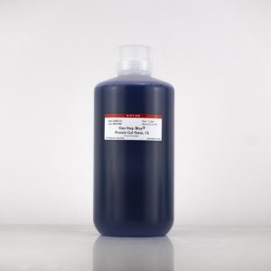
Gels stained with One-Step Blue® can be dried just like gels stained with Coomassie. The stain will not interfere with the detection of radiolabeled proteins.
The AccuOrange™ assay is a fluorescent dye-based assay. The dye binds to proteins primarily through hydrophobic interactions. Proteins denature upon heating; the dye binds to the exposed hydrophobic pockets of the protein after cooling. The free AccuOrange™ dye is fluorogenic due to non-radioactive decay but becomes highly fluorescent due to the rigid conformation inside the pocket.
The AccuOrange™ assay more sensitive than traditional protein quantitation assays such as BCA, Bradford and Lowry, and shows superior linearity and reproducibility than the NanoOrange® protein quantitation assay (Thermo Fisher Sci.), but has low tolerance for detergents like SDS and Triton® X-100.

Content #1
Content #1
Content #1
Content #2
Content #3