New Products
 Earth-Friendly Products
 Biotium Choice Antibodies
 Special Offers
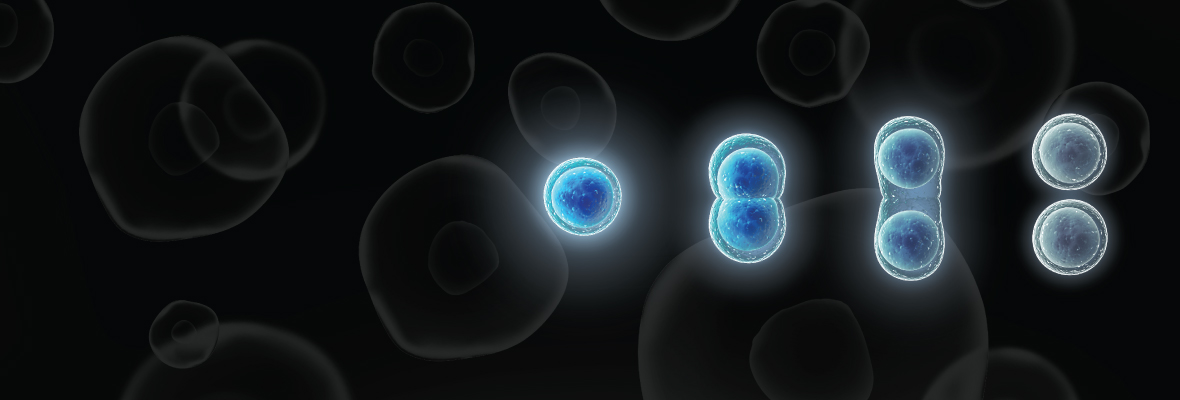
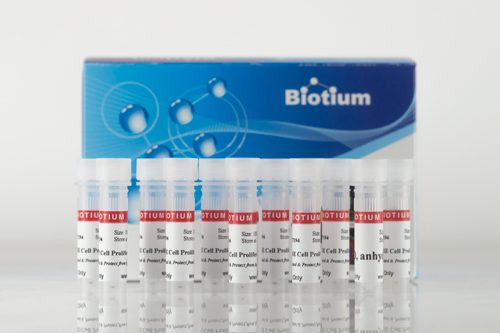
ViaFluor® SE Cell Proliferation Kits
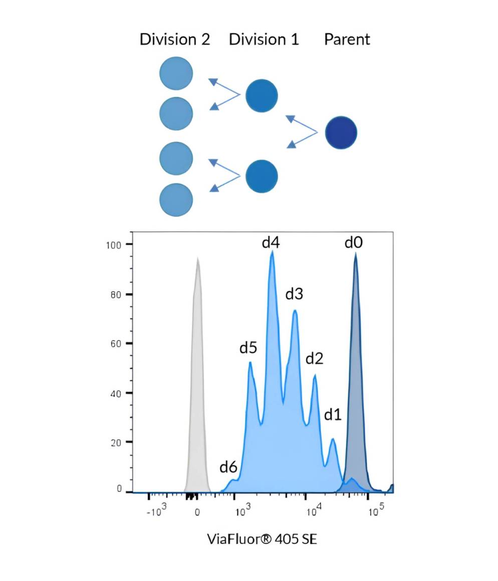
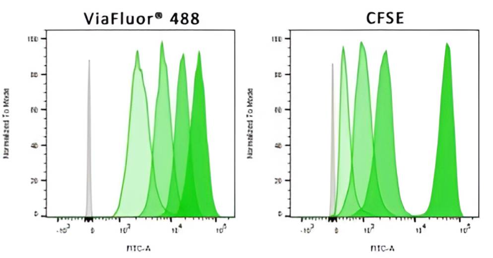
Cell Proliferation Monitoring by Flow Cytometry
Cell proliferation dyes passively enter live cells as non-fluorescent esters, which are converted to fluorescent dyes by intracellular esterases and covalently react with amine groups on intracellular proteins, forming conjugates that are retained in the cell. After staining, a single bright fluorescent population will be detected by flow cytometry, and each cell division produces progressively dimmer peaks on a flow cytometry histogram.
ViaFluor® SE Cell Proliferation Kits
CFSE (also known as CFDA-SE) has been used for many years to monitor cell proliferation by flow cytometry. While several alternatives are now available, CFSE remains widely used. Biotium offers CFSE under the tradename ViaFluor® CFSE. However, CFSE staining has several drawbacks, including leakage from the cell, cell toxicity, and bleed-through into the PE and PE-TexasRed® channels. ViaFluor® 405 SE and ViaFluor® 488 SE provide superior cell staining, fixability, and low toxicity. ViaFluor® 405 is excited with the violet laser and detected in the Pacific Blue® channel, and gives great peaks with no toxicity. ViaFluor® 488 and ViaFluor® CFSE are both detected in the FITC channel, but ViaFluor® 488 is less toxic, shows better retention in cells, more fixable, and has less bleed-through into other channels than CFSE.
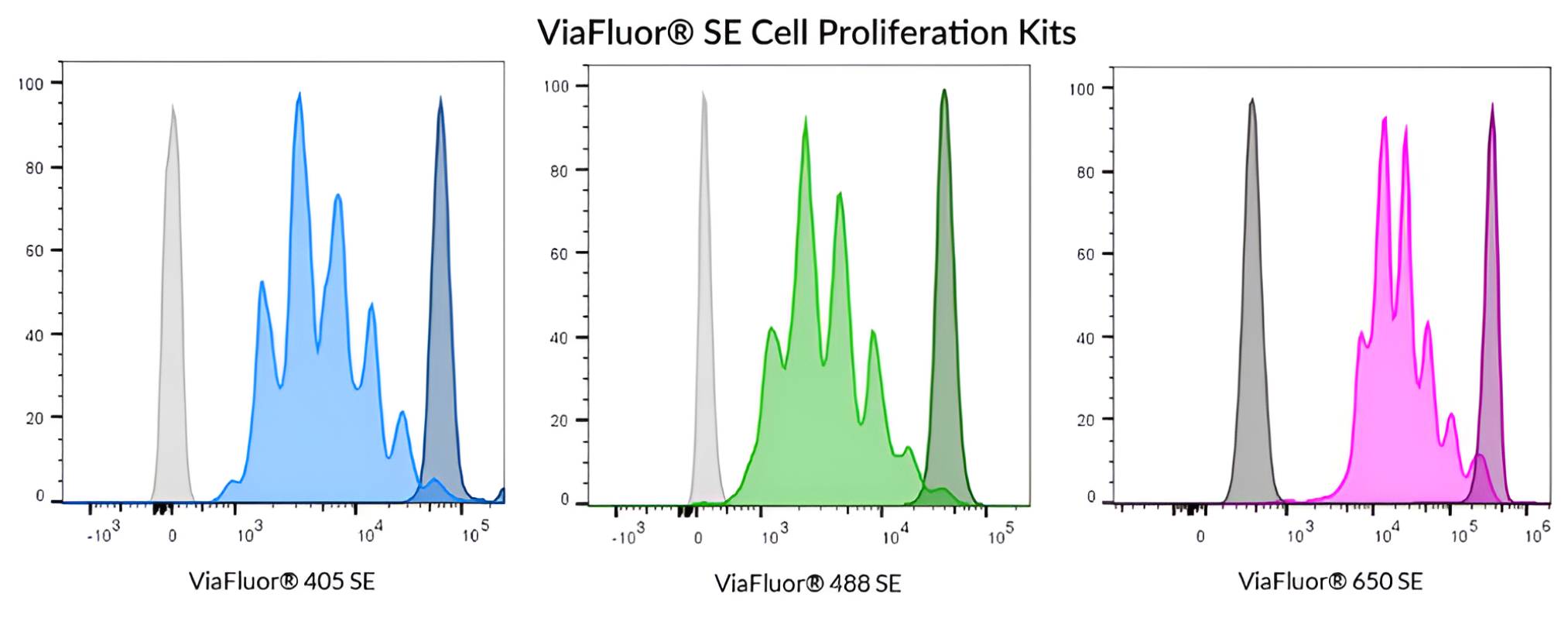
Cell Division | Catalog No. | Ex/Em (nm) | Flow detection | Features |
|---|---|---|---|---|
| ViaFluor® 405 SE Cell Proliferation Kit | 30068 | 387/446 | Pacific Blue® | • ViaFluor® 405 SE replaces CellTrace™ Violet |
| ViaFluor® 488 SE Cell Proliferation Kit | 30086 | 495/524 | FITC | • ViaFluor® 488 SE is a unique, improved green dye to replace CFSE |
| ViaFluor® 650 SE Cell Proliferation Kit | 30139 | 653/682 | APC | • ViaFluor® 650 SE is an improved and cost-effective alternative to CellTrace™ Far Red |
| ViaFluor® CFSE Cell Proliferation Kit | 30050 | 495/519 | FITC | • Classic cell division tracing dye, see our ViaFluor® 488 SE for an improved alternative |
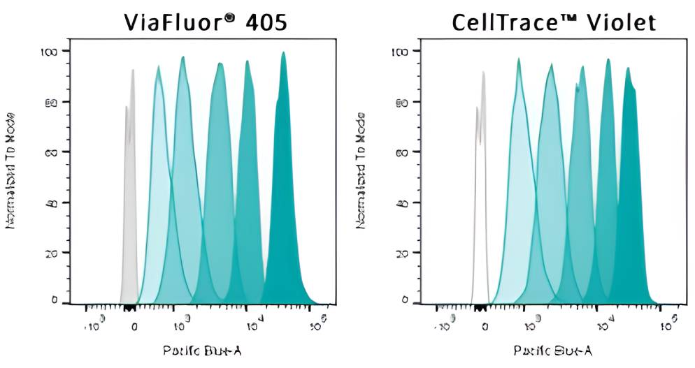
ViaFluor® 405 for the Violet Laser
A High-Performance Cell Proliferation Dye
ViaFluor® 405 is an excellent cell proliferation dye. Unlike some other dyes, it is non-toxic and does not interfere with cellular processes, such as cell cycle progression or T cell activation. The signal is bright and stable, giving sharp peaks that allow you to track cell division cycles by flow cytometry. When compared to the cell proliferation dye CellTrace™ Violet, they look identical in terms of staining pattern and lack of toxicity.
View Product Page

ViaFluor® 488: An Improved CFSE Alternative
ViaFluor® 488 for the FITC Channel
Although CFSE remains widely used, Biotium recognized that CFSE has several flaws that can interfere with experiments: it shows cell toxicity, can alter the kinetics of T cell activation, is not well retained in the cell, is not well retained after fixation, and bleeds into other detection channels. Therefore Biotium developed a new green cell proliferation dye, ViaFluor® 488, that improves on all of these properties.
ViaFluor® 488 gives nice sharp peaks for each cell division and does not show any leakage after washing, unlike CFSE. ViaFluor® 488 is retained in the cell after fixation and permeabilization, allowing samples to be immunostained or stored. ViaFluor® 488 also shows less bleed-through into other detection channels, such as PE or PE-TexasRed®, allowing more co-staining options. Since ViaFluor® 488 is detected in the same channel as CFSE, current users of CFSE can easily change to include ViaFluor® 488 in their flow panel design.
View Product Page
ViaFluor® 650 for the APC Channel
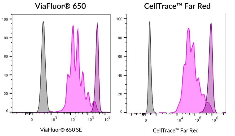
Sharper, More Distinct Peaks than CellTrace™ Far Red
ViaFluor® 650 was designed as an improved and cost-effective alternative to CellTrace™ Far Red for monitoring cell proliferation in the APC channel.
ViaFluor® 650 provides sharper proliferation peaks than CellTrace™ Far Red, allowing more robust cell division tracking. In addition, each vial of ViaFluor® 650 contains enough dye to stain a much larger volume of cells, leading to significant cost savings.
View Product Page
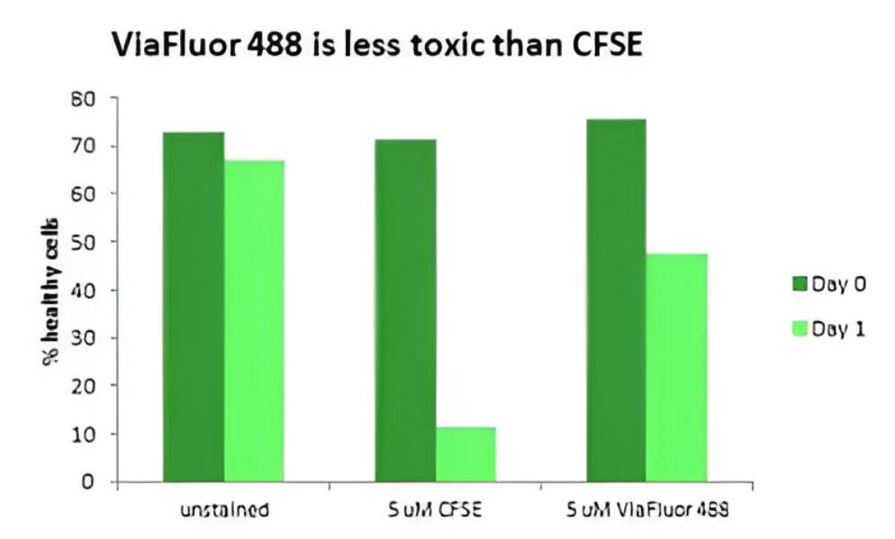
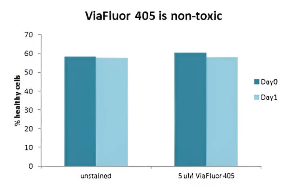
Reduced Cell Toxicity
New Dyes with Reduced Toxicity Compared to CFSE
Many cell proliferation dyes on the market are toxic to cells when used in the typical assay concentration range of 1-5 uM. This is especially problematic for proliferation assays that often last for many days. We have found that some of these dyes cause cell death and/or cell cycle arrest. The classic proliferation dye CFSE has significant toxicity at 5 uM and moderate toxicity at 1 uM, typical dye concentrations for cell proliferation assays. At Biotium we have developed several new cell proliferation dyes specifically designed to be less toxic to cells, for superior performance.
ViaFluor® 488 SE is a green cell proliferation dye, designed as an improvement to CFSE. ViaFluor® 488 has many advantages over CFSE, including reduced cell toxicity. When used at 5 uM, CFSE is extremely harmful to cells, while in the same experiment ViaFluor® 488 is much less destructive. At 1 uM, we haven’t seen any toxicity at all with ViaFluor® 488.
ViaFluor® 405 SE does not show any toxicity when used at 5 uM in either cultured Jurkat cells or PBMCs. ViaFluor® 405 is equal to CellTrace™ Violet in terms of lack of toxicity and great peaks in flow cytometry.
Improved Fixability
No Signal Loss After Fixation or Incubation
One known problem with CFSE is that it is not well-retained by cells. After staining cells with CFSE and incubating overnight, you find a wide gap between the Day 0 and Day 1 peaks. This is not just due to signal being diluted by cell division, since other proliferation dyes, like ViaFluor® 405 and ViaFluor® 488, do not have this wide gap. This is likely due to poor reactivity of the cleaved version of the CFSE dye.
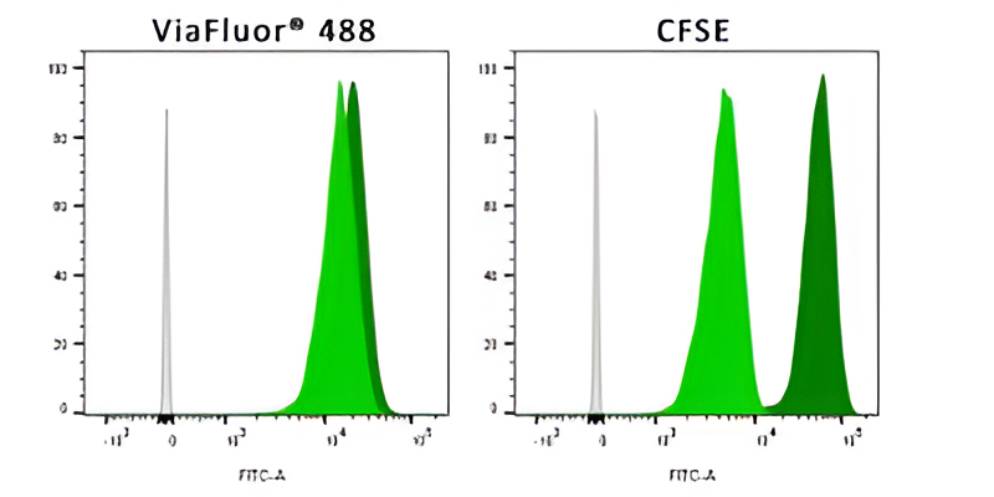
Additionally, the poor reactivity of the cleaved version of the CFSE dye makes the dye poorly fixable. CFSE exhibits a significant loss of signal after fixation, permeabilization, and washing. Unlike CFSE, the improved green proliferation dye ViaFluor® 488 is extremely well-retained in cells after incubation or fixation. The ability to perform fixation and permeabilization without loss of signal allows you to combine the cell proliferation assay with immunostaining and many other cellular assays.
Less Bleed-Through
ViaFluor® 488 Has Less Bleed-Through than CFSE
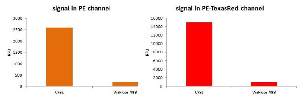
Another issue with CFSE is substantial bleed-through from the FITC channel into nearby longer wavelength detection channels, such as PE and PE-TexasRed®. This can present a problem when designing multi-color flow experiments. Biotium’s green ViaFluor® 488 proliferation dye has drastically reduced spillover into nearby channels, thus allowing it to be used together with other dyes such as R-PE or CF®594.
Pacific Blue and Texas Red are registered trademarks of Thermo Fisher Scientific; CellTrace is a trademark of Thermo Fisher Scientific.