 New Products
 Earth-Friendly Products
 Biotium Choice Antibodies
 Special Offers


 Powered by Bioz


Content #1
Content #1
Content #1
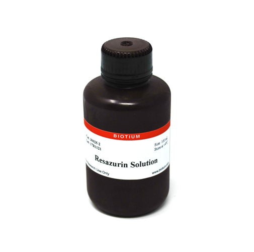
A sensitive, safe and cost-effective high-throughput homogeneous assay of cell viability by fluorescence or absorbance measurement.
Resazurin Cell Viability Assay offers a simple, rapid, reliable, sensitive, safe and cost-effective measurement of cell viability. This assay has excellent performance compared to other resazurin-based cell proliferation kits such as alamarBlue®, PrestoBlue®, or CellTiter-Blue®.
Resazurin detects cell metabolic by converting from a nonfluorescent dye to the highly red fluorescent dye resorufin in response to chemical reduction of growth medium resulting from cell growth. Continued cell growth maintains a reduced environment while inhibition of growth maintains an oxidized environment. Reduction related to growth causes the redox indicator to change from the oxidized form (nonfluorescent, blue) form to the reduced form (fluorescent, red).
Reduced resazurin can be detected using fluorescence (Ex/Em 530-560/590 nm) or absorbance at 570 nm. The fluorescent or colorimetric signal is proportional to the number of living cells in the sample. Results can obtained between 1-24 hours after adding substrate. Resazurin solution is supplied ready-to-use in 25 mL (2500 assay) or 100 mL (10,000 assay) sizes.
See our full selection of Cell Viability & Apoptosis Assays.
1. Arterioscler, Thromb, Vasc Biol (2010) 30(10), 1940–1948. DOI:10.1161/ATVBAHA.110.205997
2. J Clin Endocrinol Metab, October 2010, 95(10):E181–E191 doi: 10.1210/jc.2010-0581
3. Nucleic Acids Res (2010) 38(22), 8131–8140. doi:10.1093/nar/gkq697
4. Scand J Clin Lab Invest. (2010) 70(7), 512-8. doi: 10.3109/00365513.2010.521255
5. Tissue Eng A (2011) 17(1-2), 37-44. http://doi.org/10.1089/ten.tea.2010.0188
6. Biomaterials (2011) 32(1), 128-36. doi: 10.1016/j.biomaterials.2010.09.006.
7. Food Chem (2012) 135(3), 1533-1538. https://doi.org/10.1016/j.foodchem.2012.06.058
8. Drug Des Dev Therapy (2013) 7, 529–544. http://dx.doi.org/10.2147/DDDT.S45162
9. Langmuir (2013) 29(46), 14254-64. doi: 10.1021/la403533r
10. PLoS ONE (2013) 8(10), e76264. doi:10.1371/journal.pone.0076264
11. ACS Nano (2015) 9(2), 1219-1235. https://doi.org/10.1021/nn504890z
12. Antimicrob Agents Chemother (2016) 60(7), 4183–4196. doi:10.1128/AAC.03021-15
13. Curr Prot Toxicol (2016) 68, 2.24.1-2.24.15. doi: 10.1002/cptx.1
14. J Biol Chem (2016) 291, 21869-21879. doi: 10.1074/jbc.M115.712166
15. Curr Prot Stem Cell Biol (2018) 26(1), 5C.3.1-5C.3.19. DOI:10.1002/9780470151808.sc05c03s26
16. Int J Biol Macromol (2018) 113(1), 132-141. https://doi.org/10.1016/j.ijbiomac.2018.02.069
Medium with phenol red is compatible with the resazurin assay. Phenol red does not interfere with the resazurin reaction nor does it affect detection.
AlamarBlue® contains resazurin and additional compounds to prevent the over-reduction of resazurin to a non-fluorescent product. These additives also slow the rate of generation of the fluorescent product. Consequently, the alamarBlue® assay requires longer incubation times compared to resazurin.
Resazurin is reduced by cells to the fluorescent product resorufin. Resorufin can be reduced further to a non-fluorescent compound. Therefore the densest wells may have lowest fluorescence due to over-reduction of the substrate. Please see the product information sheet for more details. The kit protocol provides general guidelines and may need to be optimized empirically for your experimental system. You may need to vary cell density or assay incubation time to ensure that your samples fall in the linear range of the kit.
