Photoacoustic imaging (PAI) is a non-invasive, non-ionizing, high-resolution imaging technique which combines optical absorption / excitation (from endogenous chromophores such as hemoglobin or melanin, or exogenous contrast agents) with ultrasound detection. Due to the lower scattering of ultrasonic waves, PAI can provide high-resolution optical-contrast imaging, showing real promise as an alternative to MRI and CT scans.
With growing applications in areas of molecular imaging, oncology, ophthalmology, cardiology, and neurology, a recent publication characterizes a portable LED-based photoacoustic imaging system for commercial applications. By replacing lasers with multi-wavelength LEDs as the optical excitation source, the authors were able to make the imaging system compact, portable and affordable for broad clinical use. In vivo retinal vessel monitoring, as well as imaging of human mesenchymal stem cells labeled with a near-IR contrast agent (DiR) was demonstrated in living mice.
To read the original article, click here.
Hariri A, Lemaster J, Wang J, Jeevarathinam AS, Chao DL, Jokerst JV. The characterization of an economic and portable LED-based photoacoustic imaging system to facilitate molecular imaging. Photoacoustics. 2017 Nov 26;9:10-20. doi:10.1016/j.pacs.2017.11.001.
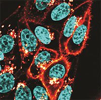
HeLa cells stained with Biotium’s near-IR cytoplasmic membrane dye CellBrite™ NIR680. Nuclei were counterstained with Hoechst 33342. Image acquired on a Zeiss LSM 700 confocal microscope.
Learn more about Biotium’s near-IR stains and CF® dye conjugates for in vivo imaging.