First reported in 2015, migrasomes are a recently discovered type of microscale extracellular vesicle (EV) formed at the rear of migrating cells. Like other types of EVs, are enriched in certain molecular cargo, such as growth factors, cytokines, and chemokines (Jiang, 2023). However, their size, production, and mechanisms of cargo packaging are distinct from other types of EVs. Reported functions of migrasomes include mediating early organ morphogenesis, disposal of damaged mitochondria, and transporting mRNA and proteins. Additionally, mounting evidence demonstrates that migrasomes mediate various pathological processes. Multipotent mesenchymal stromal cells (MSCs) are important precursor cells known to support tissue homeostasis through a variety of cell-cell interactions. In bone marrow, MSCs promote hematopoiesis, and their activity can enhance cancer progression. The full communication repertoire of MSCs remains to be discovered, and migrasomes from MSCs had not previously been studied, although their involvement would be in line with other MSC traits.
Deniz et al. elegantly demonstrate the production of migrasomes by MSCs isolated from human bone marrow in their 2023 Cell Communication and Signaling paper and go on to further show their role in intercellular communication and impact on hematopoietic cells. The team used a correlative approach using a panel of antibodies and cellular stains along with techniques including SEM, confocal, and TIRF microscopy to analyze the behavior of cell types, including primary human MSCs and hematopoietic stem and progenitor cells (HSPCs) under various scenarios. They chose Biotium’s CF®488A or CF®640R conjugated WGA to outline migrasomes in nearly all fluorescent microscopy experiments, most often in combination with anti-CD marker antibodies to characterize the composition of the migrasome surface and protein contents (see Figure ). Several striking videos demonstrate CD marker staining patterns in 3D or show time-lapse experiments demonstrating the effects of MSC-produced migrasomes on the migration of other cell types. Their data showed that MSC-produced migrasomes, which contained abundant MSC markers, most notably upregulated leukocyte cell adhesion molecules. Co-culture experiments with leukemic and hematopoietic progenitor cells then showed that MSC-associated migrasomes can determine the migration of other cell lines. While this initial data suggests a possible role for MSC-migrasome-based communication during the progression of certain types of cancer, further studies are needed to fully determine the biological aspects of migrasomes in the healthy and diseased bone marrow environment.
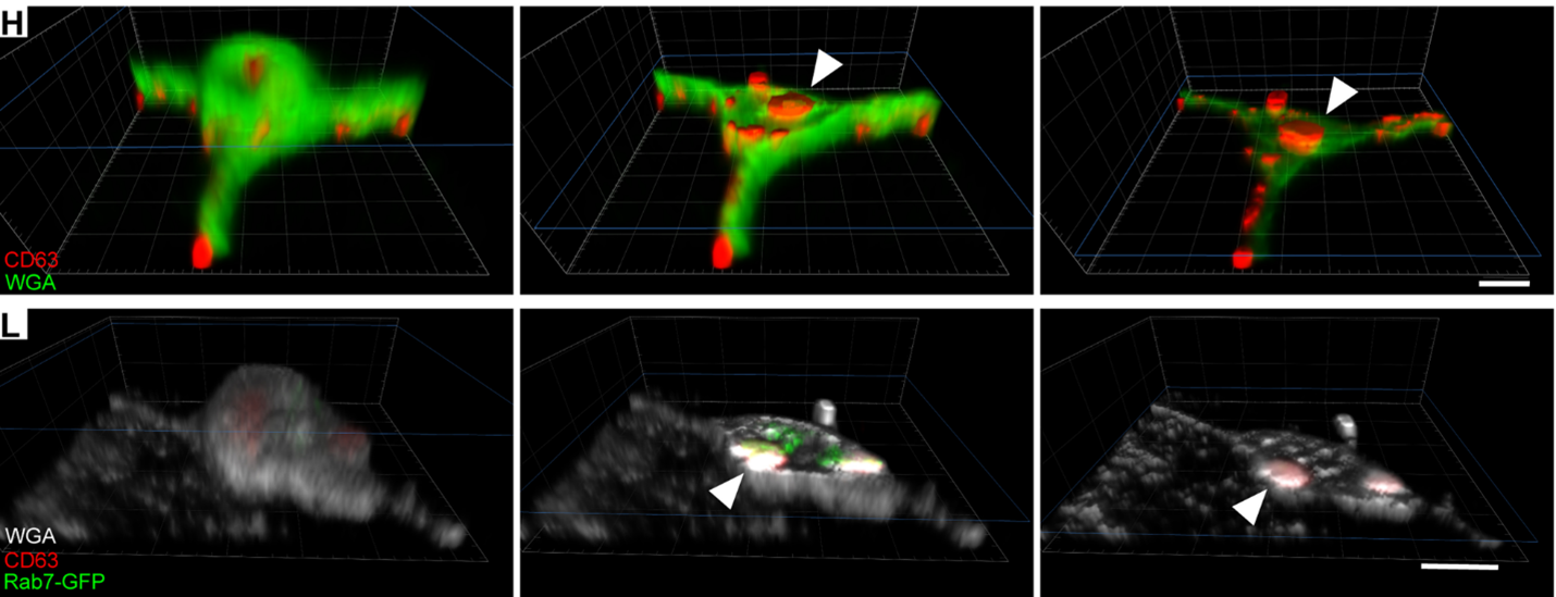
Figure: Migrasomes produced by MSCs exhibit distinctive cell surface marker profiles and transfer intracellular vesicles. Transiently transfected Rab7-GFP MSCs were immunolabeled for CD63 and stained with fluorophore-conjugated WGA. H) A 3D rendering of a migrasome from a PFA-fixed and saponin-permeabilized MSC cell immunolabeled for CD63 (red) and stained with Biotium’s CF® Dye-conjugated WGA (green). L) A 3D rendering of a migrasome from a PFA-fixed and saponin-permeabilized cell expressing Rab7-GFP (green), immunolabeled for CD63 (red) and stained with Biotium’s fluorophore-conjugated WGA (white). Slicing through the z-levels highlights the presence of cytoplasmic CD63 and the late endosomal marker Rab7 (L, arrowhead). Credit: Modified from Deniz, I.A. et al. https://doi.org/10.1186/s12964-022-01028-6 reproduced under the Creative Commons license (CC BY 4.0).
Videos:
Learn more about Biotium’s wide selection of cellular stains and other optimized solutions for your immunofluorescence microscopy workflow. We also carry a selection of products for extracellular vesicle (EV) research including three varieties of unique stains for EVs: ExoBrite™ CTB EV Staining Kits, ExoBrite™ Annexin EV Staining Kits, and ExoBrite™ WGA EV Staining Kits.
Full citation
Deniz, I. A., Karbanová, J., Wobus, M., Bornhäuser, M., Wimberger, P., Kuhlmann, J. D., & Corbeil, D. (2023). Mesenchymal stromal cell-associated migrasomes: a new source of chemoattractant for cells of hematopoietic origin. Cell Communication and Signaling, 21(1), 1-17. https://doi.org/10.1186/s12964-022-01028-6
References
Jiang, Y., Liu, X., Ye, J., Ma, Y., Mao, J., Feng, D., & Wang, X. (2023). Migrasomes, a new mode of intercellular communication. Cell Communication and Signaling, 21(1), 1-11.