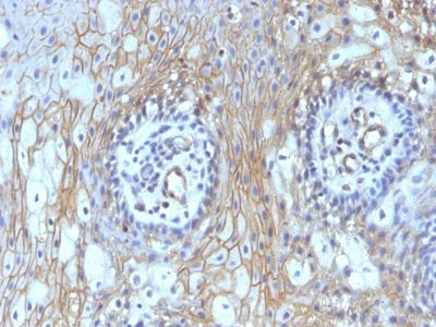
CD59 Monoclonal Mouse Antibody (BRA-10G)

Please fill in the inquiry form and we will contact you shortly.
Wishlist updated! View wishlist
 Powered by Bioz
Product Description
Reacts with human CD59, a 20 kDa glycosyl phosphatidyl-inositol (GPI)-anchored cell surface protein. CD59 regulates complement-mediated cell lysis, and it is involved in lymphocyte signal transduction. This protein is a potent inhibitor of the complement membrane attack complex, whereby it binds complement C8 and/or C9 during the assembly of this complex, thereby inhibiting the incorporation of multiple copies of C9 into the complex, which is necessary for osmolytic pore formation. CD59 is widely distributed on cells in all tissues. It inhibits formation of MAC, thus protecting cells from complement-mediated lysis. The expression of CD59 on erythrocytes is important for their survival. Genetic defects in GPI-anchor attachment, that cause a reduction or loss of CD59 and CD55 on erythrocytes produce the symptoms of the disease paroxysmal hemoglobinuria (PNH). This MAb is useful for study on GPI-anchored proteins, PNH and CD59 functions.
Primary antibodies are available purified, or with a selection of fluorescent CF® dyes and other labels. CF® dyes offer exceptional brightness and photostability. See the CF® Dye Brochure for more information. Note: Conjugates of blue fluorescent dyes like CF®405S and CF®405M are not recommended for detecting low abundance targets, because blue dyes have lower fluorescence and can give higher non-specific background than other dye colors.
Catalog number key for antibody number 0181, Anti-CD59 (BRA-10G)
| Antibody # prefix | Conjugation | Ex/Em (nm) | Laser line | Detection channel | Dye Features |
|---|---|---|---|---|---|
| BNC04 | CF®405S | 404/431 | 405 | DAPI (microscopy), AF405 | CF®405S Features |
| BNC88 | CF®488A | 490/515 | 488 | GFP, FITC | CF®488A Features |
| BNC68 | CF®568 | 562/583 | 532, 561 | RFP, TRITC | CF®568 Features |
| BNC94 | CF®594 | 593/614 | 561 | Texas Red® | CF®594 Features |
| BNC40 | CF®640R | 642/662 | 633-640 | Cy®5 | CF®640R Features |
| BNC47 | CF®647 | 650/665 | 633-640 | Cy®5 | CF®647 Features |
| BNC74 | CF®740 | 742/767 | 633-685 | 775/50 | CF®740 Features |
| BNCB | Biotin | N/A | N/A | N/A | |
| BNUB | Purified | N/A | N/A | N/A | |
| BNUM | Purified, BSA-free | N/A | N/A | N/A |
Note: Listed references are for this antibody clone sold by Biotium and other suppliers.