Menu
biotium logo
Antibody Finder
Menu
Hero Image

Membrane & Cell Surface Stains

Find the right dye for your application
Researchers frequently wish to label the cell surface or plasma membrane to visualize cellular boundaries or cell morphology in multi-color fluorescence imaging... See More

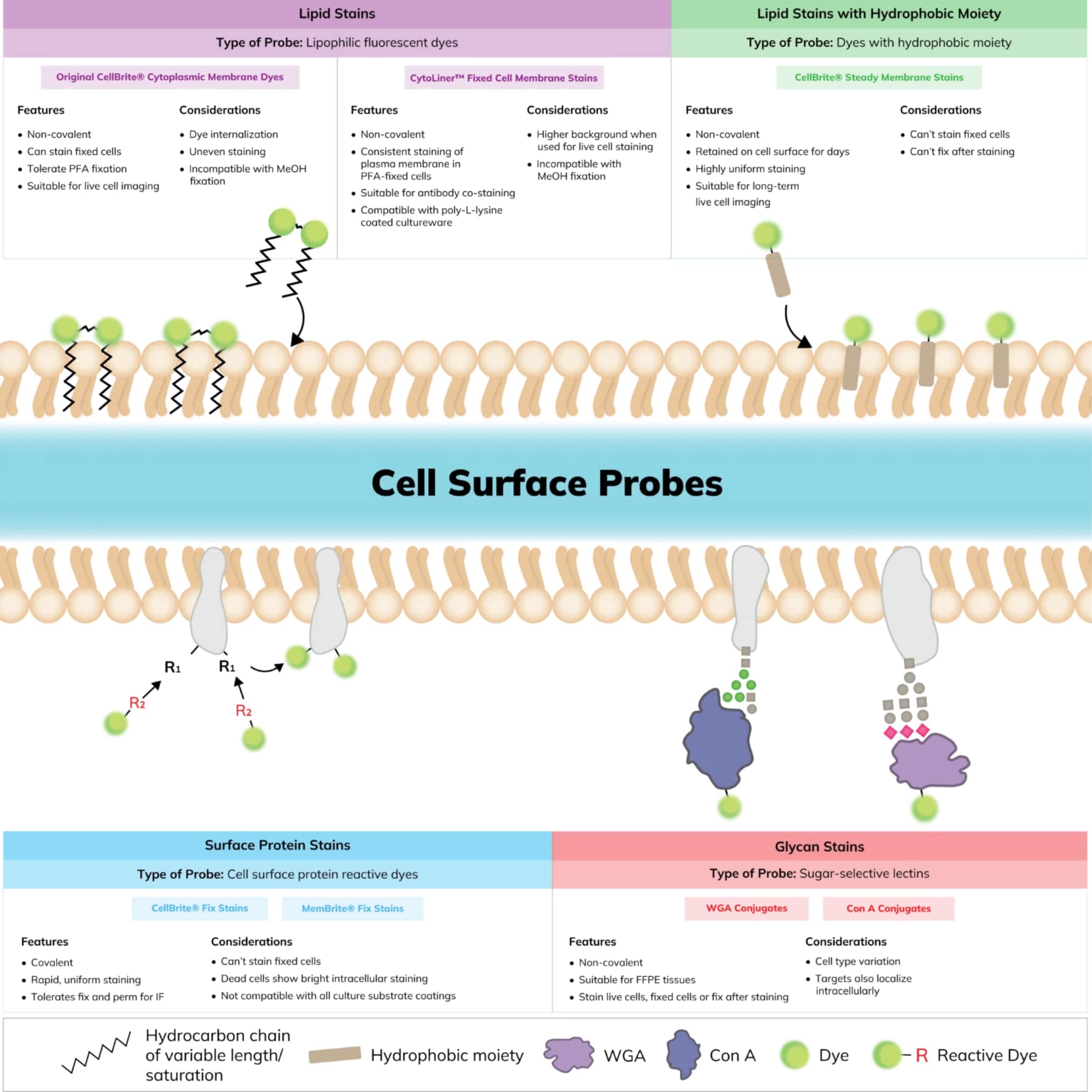
Find a Membrane or Surface Stain for Your Workflow

Researchers frequently use a plasma membrane dye to label cell surfaces or to visualize cellular boundaries or cell morphology in multi-color fluorescence imaging. Biotium offers a number of traditional and novel membrane dyes that are designed for convenient labeling of live cells, fixed cells, or for fixation and subsequent immunostaining. Many of our stains are available in numerous colors and can be paired with other cellular stains. See below to find the right stain for you based on your application and experimental details.

A. Stain live cells
  1. Stain live cells
  2. Wash1
  3. Image live cells

Recommended products:

  • CellBrite® Steady Membrane Stains1
  • 1Washing is optional for CellBrite® Steady Stains when imaged by confocal. The stains can be used to image cell surface in culture for days.
B. Stain then fix
  1. Stain live cells
  2. Fix cells
  3. (Optional) Proceed to additional staining
C. Stain, fix, & permeabilize
  1. Stian live cells
  2. Fix cells
  3. Permeabilize cells
  4. (Optional) Proceed to additional staining
D. Fix, then stain
  1. Fix cells
  2. Stain cells

Recommended products:

Compare Membrane and Cell Surface Stains

StainDye transfer
between cells
Non-toxicStains
yeast
Stains
bacteria
Stain after fixing2Fix after
staining
Tolerates detergent /
MeOH fix?
Color
selection
Pros & Cons
CytoLiner™ Fixed Cell Membrane StainsYesYes1YesNoYes3Yes5No66 colors
(blue to NIR)
Pro: Specifically formulated for bright & uniform membrane staining in fixed cells
Pro: Suitable for antibody co-staining
Con: High background in live cells
CellBrite® Steady
Membrane Staining Kits
YesYesNoNoNoNoNo8 colors
(blue to NIR)
Pro: Image live cell surface for days
Pro: Enhancer masks intracellular staining
Pro: Fast, even staining in medium
Con: Does not tolerate fixation
GlycoLiner™ Cell Surface Glycoprotein Labeling KitsMinimalYes1YesGram+ Yes
Gram- Weak
NoYesYes5 colors
(blue to NIR)
Pro: Rapid, uniform staining
Pro: Fix & perm cells for IF
Pro: Less cytoplasmic background in dead cells
Con: Can't stain fixed cells
CellBrite® Fix
Membrane Stains
MinimalYes1YesYesNoYesYesGreen,
red, &
far-red
Pro: Rapid, uniform staining
Pro: Wash after stain optional
Pro: Fix & perm cells for IF
Pro: Stains yeast & bacteria
Con: Can't stain fixed cells
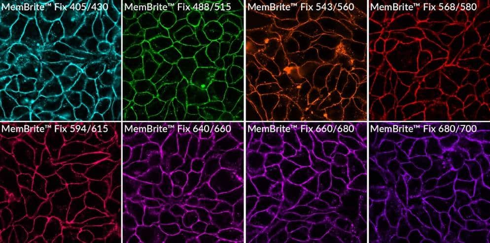
MemBrite® Fix
Cell Surface Staining Kits
MinimalYes1YesGram+
only
NoYesYes8 colors
(blue to NIR)
Pro: Rapid, uniform staining
Pro: Fix & perm cells for IF
Pro: Great color selection
Pro: Stains yeast
Con: Can't stain fixed cells
CF® Dye
WGA Conjugates
PossiblePossibly toxic4Bud scarsFluorescent
Gram stain
YesYesYes13 colors
(UV to NIR)
Pro: Fix before or after staining
Pro: Fluorescent Gram stain
Pro: Yeast bud scar stain
Pro: Great color selection
Con: May vary among cell types
CF® Dye
Con A Conjugates
PossiblePossibly toxic4YesReported to
stain biofilms
YesYesYes10 colors
(UV to NIR)
Pro: Fix before or after staining
Pro: Great color selection
Pro: Stains yeast
Con: May vary among cell types
CellBrite® Cytoplasmic
Membrane Dyes
MinimalYes1NoNoYes3Yes3No8 colors
(blue to NIR)
Pro: Fix before or after staining3
Con: Uneven live cell staining
Con: Poor tolerance for MeOH/detergent
1. Membrane and cell surface labels will be internalized by endocytosis in live cells over time.
2. Intracellular membranes may be labeled in fixed cells.
3. Formaldehyde fixation only.
4. Lectins may be toxic or stimulatory to live cells depending on cell type.
5. CytoLiner™ Fixed Cell Membrane Stains may have higher background if used to stain live cells before fixation than when used to stain cells after fixation.
6. Mild permeabilization with 0.1% Triton® X-100 is recommended, see product information sheet for more info.

Which Membrane Stain is Right for Your Experiment?

Watch our video where Technical Applications Scientist II, Jacqueline Steenhuis PhD answers your top questions about Biotium’s various membrane stains for fluorescence microscopy.

For additional support or product recommendations, contact us at techsupport@biotium.com.

Live MCF7 cells were stained with CellBrite® Steady 650 plus Enhancer in complete culture medium for 48 hours and imaged by confocal microscopy without a wash s... See More
NucSpot® 470 Nuclear Stain, 1000X in DMSO
HeLa cells stained with CellBrite® Fix 555 (red), then MeOH-fixed and stained with CF®640R Mitochondrial Marker mAb clone 113-1 (cyan) and NucSpot® 470 (green).... See More
S. cerevisiae were stained separately with MemBrite® Fix 488/515 (green), MemBrite® Fix 568/580 (red), and MemBrite® Fix 640/660 (magenta) then mixed tog... See More
Live HeLa cells stained with CF® 594 Con A (red) and Hoechst 33342 (blue) for 10 minutes at 37°C, then washed and imaged.
MemBrite® Fix labels cells more evenly than traditional membrane stains, and tolerates permeabilization. HeLa cells were labeled with DiI or MemBrite® Fix 568/5... See More
MemBrite® Fix kits offer a wide selection of dye colors, with excellent tolerance for common fixatives like PFA and methanol. Cells were labeled with the indica... See More

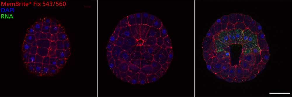
Stain Cell Surfaces in Whole Organisms

MemBrite® Fix stains have been used to label cell surfaces in whole organisms. In the following figures provided by Patrick Lemaire’s lab of CRBM (Centre de Recherche en Biologie cellulaire de Montepellier), MemBrite® Fix 543/560 was used to stain whole embryos of the tunicate Phallusia mammillata followed by downstream hybridization chain reaction fluorescent in situ hybridization (HCR-FISH) for detection of a RNA transcript. Cell boundaries were clearly labeled through the entirety of the Z-stack from the animal hemisphere to the vegetal hemisphere of the embryo.

Embryos of the tunicate Phallusia mammillata were stained using MemBrite® Fix 543/560 (red) at stage 12: mid gastrula, followed by downstream HCR-FISH.... See More
Embryo of the tunicate Phallusia mammillata stained with MemBrite® Fix 543/560 (red) at stage 12: mid gastrula and mounted with DAPI (blue). Images pro... See More

FAQs

CellBrite® Cytoplasmic Membrane Stains are lipophilic dyes for simple, non-toxic, stable labeling of membranes in live or fixed cells. Cells can be fixed with formaldehyde before or after CellBrite® staining. But the staining has poor tolerance for permeabilization or methanol fixation, so CellBrite® staining is not easily combined with intracellular immunofluorescence (IF) staining. The dyes also do not stain bacteria or yeast. CellBrite® NIR Dyes are CellBrite® Dyes with near-infrared fluorescence compatible with small animal NIR imaging systems.

CellBrite® Fix and MemBrite® Fix stains were developed to overcome some of these shortcomings. They are novel covalent stains that can be fixed and permeabilized for IF staining. CellBrite® Fix Membrane Stains are fluorogenic reactive membrane dyes that rapidly accumulate at the plasma membrane. When they incorporate into lipids, they become fluorescent, and at the same time react covalently with membrane proteins for stable labeling. Staining takes only 15 minutes in a single step with no wash. CellBrite® Fix stains mammalian cells, yeast, and bacteria. CellBrite® Fix reacts with plates coated with poly-L-lysine, collagen, gelatin, or other proteins, resulting in high background. The dyes tend to have high background on uncoated cell culture surfaces as well. Imaging cells by confocal microscopy can reduce interference from out-of-plane background fluorescence.

MemBrite® Fix Cell Surface Stains do not bind lipids, but label cell surface proteins. MemBrite® Fix requires a two-step staining protocol with washing, but offers a more extensive choice of dye colors than CellBrite® Fix. MemBrite® Fix also can be used to stain yeast and Gram-positive bacteria. But unlike original CellBrite® Dyes and lectins, CellBrite® Fix and MemBrite® Fix cannot be used on cells that are already fixed. While we do not expect MemBrite® Fix stains to react with poly-L-lysine coated surfaces, we have seen high background with these types of plates and with uncoated cell culture surfaces. To circumvent this issue, we recommend imaging cells by confocal microscopy to reduce out-of-plane background fluorescence. The stains will react with surfaces treated with collagen, gelatin, fibronectin, or other extracellular matrix protein coatings.

GlycoLiner™ Cell Surface Glycoprotein Labeling Kits are designed for covalent labeling of glycoproteins on the surface of live cells. Like CellBrite® Fix and MemBrite® Fix, staining can withstand fixation and permeabilization. GlycoLiner™ also offers less cytoplasmic background in dead cells than CellBrite® Fix and MemBrite® Fix, allowing easier imaging of cell surfaces. GlycoLiner™ can also be used to stain yeast and Gram-positive bacteria. GlycoLiner™ is not expected to react with poly-L-lysine-coated surfaces, however, glycosylated matrix components like collagen or proteoglycans in complex matrices like Matrigel® may be labeled.

CytoLiner™ Fixed Cell Membrane Stains are novel lipophilic fluorescent dyes developed for selective plasma membrane staining in fixed cells and are suitable for downstream immunofluorescence staining protocols. Please note, staining with CytoLiner™ Dyes is not compatible with cells fixed using solvents like methanol or acetone, or with paraffin-embedded samples, because these treatments will remove the lipids from cells, which are required for CytoLiner™ staining. For co-staining with antibodies, we recommend staining with CytoLiner™ Dye first, then blocking with 2% fish gelatin in PBS, followed by antibody incubation in the same buffer. Blocking with BSA or serum is not recommended. CytoLiner™ staining is compatible with poly-L-lysine coated culture surfaces and Transwell® permeable supports. For best results, confocal microscopy is recommended for imaging fluorescent staining of Transwell® supports to avoid background from the filter material.

To select a dye that’s right for your application, see our Membrane and Cell Surface Stains Comparison, or download our Membrane & Surface Stains Brochure.

CellBrite® Cytoplasmic Membrane Dyes are too prone to aggregation to efficiently stain EVs. Some of the CellBrite® Fix, MemBrite® Fix, and CellBrite® Steady dye options have been reported for this application, however we do not recommend them. For optimal staining of exosome membranes we recommend our ExoBrite™ True EV Membrane Stains, which are novel lipophilic membrane dyes specifically designed and optimized for efficient staining of EV membranes with minimal dye aggregation. See our Extracellular Vesicle Research page for more information about our complete line of EV stains and antibodies.

Biotium’s CellBrite® Cytoplasmic Membrane Dyes are dye delivery solutions that can be added directly to normal culture media to uniformly label suspended or adherent cells in culture. The PKH dyes are structurally related dyes for cell membrane labeling. But unlike CellBrite®, labeling with PKH dyes requires multiple steps and subjects cells to an iso-osmotic mannitol loading medium that can negatively affect cell membrane integrity and viability.

CellBrite® Fix dyes also feature rapid and simple labeling in isotonic buffer. Cells can be fixed and permeabilized after labeling with CellBrite® Fix, unlike original CellBrite® dyes or PKH dyes, which don’t tolerate detergent.

View more FAQs
Can't find your answer?
Talk to an Expert