NucView® Caspase-3 Enzyme Substrates
Fluorescent caspase-3/7 substrates for detecting apoptosis in intact cells by confocal microscopy, flow cytometry, or live cell imaging.


Please fill in the inquiry form and we will contact you shortly.
Wishlist updated! View wishlist
 Powered by Bioz
Product Description
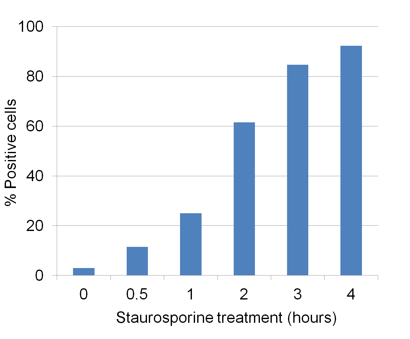
NucView® Caspase-3 Substrates are a convenient tool for detecting apoptosis in intact cells based on caspase-3/7 activity using confocal microscopy, flow cytometry, or live cell imaging.
- Rapid, no-wash, endpoint or real-time assays
- Non-toxic, allowing multi-day experiments to be performed
- Available in green, blue, or orange fluorescence
- For flow cytometry, microscopy or live cell imaging systems
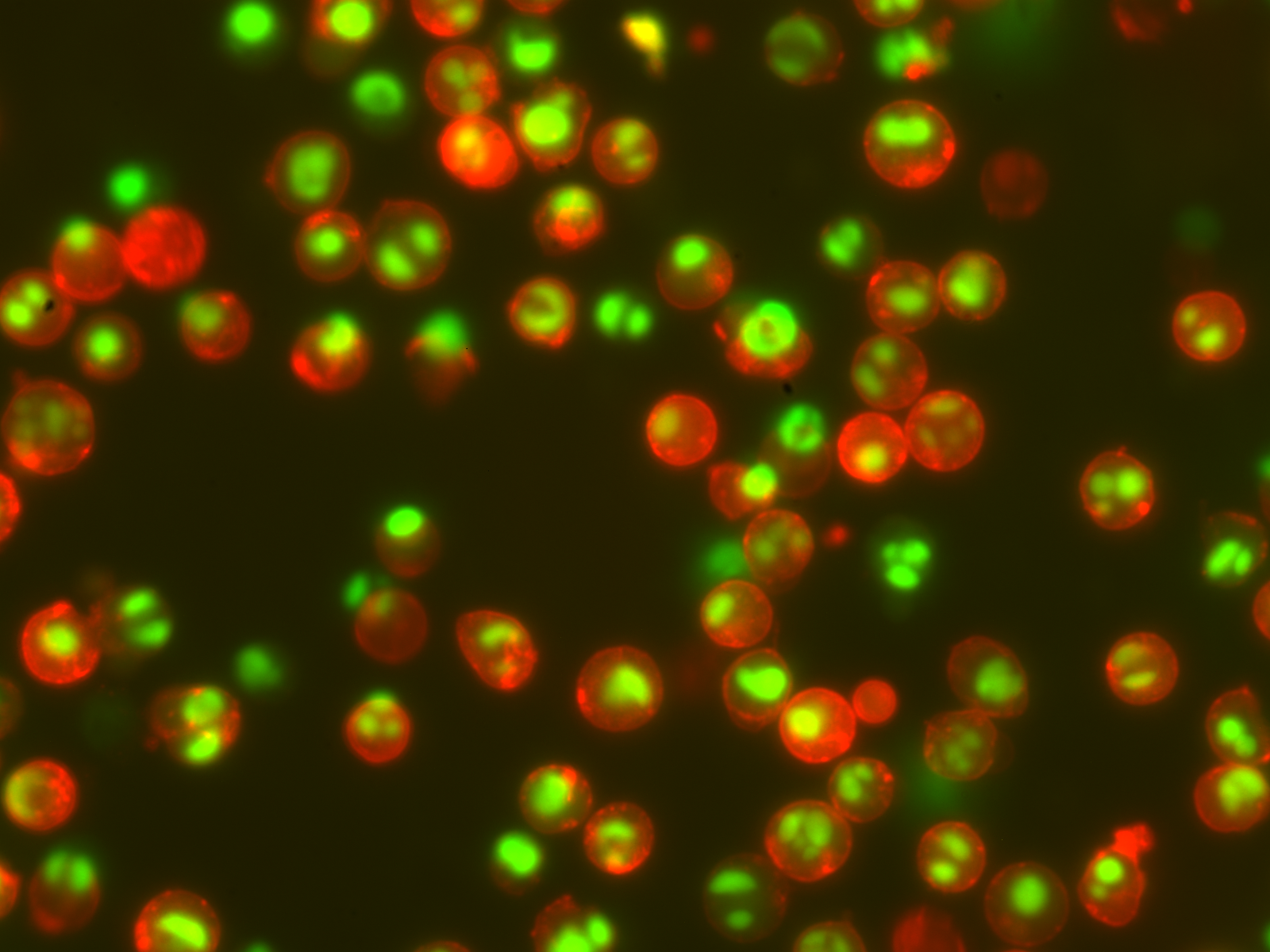
- Dual detection of caspase activity and nuclear morphology
- Formaldehyde fixable
About NucView®
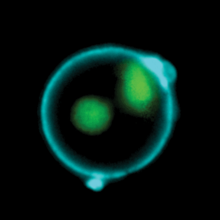
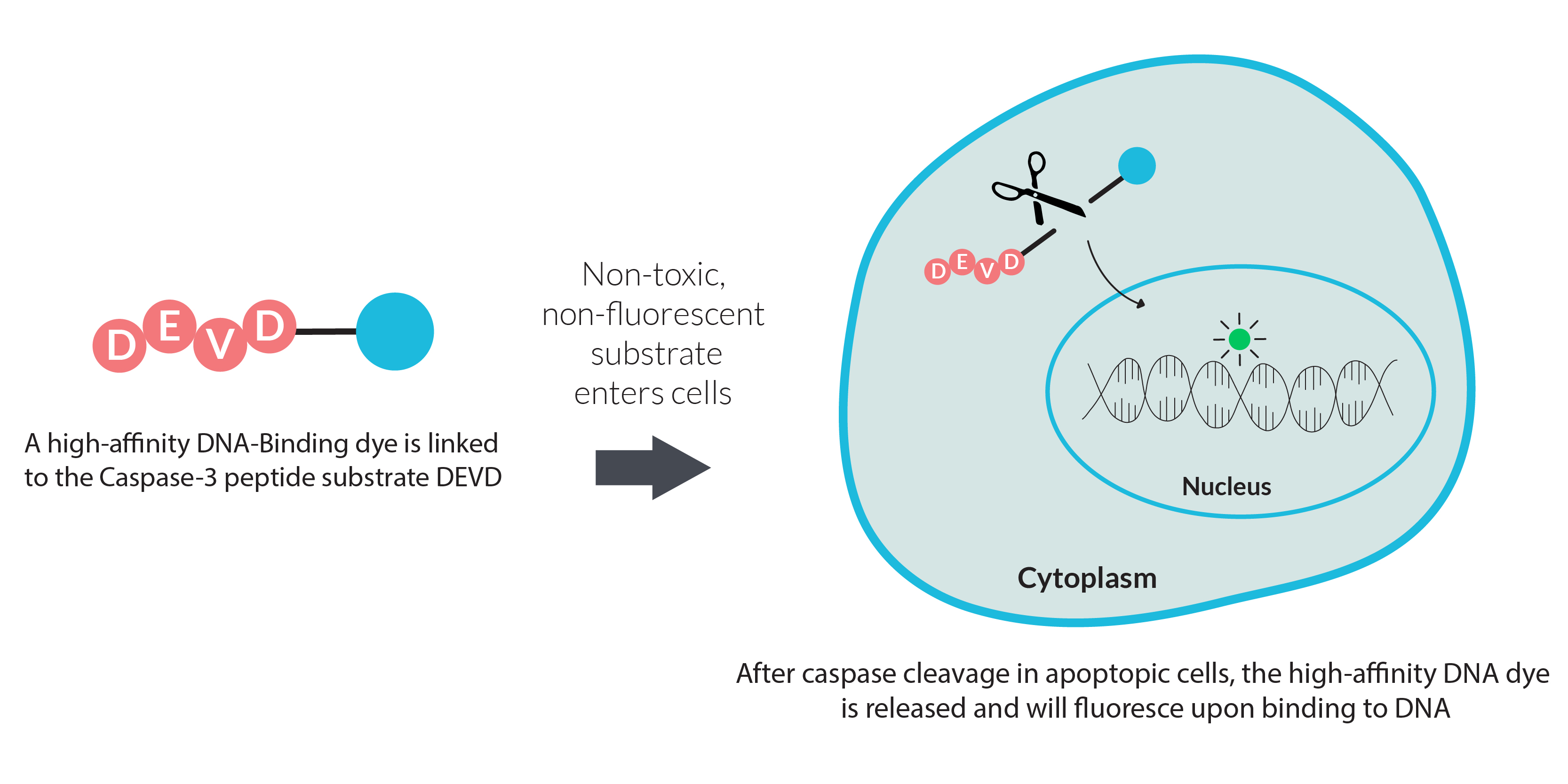
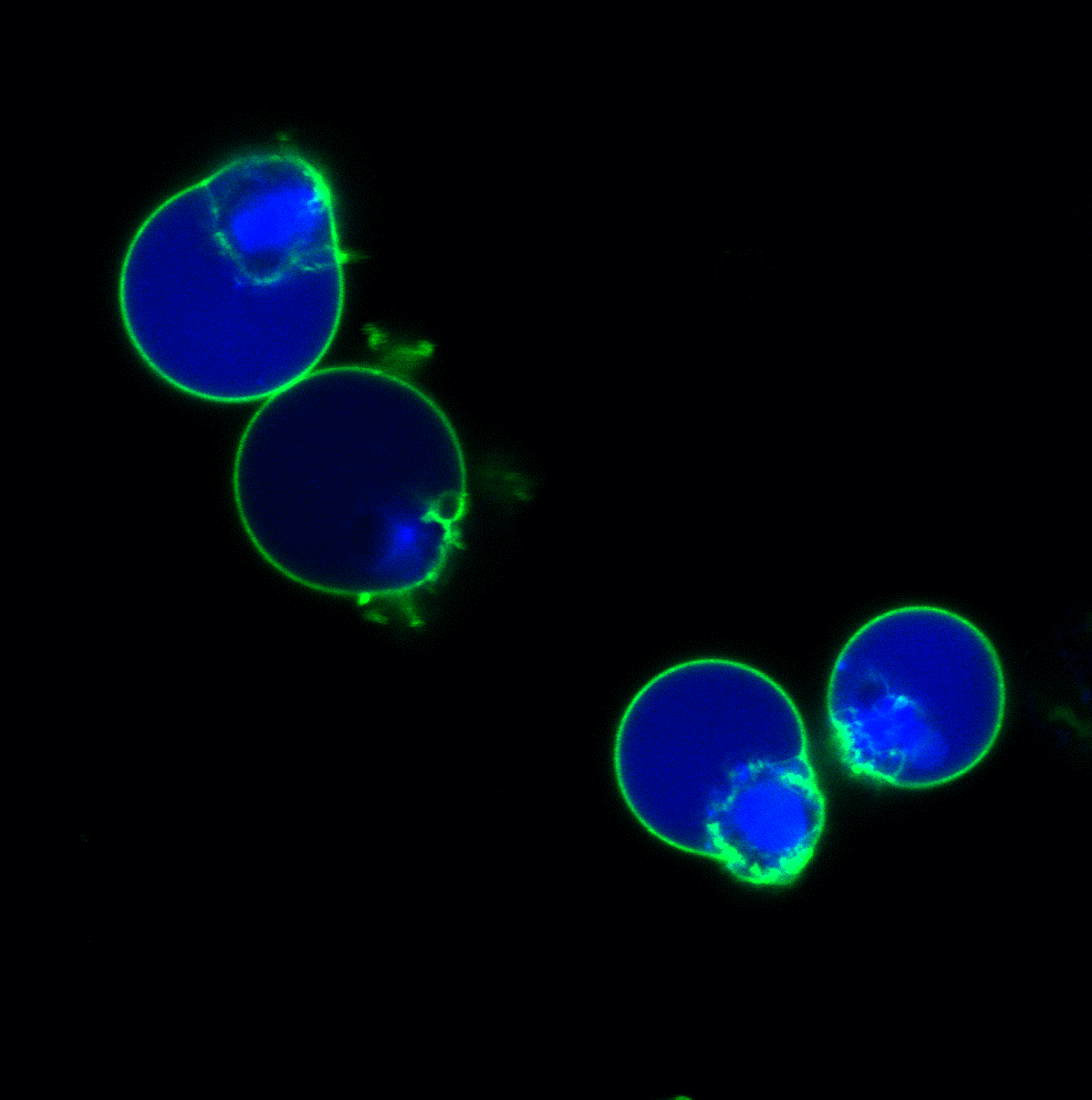
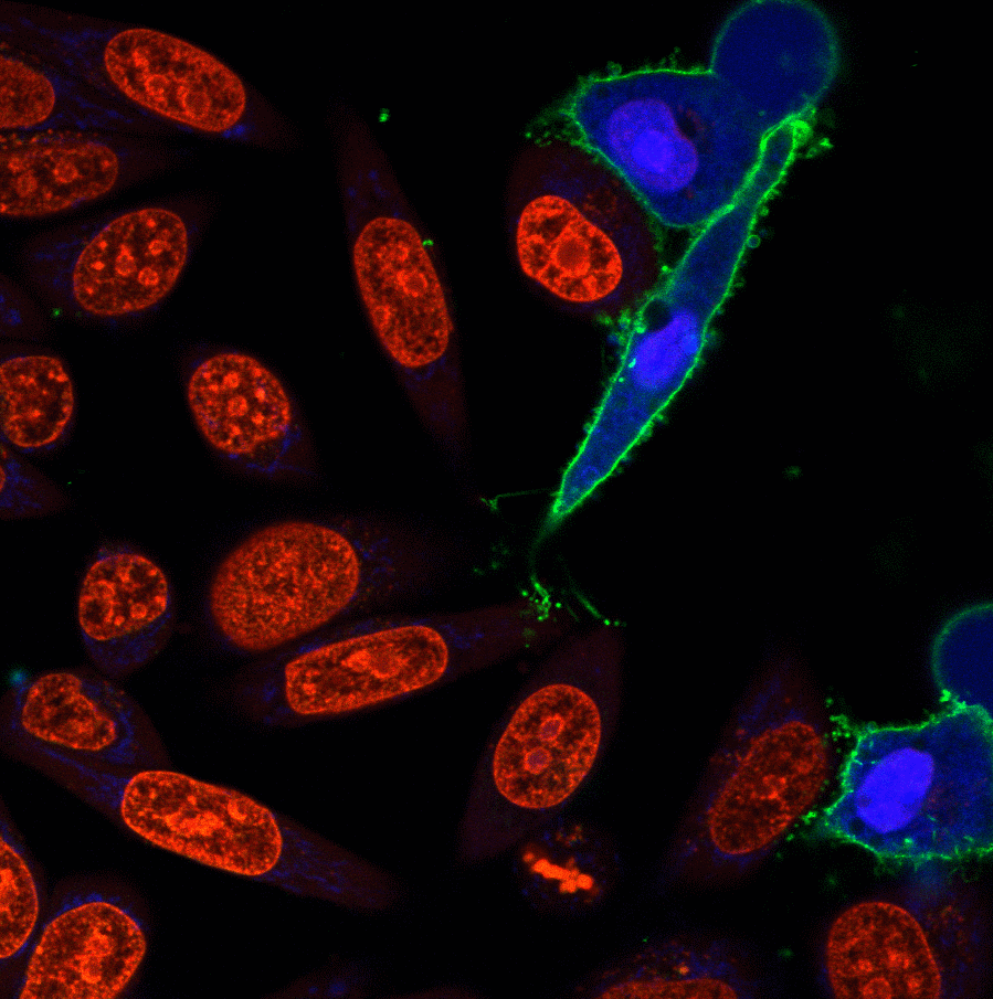
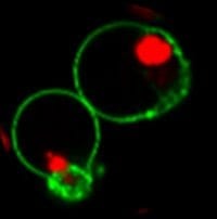
NucView® substrates consist of a fluorogenic DNA dye coupled to the caspase-3/7 DEVD recognition sequence. The substrate, which is initially non-fluorescent, penetrates the plasma membrane and enters the cytoplasm. In apoptotic cells, caspase-3/7 cleaves the substrate, releasing the high-affinity DNA dye, which migrates to the cell nucleus and stains DNA with fluorescence. Thus, NucView caspase-3 substrates are bifunctional, allowing detection of caspase-3/7 activity and visualization of morphological changes in the nucleus during apoptosis.
In contrast to other fluorogenic caspase substrates or fluorescent caspase inhibitor based (FLICA) assays, NucView® caspase-3 substrates can be used to detect caspase-3/7 activity within individual intact cells without inhibiting apoptosis progression. Staining is compatible with subsequent fixation and permeabilization for immunostaining.
NucView® substrates are offered as solutions in DMSO or phosphate-buffered saline (PBS). The substrates in PBS are formulated for use in cells that are sensitive to DMSO toxicity. In non-DMSO sensitive cell types, adding DMSO during the substrate incubation may enhance NucView® staining.
To learn about the advantages of monitoring apoptosis using NucView® caspase-3 substrates, visit the NucView® Technology Page.
NucView® Caspase-3 Substrates and Kits | Catalog No. | Features |
|---|---|---|
| NucView® 405 Caspase-3 Substrate, 1 mM in DMSO | 10405 | Blue fluorescence for flow cytometry in the Pacific Blue® channel or microscopy with the 405 nm laser |
| NucView® 405 Caspase-3 Substrate, 1 mM in PBS | 10407 | NucView® 405 substrate in PBS, for DMSO-sensitive cell types |
| NucView® 488 Caspase-3 Substrate, 1 mM in DMSO | 10402 | Green fluorescent substrate validated in more than 100 cell types and 200 publications |
| NucView® 488 Caspase-3 Substrate, 1 mM in PBS | 10403 | NucView® 488 substrate in PBS, for DMSO-sensitive cell types |
| NucView® 530 Caspase-3 Substrate, 1 mM in DMSO | 10406 | Orange fluorescence for microscopy in the Cy®3 channel or flow cytometry in the R-PE channel |
| NucView® 530 Caspase-3 Substrate, 1 mM in PBS | 10408 | NucView® 530 substrate in PBS, for DMSO-sensitive cell types |
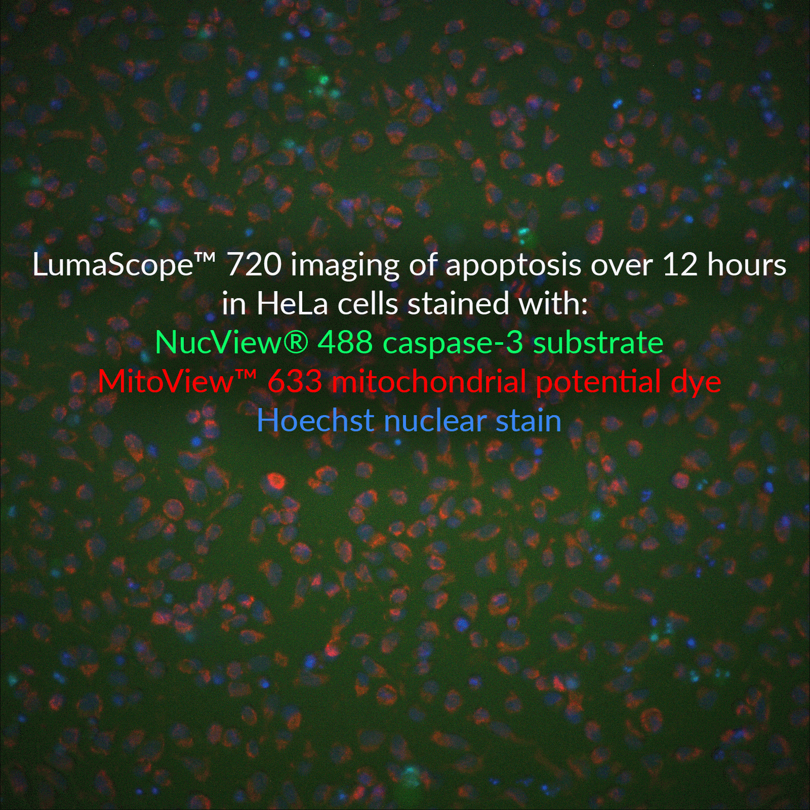
| NucView® 488 and MitoView™ 633 Apotosis Detection Kit | 30062 | NucView® 488 and far-red fluorescent MitoView™ 633 for the Cy®5 channel |
| NucView® 488 and RedDot™ 2 Apoptosis & Necrosis Kit | 30072 | NucView® 488 and far-red dead cell DNA dye RedDot™ 2 for the Cy®5 channel |
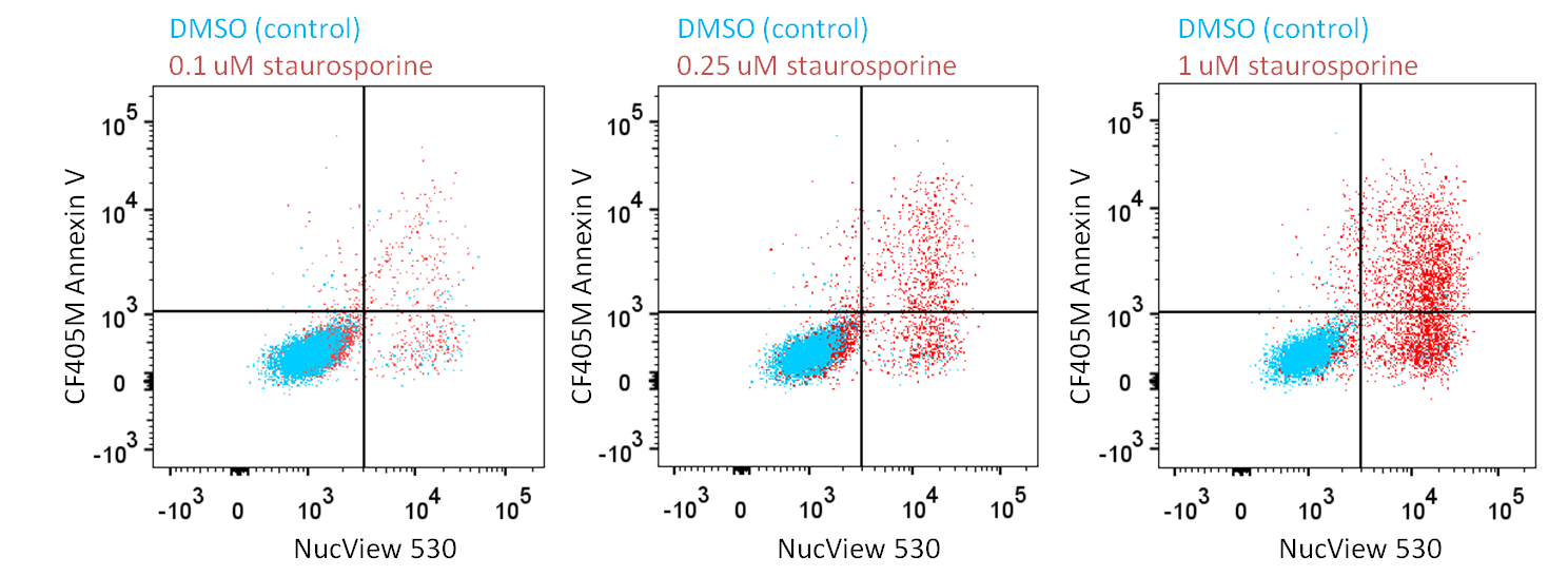
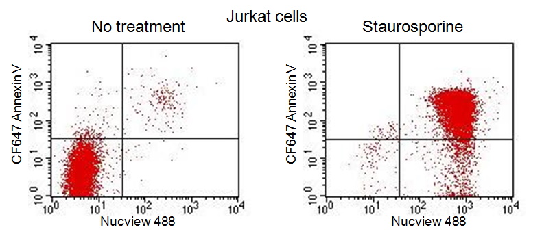
| Dual Apoptosis Assay with NucView® 488 and CF®594 Annexin V | 30067 | NucView® 488 and red fluorescent Annexin V apoptosis probe |
| Dual Apoptosis Assay with NucView® 488 and CF®640R Annexin V | 30073 | NucView® 488 and far-red fluorescent Annexin V apoptosis probe |
Choose from Blue, Green, or Orange Fluorescence
NucView® 488
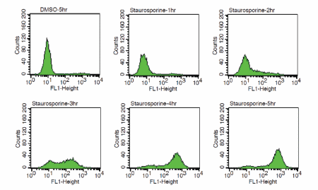
NucView® 488 Caspase-3 Substrate stains apoptotic cell nuclei with green fluorescence, for detection in the FITC channel in fluorescence microscopy or flow cytometry. The dye is excited by the 488 nm laser line. NucView® 488 can be used for multi-color imaging with blue and far-red fluorescent probes. NucView® 488 is the original NucView® probe, and has been validated in over 200 publications and more than 100 cell types.
NucView® 405
NucView® 405 Caspase-3 Substrate stains apoptotic cell nuclei with blue fluorescence, for detection in the DAPI channel by confocal microscopy, or by flow cytometry in the Pacific Blue® channel. The dye is excited by the 405 nm laser line. NucView® 405 is ideal for caspase-3 detection in multi-color applications for researchers who wish to reserve the green fluorescence channel for other detection reagents.
Note: NucView® 405 signal may be difficult to observe using epifluorescence microscopy. Confocal microscopy using 405 nm laser excitation is recommended for imaging.
NucView® 530
NucView® 530 Caspase-3 Substrate stains apoptotic cell nuclei with orange fluorescence, for detection in the Cy®3 channel by fluorescence microscopy, or the PE channel by flow cytometry. The dye is excited by the 488 nm laser line. NucView® 530 can be used for multi-color imaging with blue, green, or far-red fluorescent probes.
Note: When excited by the 488 nm laser line, NucView® 530 also fluoresces in the FITC channel, and therefore cannot be analyzed together with green probes by flow cytometry.
References
Download list of curated NucView® references and validated cell lines.