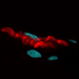
The study of skeletal muscle structure and function typically relies on invasive in vitro and ex vivo investigations in mammalian models. In a recent article in IntraVital, Mercier et al. used our green fluorescent CF™488A-conjugated α-bungarotoxin to demonstrate a novel, non-invasive method for analysis of murine intracellular skeletal muscle organization. By using a combination of two-photon excitation-microscopy and second harmonic generation, researchers simultaneously detected blood vessels stained with Evans Blue, neuromuscular junctions stained with CF™488 α-bungarotoxin, as well as sarcomeres and collagen fibers, and were able to reconstruct the 3D architecture of the neuromuscular system at an intracellular resolution up to 150 micrometers. These methods could facilitate advances in our understanding of neuromuscular disease.
To read the original article, click here.
Mercier L, et al. In vivo imaging of skeletal muscle in mice highlights muscle defects in a model of myotubular myopathy. IntraVital. 2016 Apr 6;5(1):e1168553. doi: 10.1080/21659087.2016.1168553.
Biotium has an extensive list of reactive CF™ dyes and dye conjugates spanning the visible and near-infrared spectrum for labeling biomolecules, including CF™488A and CF™594, which have been utilized for 2-photon microscopy. For more information on our CF™ dyes, click here.