 New Products
 Earth-Friendly Products
 Biotium Choice Antibodies
 Special Offers


Content #1
Content #1
Content #1
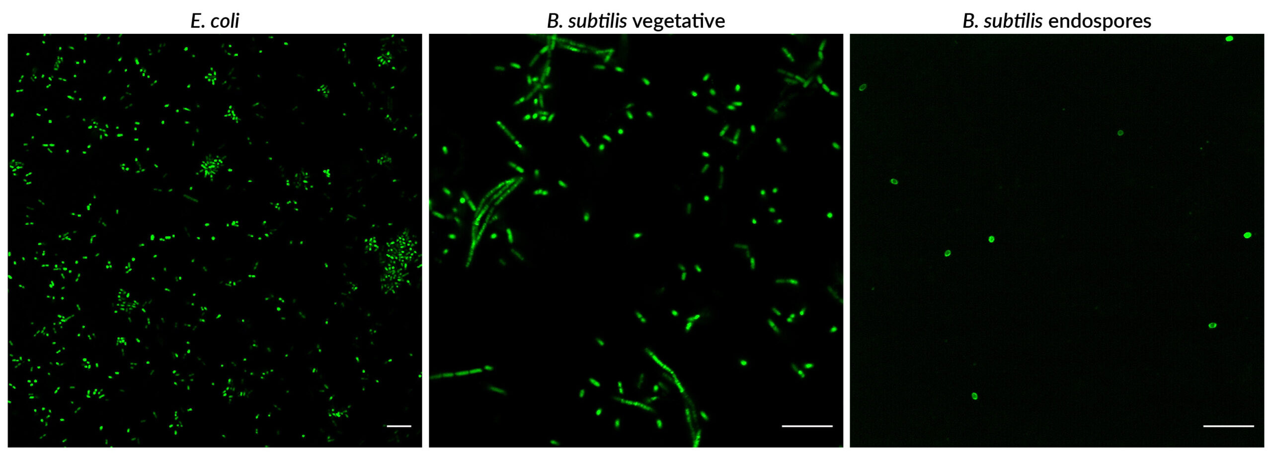
Fluorescent dyes optimized for staining endospores. The dyes also stain both live and dead bacteria of gram-positive and gram-negative strains. In B. subtilis, the dyes stain both vegetative cells and endospores and have been validated for detection by fluorescence microscopy and flow cytometry.
BactoSpore™ Stains are fluorescent dyes optimized for staining endospores. The dyes also stain both live and dead bacteria of gram-positive and gram-negative strains. In B. subtilis, the dyes stain both vegetative cells and endospores and have been validated for detection by fluorescence microscopy and flow cytometry.
Bacterial endospores are tough dormant structures formed by certain strains of bacteria including Bacillus and Clostridium species in response to nutrient deprivation and other stressors. Endospore formation allows these bacteria to survive in a non-replicative state until growth conditions improve, at which point the spores can germinate to allow vegetative cell replication. Endospores provide a reservoir of potentially infectious bacteria that are resistant to disinfectants, heat, and other decontamination treatments. In addition, the spore coat is highly impermeant and resistant to staining with bacterial detection reagents, making it difficult to study endospore formation and inactivation.
BactoSpore™ stains were developed to tackle the challenge of endospore detection by offering bright staining of endospores as well as live and dead bacteria. BactoSpore™ 485/500 Membrane Stain is a green fluorescent lipophilic membrane dye for the FITC channel. BactoSpore™ 488/540 Nucleic Acid Stain is a yellow fluorescent nucleic acid dye that is detectable in both the FITC channel and the PE channel for flow cytometry.
| Product Name | Ex/Em (nm) | Detection Channel | Size | Catalog No. |
|---|---|---|---|---|
| BactoSpore™ 485/500 Membrane Stain, 500X in EtOH | 484/504 | FITC | 20 uL | 40119-T |
| 100 uL | 40119 | |||
| BactoSpore™ 488/540 Nucleic Acid Stain, 500X in DMSO | 488/536 (with DNA) | FITC or PE Channel | 20 uL | 40120-T |
| 100 uL | 40120 |
BactoSpore™ 485/500 Membrane Stain
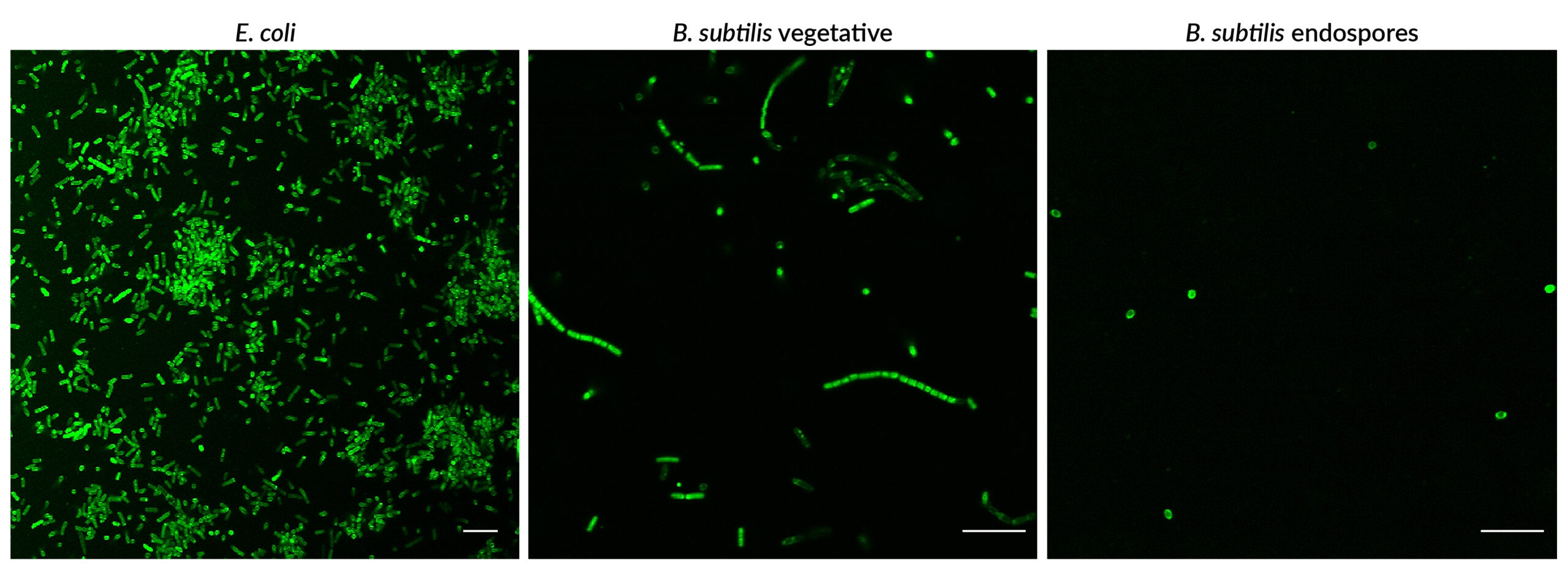
BactoSpore™ 485/500 Membrane Stain labels gram-negative bacteria, gram-positive bacteria, and endospores. E. coli or B. subtilis vegetative cultures were grown in tryptic soy broth (TSB) overnight. B. subtilis were induced to form endospores by growing cultures in Difco sporulation medium (DSM) for 7 days. Cultures were washed in PBS and stained with 1X BactoSpore™ 485/500 Membrane Stain in PBS for 30 minutes at room temperature, then imaged by confocal microscopy in the FITC channel. Scale bars: 10 um.
BactoSpore™ 488/540 Nucleic Acid Stain
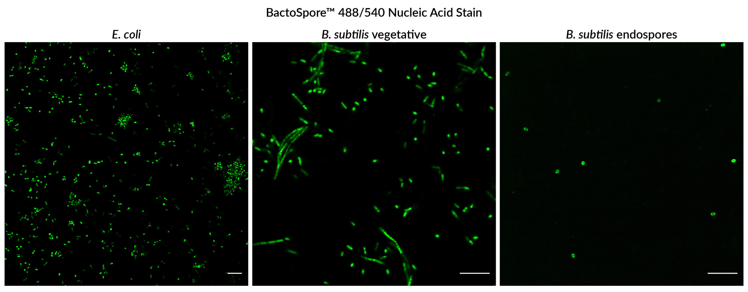
BactoSpore™ 488/540 Nucleic Acid Stain labels gram-negative bacteria, gram-positive bacteria, and endospores. E. coli or B. subtilis vegetative cultures were grown in tryptic soy broth (TSB) overnight. B. subtilis were induced to form endospores by growing cultures in Difco sporulation medium (DSM) for 7 days. Cultures were washed in PBS and stained with 1X BactoSpore™ 488/540 Nucleic Acid Stain in PBS for 30 minutes at room temperature, then imaged by confocal microscopy in the FITC channel. Scale bars: 10 um.
View Biotium's full selection of microbiology stains, including BactoView™ Live and BactoView™ Dead stains for staining live and dead bacteria, respectively. Biotium also offers BactoView™ Viability Kits, which include a choice of red or far-red BactoView™ Dead Stain for dead bacteria and BactoView™ Viability Green Counterstain to stain all bacteria. BactoView™ Dead Stains also can be combined with fluorescent Gram stains like our CF® Dye WGA Conjugates.
To date, we have not identified a fluorescent cellular stain that will detect bacteria but not mammalian cells with high specificity, or vice versa. While some mammalian cell stains show weak staining of bacteria, they usually do show some signal, and will frequently stain dead bacteria more intensely than live bacteria.
We offer a selection of antibodies for specific bacterial antigens, which potentially have applications for differential staining of bacteria vs. mammalian cells, but we have not validated them in co-culture models.
Also see our Viability PCR Technology Page to learn about how PMA dye can be used for highly specific detection of microbial cell viability in complex samples.
CellBrite® and MemBrite® Stains were originally developed for staining mammalian cells in culture, but some of the stains also have been validated for other organisms and applications. For dyes to stain yeast or bacteria membranes, see Cellular Stains in Different Organisms. For information on staining other organisms or cell types, please see our Tech Tip: Researching Applications for Membrane Dyes.
The CellBrite® Cytoplasmic Membrane Dyes do not stain bacteria. The reactive CellBrite® Fix dyes stain both gram-positive and gram-negative bacteria, while the MemBrite® Fix dyes stain only gram-positive bacteria. However we have not tested these dyes for cell division tracking in bacteria.
There is literature describing the use of CFSE to track bacterial cell division, the ViaFluor® SE cell proliferation dyes are likely to work in a similar manner, but we have not tested this.
See our Cellular Stains Table for a comprehensive list of cellular stains with their ability to stain various cell types.
