 New Products
 Earth-Friendly Products
 Biotium Choice Antibodies
 Special Offers


Content #1
Content #1
Content #1
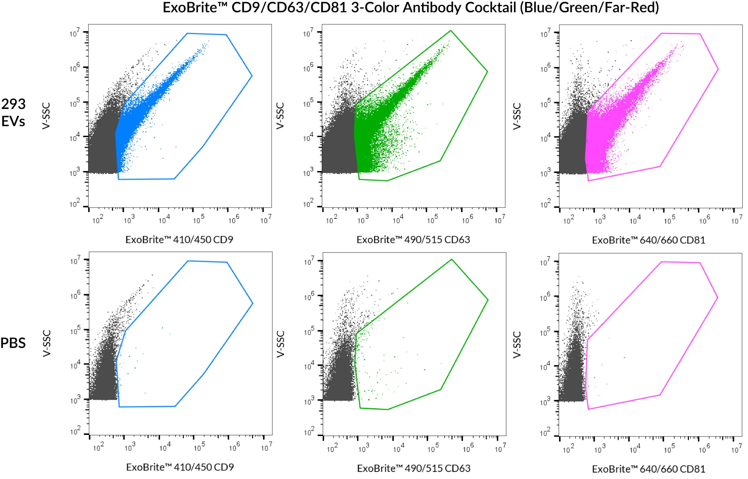
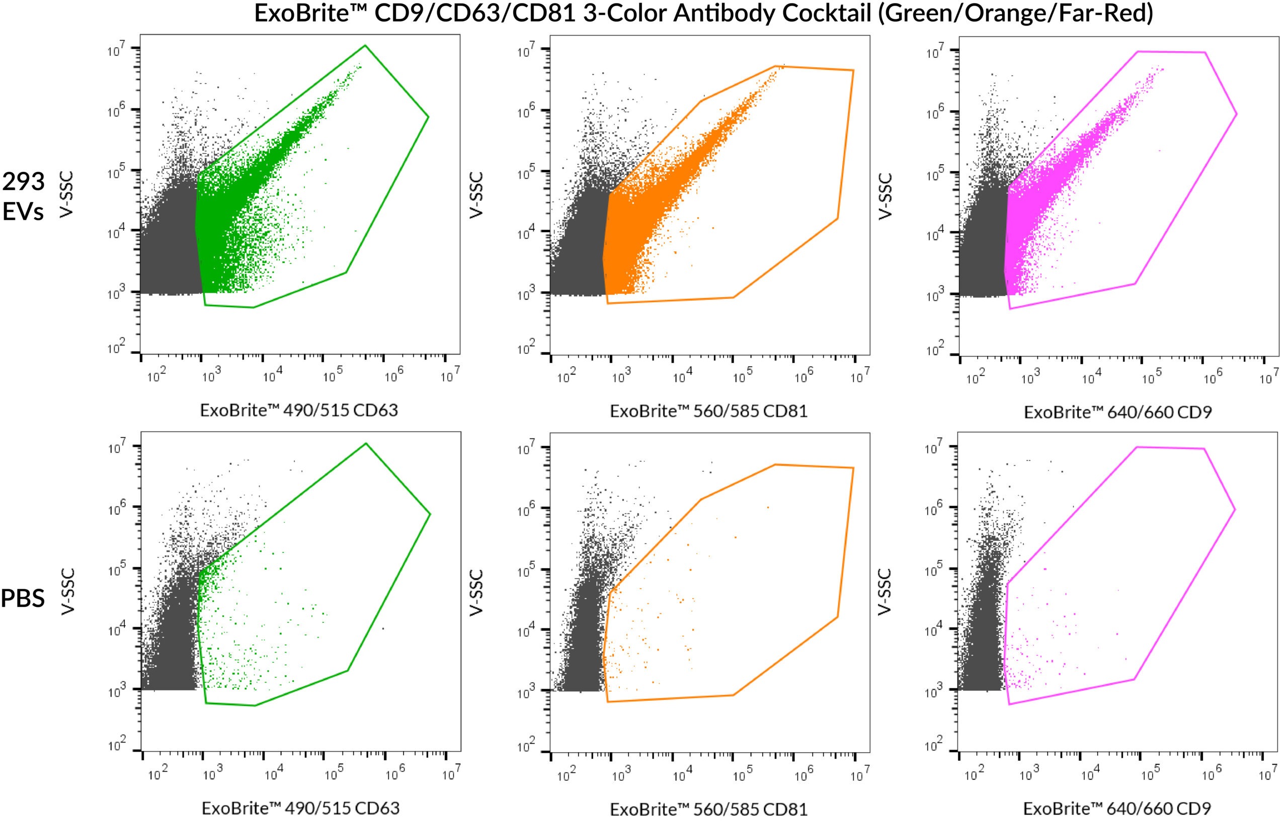
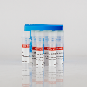
Antibody mixtures developed with optimal brightness and signal-to-noise for phenotyping EV samples. Available in blue/green/far-red or green/orange/far-red.
The ExoBrite™ CD9/CD63/CD81 3-Color Antibody Cocktails were developed for phenotyping EV samples by flow cytometry. The mixtures have been meticulously optimized for optimal brightness and signal-to-noise. The cocktails are available in blue/green/far-red or green/orange/far-red.
Skip the optimization, get results faster
The most commonly used markers for extracellular vesicle (EV) detection are the three tetraspanin proteins CD9, CD63, and CD81. Depending on the source of the EVs, each of these proteins will be present at varying levels. The ExoBrite CD9/CD63/CD81 3-Color Antibody Cocktails were designed for fast, reproducible phenotyping of an EV sample by flow cytometry. Each individual antibody in the mixture has been validated to have the same coverage as its respective ExoBrite™ Flow Antibody, ensuring accurate results.
In addition, like all ExoBrite™ Flow antibodies, the ExoBrite™ cocktails are provided in a proprietary buffer formulation for reduced antibody aggregation and brighter EV staining for optimal accuracy and signal-to-noise. The cocktails can be used on their own, for tetraspanin profiling, or combined with other antibodies of interest for more multi-target analyses.
EV antibodies you can trust
Other commercially available antibodies for tetraspanin proteins CD9, CD63, and CD81 are generally not validated for isolated EVs and may require tedious optimization for your EV prep and staining protocol. The antibodies and dye labels of ExoBrite™ Flow Antibody Conjugates were carefully selected and validated for robust detection of isolated EVs. See the table below for a full list of available ExoBrite™ Flow Antibody Conjugates. ExoBrite™ Isotype Control Flow Antibodies, which have been found to not react with any target in human cells and have the same isotype as the tetraspanin antibodies, are also available as a negative control.
Accelerate your EV research with ExoBrite™ EV stains
For general EV staining, Biotium's ExoBrite™ True EV Membrane Stains offer unparalleled coverage of EVs in a sample and address issues of dye aggregation often seen with PKH and other common membrane dyes. Biotium also offers optimized ExoBrite™ EV Surface stains conjugated to cholera toxin B (CTB), wheat germ agglutinin (WGA), and Annexin V. These stains are specially formulated for bright and specific detection of isolated EVs by flow cytometry. These ExoBrite™ EV Surface stains may also be combined with antibody staining, for multi-parameter analysis.
Biotium also offers conjugated ExoBrite™ Western Antibodies against CD9, CD63, and CD81 designed for optimal detection in EV extracts by fluorescent western blot.
Note: In our testing, we have found that ExoBrite™ 490/515 dye may bind to streptavidin coated surfaces or beads if free biotin binding sites are not blocked. We recommend performing a biotin blocking step after binding your biotinylated capture antibody to streptavidin beads or surfaces when using ExoBrite™ 490/515 conjugates. Alternatively, consider using a different ExoBrite™ dye for staining EVs captured on streptavidin beads or surfaces.
| Cocktail | Ex/Em | Detection Channel | Size | Catalog No. |
|---|---|---|---|---|
| ExoBrite™ CD9/CD63/CD81 Single-Color Antibody Cocktail (Green) | 490/516 nm | FITC | 25 tests | P030-125 |
| 100 tests | P030-500 | |||
| ExoBrite™ CD9/CD63/CD81 Single-Color Antibody Cocktail (Orange) | 562/584 nm | PE | 25 tests | P031-125 |
| 100 tests | P031-500 | |||
| ExoBrite™ CD9/CD63/CD81 Single-Color Antibody Cocktail (Far-Red) | 642/663 nm | APC | 25 tests | P032-125 |
| 100 tests | P032-500 | |||
| ExoBrite™ CD9/CD63/CD81 3-Color Antibody Cocktail (Blue/Green/Far-Red) | 411/452 nm 490/516 nm 642/663 nm | Pacific Blue® FITC APC | 25 tests | P028-125 |
| 100 tests | P028-500 | |||
| ExoBrite™ CD9/CD63/CD81 3-Color Antibody Cocktail (Green/Orange/Far-Red) | 490/516 nm 562/584 nm 642/663 nm | FITC PE APC | 25 tests | P029-125 |
| 100 tests | P029-500 |
While early studies of EVs attempted to use first-generation membrane dyes like DiI or PKH to stain EVs, more recently this class of dyes has been found to be largely unsuitable for EV staining due to their high degree of aggregation. Dye aggregation not only generates nonspecific particles that are indistinguishable from EVs in flow cytometry, but also results in poor EV labeling efficiency. Biotium developed the ExoBrite™ True EV Membrane Stains in response to our customers difficulties with using traditional membrane dyes to stain EVs. See our Literature Digest for more information.
We strongly recommend our ExoBrite™ Flow Antibody Conjugates for staining both purified or bead-bound EVs. The antibodies are validated and optimized to offer bright signal and low background. They are available against human or mouse CD9, CD63, and CD81 tetraspanin proteins.
Yes, EVs can be stained simultaneously with an ExoBrite™ True EV Membrane Stain and a fluorescent antibody.
With purified EVs, we have seen good results when EVs were stained in 500 mL of 1X ExoBrite™ plus 1 ug/mL fluorescent antibody. Please view our Product Information Sheet for detailed protocols.

Content #1
Content #1
Content #1
Content #2
Content #3