Lumitein™ Protein Gel Stain 1X
A luminescent dye designed for detecting proteins in SDS polyacrylamide (SDS-PAGE) gels. As sensitive as silver stain.
Please fill in the inquiry form and we will contact you shortly.
Wishlist updated! View wishlist
 Powered by Bioz
Product Description
Lumitein™ is a luminescent dye designed for detecting proteins in SDS polyacrylamide (SDS-PAGE) gels, and can also be used to detect proteins in native PAGE gels after an additional SDS incubation step. The stain combines excellent sensitivity, user-friendliness and compatibility with common instruments and downstream analysis. Lumitein™ is as sensitive as silver stain, detecting 1 ng or less protein. For faster, non-toxic alternatives to Lumitein™ for protein gel staining, see our One-Step Lumitein™ and One-Step Blue™ Protein Gel Stains.
- As sensitive as silver stain, detecting < 1 ng protein.
- Simple staining procedure.
- Image with UV gel box (EtBr filter) or laser scanner.
- Wide linear detection range.
- Compatible with Downstream Analysis such as mass spec and sequencing.
- Stable at room temperature.
- 200 mL, 1 L, and 5 L sizes.
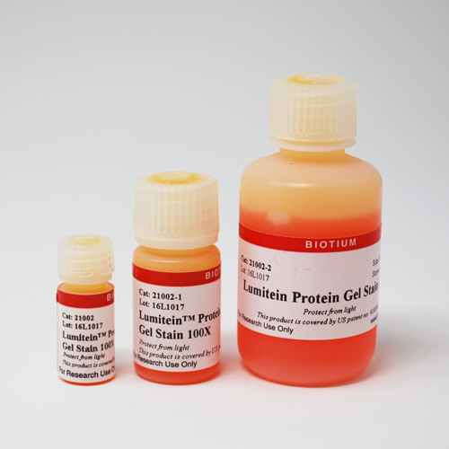
Lumitein™ Protein Gel Stain, 1X is a convenient, ready-to-use staining solution. Lumitein is also available as an economical 100X concentrated solution (catalog no. 21002).
Lumitein™ sensitivity
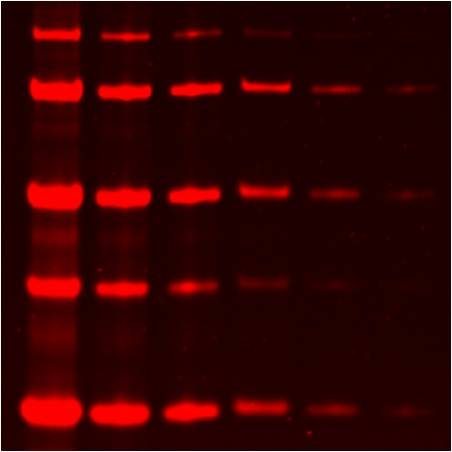
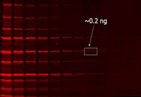
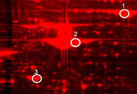
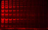
Lumitein™ is as sensitive as the best silver stain by detecting 1 ng or less protein (Figure 1). Unlike silver stain, however, Lumitein™ has a linear detection range of at least 3 orders of magnitude. It is among the simplest protein gel stain by staining protein in gels in 90 minutes or less time without a separate fixation step. Lumitein™ has an excitation spectrum that makes detection possible with either a simple UV box or a high-end laser scanner. Moreover, protein gel staining with Lumitein™ is compatible with downstream protein analyses such as mass spectrometry and Edman-based sequencing (Figure 2).
 |
 |
|
| Figure 1. Two-fold serial dilutions of protein marker were separated via SDS-PAGE and then stained with Lumitein™. Imaged with a GE Typhoon Trio using 532 nm excitation and 610BP30 emission filter. | Figure 2. 2-D gel of human liver protein lysate stained with Lumitein™. The three circled spots were picked for MS analysis by Applied Biomics, Inc. (Hayward, CA), confirming that Lumitein™ staining is fully compatible with MS analysis. | |
|
Lumitein™ vs. Coomassie
|
||
 |
 |
|
|
Lumitein™ |
Coomassie |
|
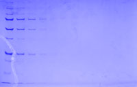
| Figure 3. PAGE gels containing electrophoretically separated protein marker (loaded in two-fold serial dilution from left to right) were stained with Lumitein™ protein gel stain (left) and Coomassie Blue (right). The Lumitein™-stained gel was imaged using a UV box equipped with EtBr filter (UVP) while the Coomassie-stained gel was imaged using a white light converter (UVP). | ||