NucSpot® Nuclear Stains
Cell membrane-impermeant, nuclear-specific counterstains. Suitable for fixed cells or staining dead cells in live cultures.


Please fill in the inquiry form and we will contact you shortly.
Wishlist updated! View wishlist
Product Description
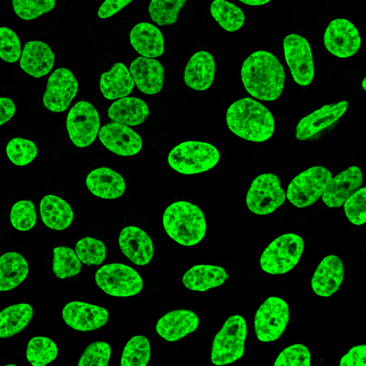
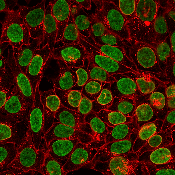
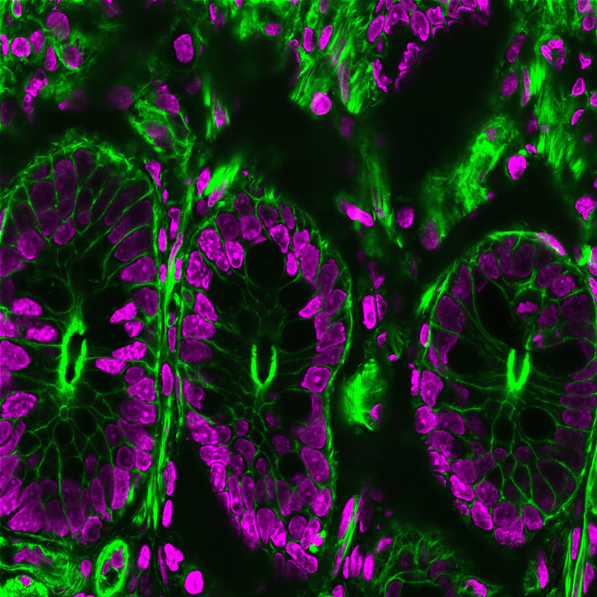
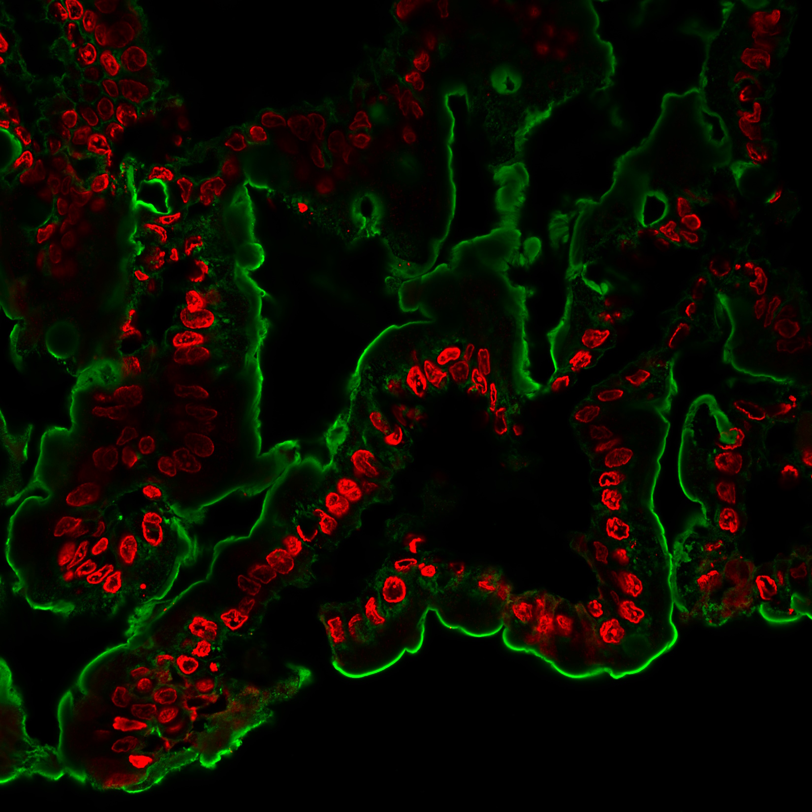
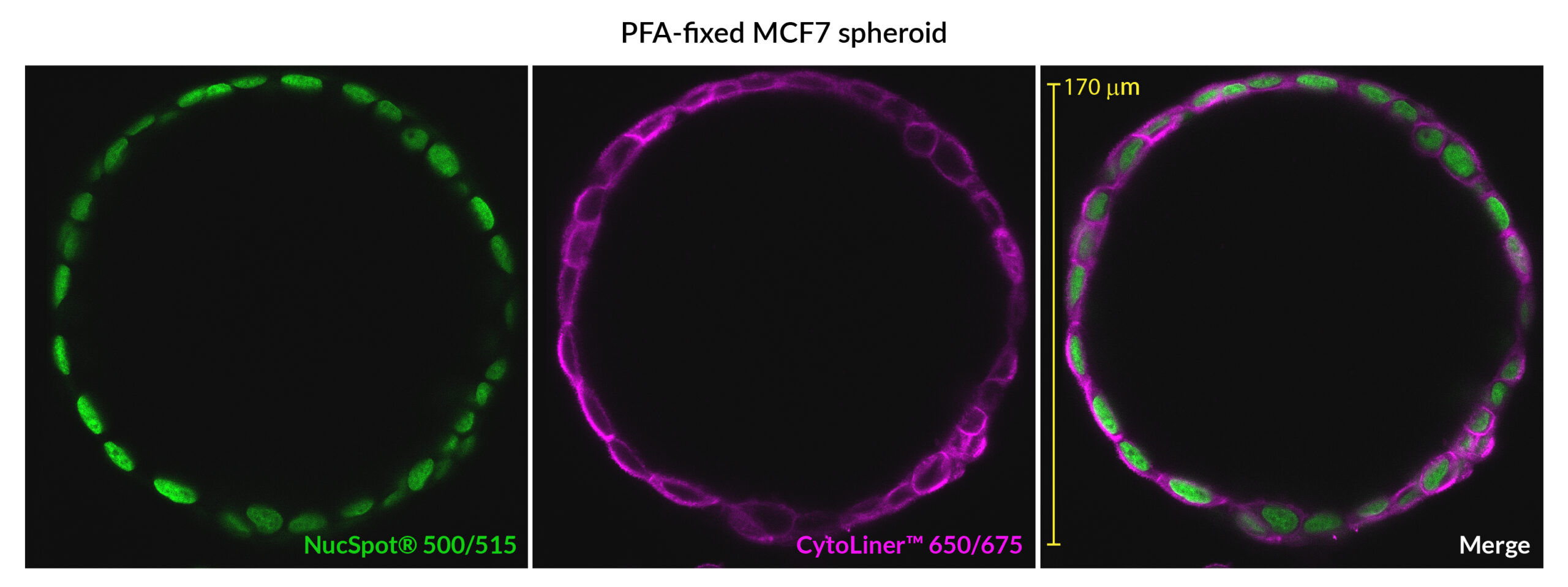
NucSpot® Nuclear Stains are cell membrane-impermeant, nuclear-specific counterstains available in a variety of colors from green to near-infrared (near-IR).
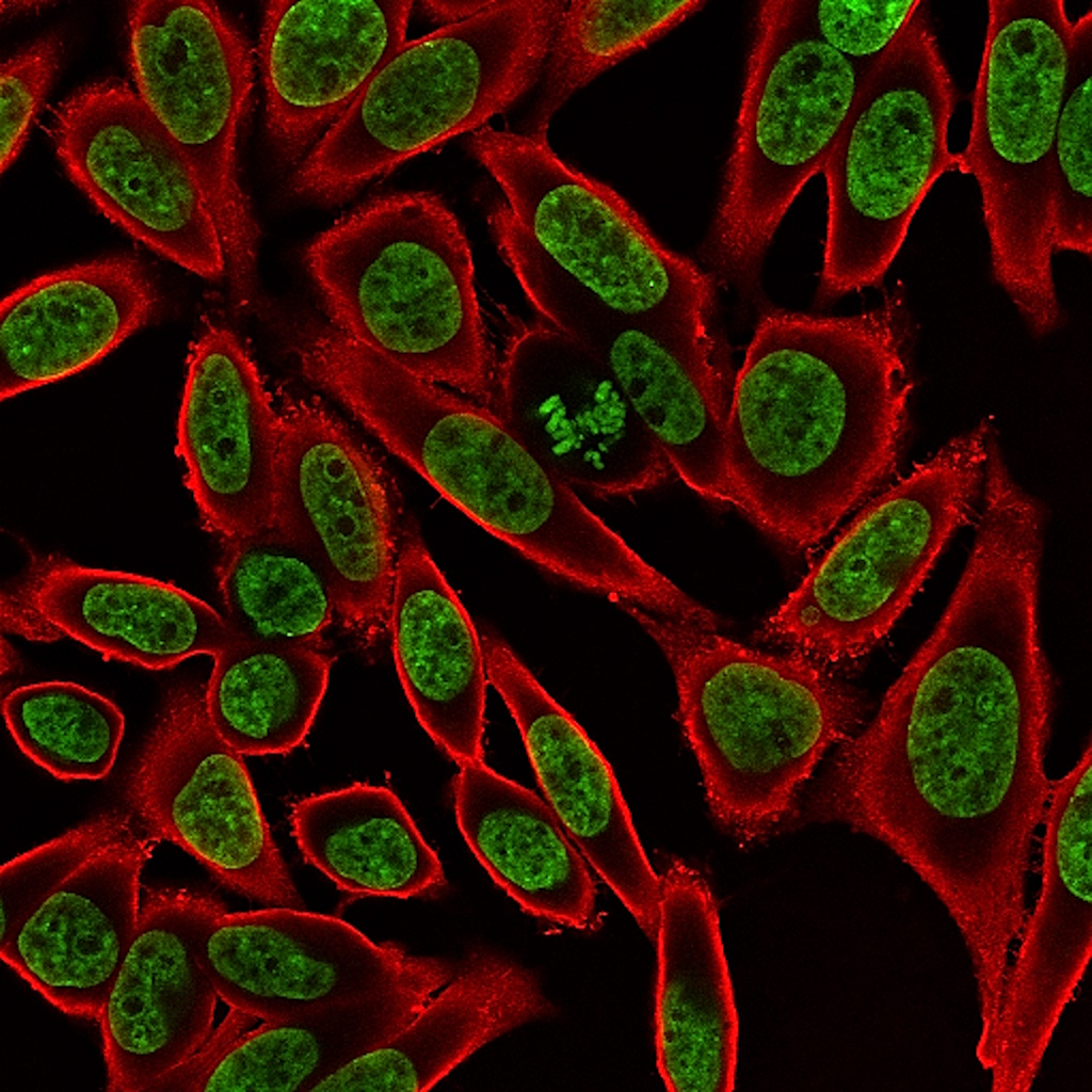
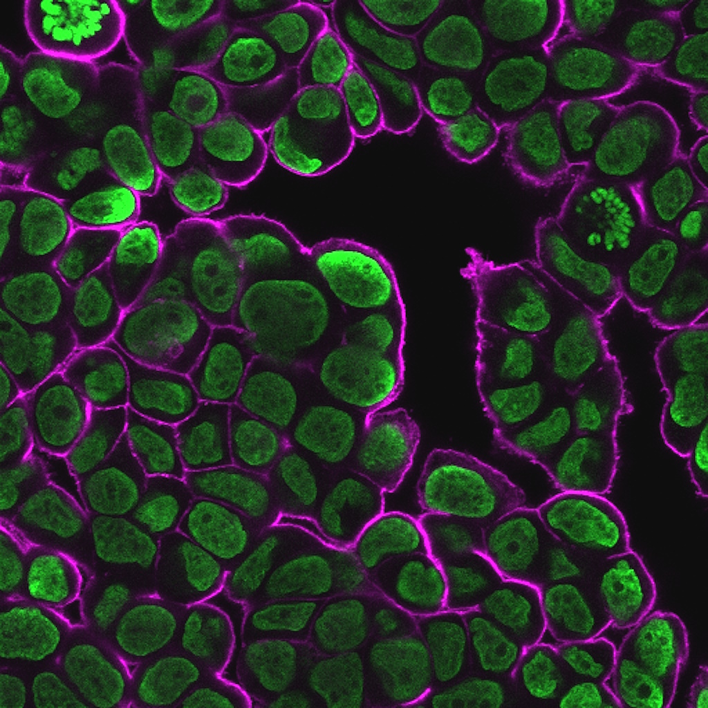
- Nuclear-specific counterstains for fixed cells
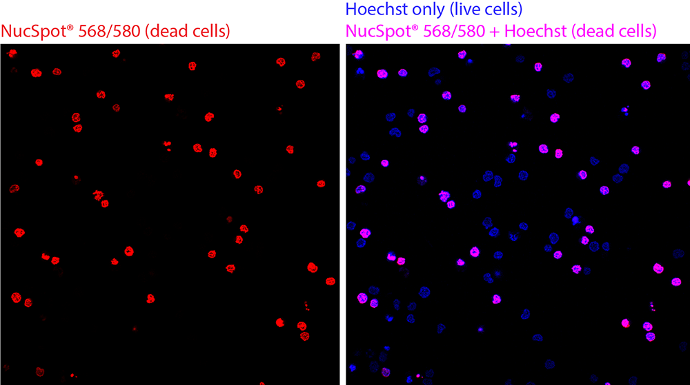
- Selectively stains dead cells in live culture
- No wash required
- Color options for FITC, Cy®3, PE, Texas Red®, Cy®5, APC, Cy®5.5, and Cy®7 channels
Commonly used blue nuclear stains, such as DAPI and Hoechst, undergo photoconversion after exposure to UV wavelengths and cause cross-talk in other channels. NucSpot® Nuclear Stains were developed to address these photoconversion issues by offering bright and nuclear-specific counterstaining in a wide selection of colors from green to near-IR. The have minimal fluorescence until they bind to DNA and can be used for no-wash nuclear staining. Unlike other nucleic acid dyes such as propidium iodide (PI), TOTO®, TO-PRO®, and similar dyes that stain both the nucleus and cytoplasm, NucSpot® Nuclear Stains selectively stain the nucleus in fixed and permeabilized cells without the need for RNase treatment. NucSpot® Nuclear Stains also can be used for selective staining of dead cells in unfixed cell cultures for analysis by flow cytometry or fluorescence imaging. Several of the dyes can be continuously incubated with cells for multi-day imaging.
NucSpot® Nuclear Stains
| Product | Ex/Em | Detection Channel | Size | Catalog No. |
|---|---|---|---|---|
| NucSpot® 500/515 | 497/513 nm | FITC* | 20 uL | 41040-T |
| 100 uL | 41040 | |||
| NucSpot® 555/570 | 559/566 nm | Cy®3 or PE* | 20 uL | 41033-T |
| 100 uL | 41033 | |||
| NucSpot® 568/580 | 572/583 nm | Cy®3 or PE* | 20 uL | 41036-T |
| 100 uL | 41036 | |||
| NucSpot® 594/615 | 603/613 nm | Texas Red® or PE-Texas Red®* | 20 uL | 41037-T |
| 100 uL | 41037 | |||
| NucSpot® 650/665 | 653/671 nm | Cy®5 or APC* | 20 uL | 41034-T |
| 100 uL | 41034 | |||
| NucSpot® 680/700 | 683/707 nm | Cy®5.5* | 20 uL | 41035-T |
| 100 uL | 41035 | |||
| NucSpot® 750/780 | 757/780 nm | Cy®7 or APC-Cy®7* | 20 uL | 41038-T |
| 100 uL | 41038 |
† NucSpot® Far-Red is designed for flow cytometry optical systems and is not recommended for fluorescence microscopy.
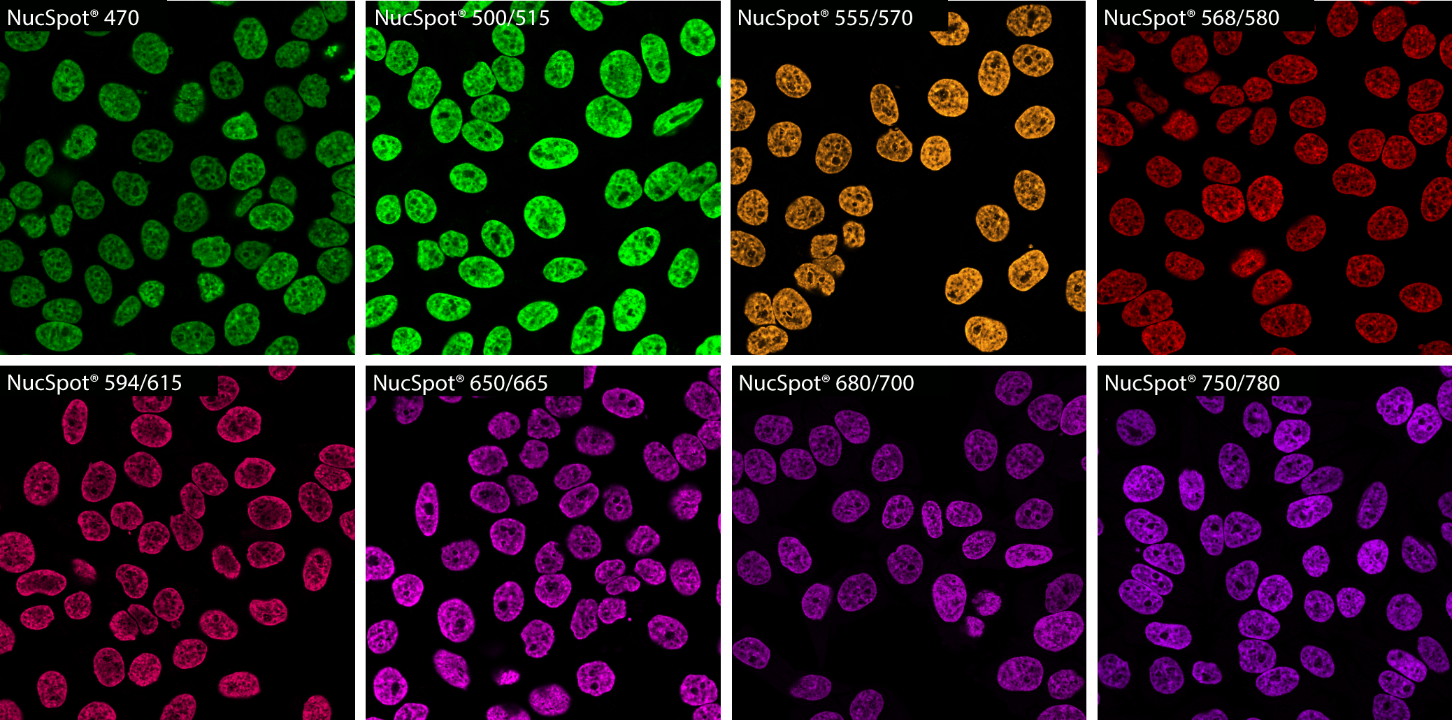
Find the Right Dye for Your Application
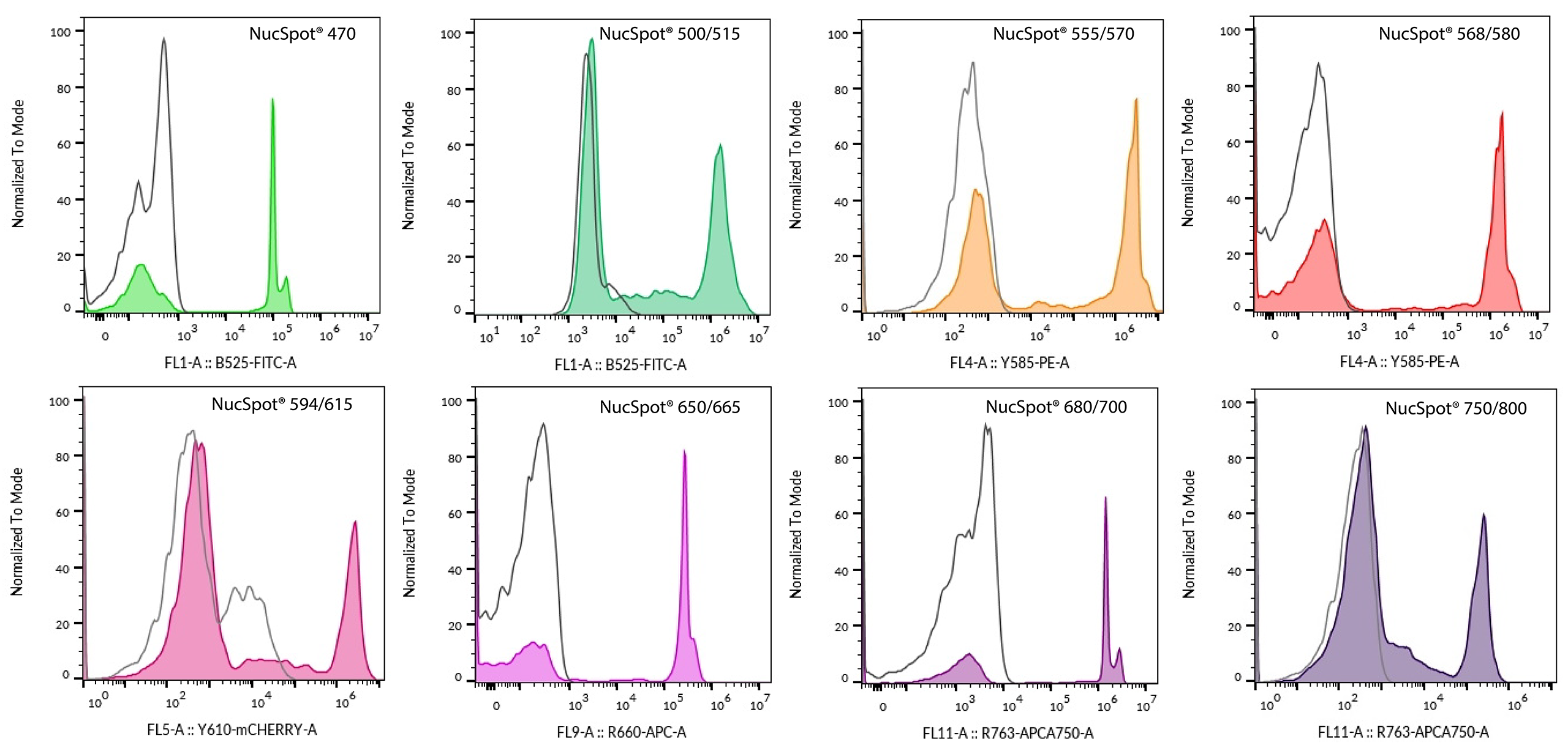
Stain Dead Cells in Live Cultures
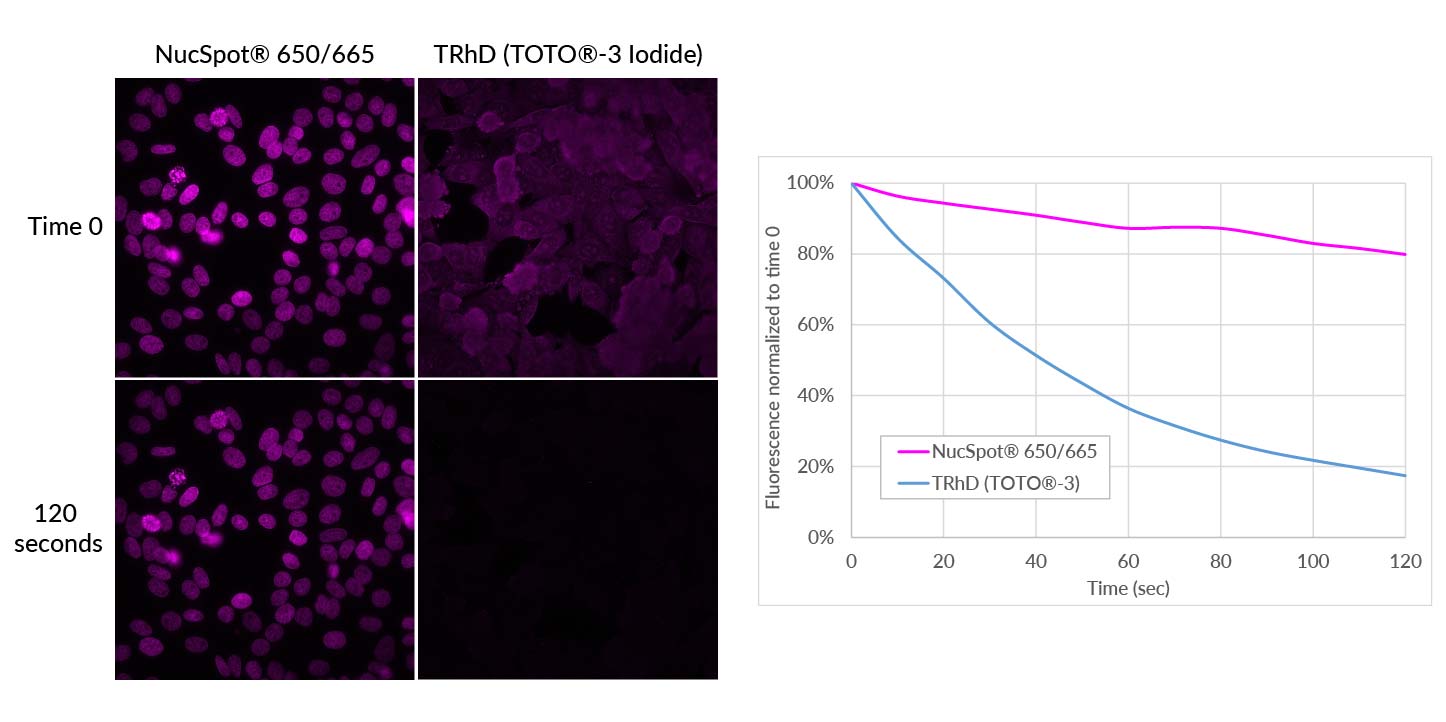
Exceptional Photostability
NucSpot® Nuclear Stains offer improved photostability over commonly used cyanine-based nuclear stains. The figure below shows how NucSpot® 650/665 is significantly more photostable than Thiazole Red Homodimer (TOTO® -3).
NucSpot® 500/515 is a green fluorescent nuclear stain for the FITC channel. In addition to bright nuclear counterstaining of fixed cells, it can also be used for multi-day live/dead staining of mammalian cells in culture. NucSpot® 500/515 is designed as an improved alternative to NucSpot® 470 which has limited use for cell viability tracking due to its instability in culture medium during long incubation periods.
NucSpot® 555/570 and NucSpot® 568/580 have orange and visible red fluorescence, respectively, and are nuclear-specific alternatives to PI and similar dyes.
NucSpot® 594/615 has deep red fluorescence for the Texas Red® channel.
NucSpot® 650/665 has far-red fluorescence with superior nuclear specificity compared to first-generation far-red nuclear stains such as RedDot™2 and Draq7™.
NucSpot® 680/700 and NucSpot® 750/780 are spectrally unique DNA stains for far-red and near-IR detection.
Biotium also offers NucSpot® Far-Red, a flow cytometry stain developed as an improved alternative to 7-AAD. It shows less bleed-through fluorescence in the PE-Texas Red® channel compared to 7-AAD and is ideal for selective detection of dead cells or DNA content analysis by flow cytometry without RNase treatment.
Learn more about our full selection of Cellular Stains for the nucleus and other organelles.