Melanoma is the most dangerous type of skin cancer, and its incidence is increasing worldwide. It develops from melanin-producing cells known as melanocytes and typically occurs in the skin, but may on rare occasions occur in the mouth, intestines, or eye (uveal melanoma). Because metastatic melanoma is highly aggressive, much research has been directed towards understanding its cellular biology. Cellular processes that can be targeted to diagnose or limit the propagation of melanoma cells have the potential for therapeutic intervention and the possibility of extracellular vesicles (EVs) being involved opens new possibilities for intervention. It has previously been shown that certain metastatic melanoma cells release small (< 150 nm) EVs classified as exosomes and ectosomes that contain cancer biomarkers. While EVs play important roles in intercellular communication, little else is known about the EVs produced by melanoma cells and the effects they might have on the tumor microenvironment.
Karbanová et al. recently reported a novel type of large EVs released by melanoma cells in their 2024 Cell Communication and Signaling paper. Because the novel EVs contained lipid droplets and were released extracellularly, the authors named them “extracellular lipidosomes.” To uncover the existence and attributes of the novel EVs, the team employed correlative flow cytometry and microscopy-based analyses using a large suite of cellular stains including Biotium’s LipidSpot ™ 488 and LipidSpot™ 610 Lipid Droplet Stains, CF®488A, CF®640R, and CF®555-conjugated wheat germ agglutinin, as well as MitoView™ Fix 640. Using this approach, they determined that a subset of EVs were produced that ranged in size from 2 to 6 μm in diameter and contained both lipid droplets and mitochondria (see Figure 1 below). They then used video microscopy to reveal that these large EVs originated from lipid droplet-enriched cell extremities during cell division (see Video link below). Further studies using LipidSpot™-labeled EVs and unstained cells showed that the EVs were taken up by other cells after their release, while silencing experiments showed that the cholesterol-binding membrane protein CD133, a widely used marker for stem cells and cancer cells, affected the cellular distribution of lipid droplets and determined the formation of the large EVs. Together, these results suggest that the release of these novel large EVs from aggressive melanoma cells and their uptake by other cells likely impact melanoma growth and dissemination.
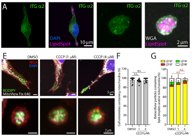
Figure 1. A) Expression extracellular lipidosomes at cell extremities. FEMX-I (melanoma cell line) cells were co-stained with integrin (ITG) antibodies, LipidSpot™ 610, and CF® Dye conjugated WGA to highlight lipid droplets and glycoconjugates. Samples were counterstained with DAPI to highlight nuclei. E-G) Extracellular lipidosomes contain mitochondria. Following treatment with CCCP (an inhibitor of mitochondrial oxidative phosphorylation, that evokes the integrated stress response) or a DMSO control FEMX-I cells and lipidosomes were stained with MitoView™ Fix 640, BODIPY™ 493/503, CF® Dye conjugated WGA, and DAPI. Mitochondria-containing and mitochondria-free cell extremities (F) and extracellular particles (G) were quantified under various conditions as indicated. CCCP treatment did not impact the integration of lipid droplets in lipidosomes Credit: Modified from Karbanová, J. et al. https://doi.org/10.1186/s12964-024-01471-7 reproduced under the Creative Commons license (CC BY 4.0).
Related Products
Learn more about Biotium’s wide selection of cellular stains and other optimized solutions for your immunofluorescence microscopy workflow. We also carry a selection of products for extracellular vesicle (EV) research including bright and sensitive stains for EVs: ExoBrite™ True EV Membrane Stains, ExoBrite™ CTB EV Staining Kits, ExoBrite™ Annexin EV Staining Kits, and ExoBrite™ WGA EV Staining Kits.