 New Products
 Earth-Friendly Products
 Biotium Choice Antibodies
 Special Offers


Content #1
Content #1
Content #1
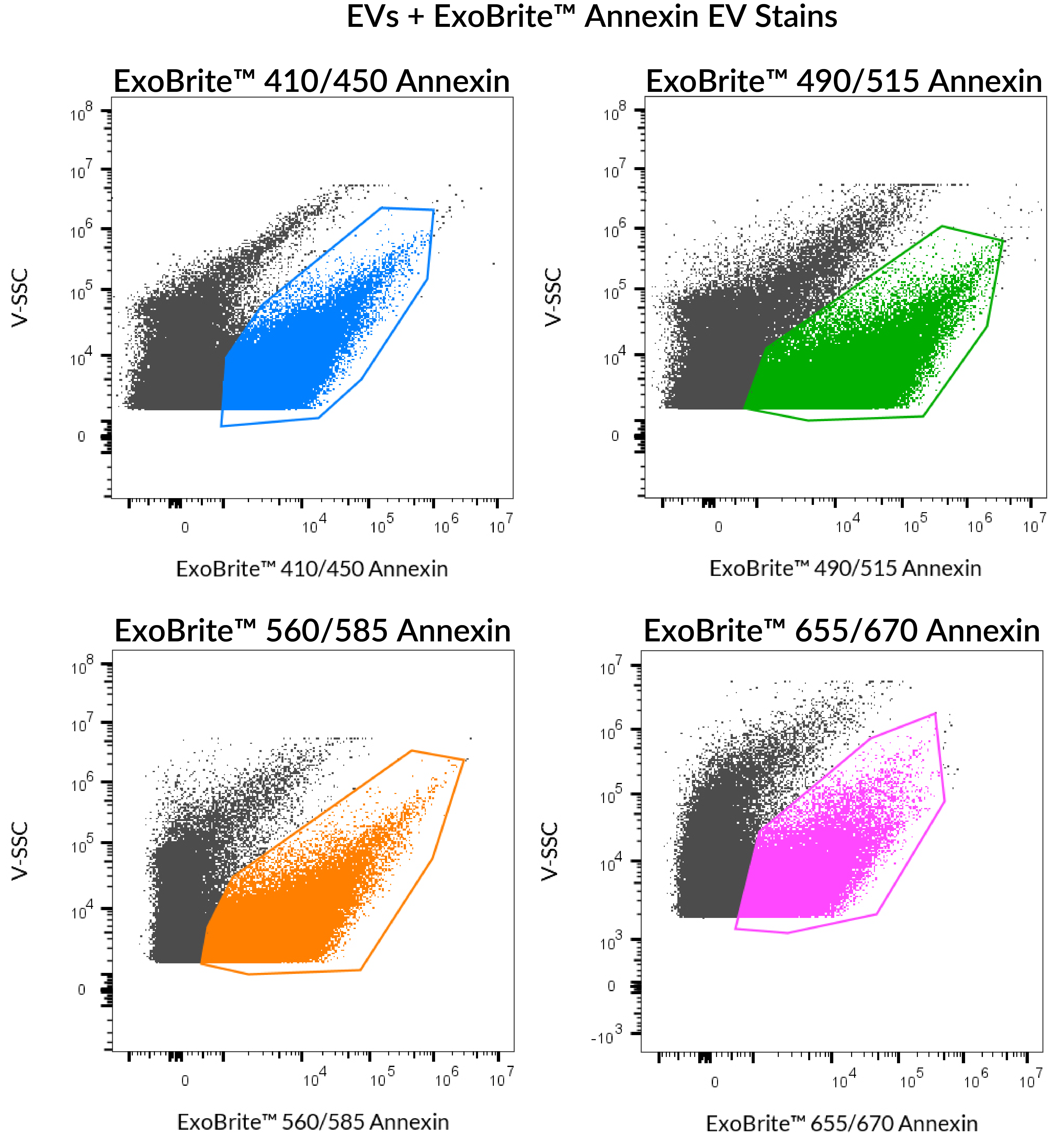
Fluorescent Annexin V conjugates that are optimized for bright and low background staining of extracellular vesicles for flow cytometry.
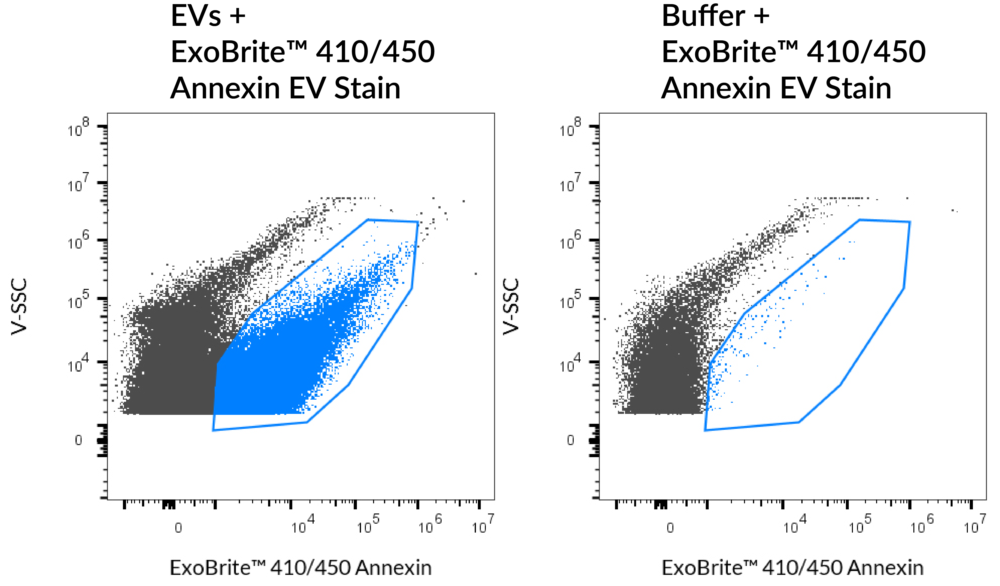
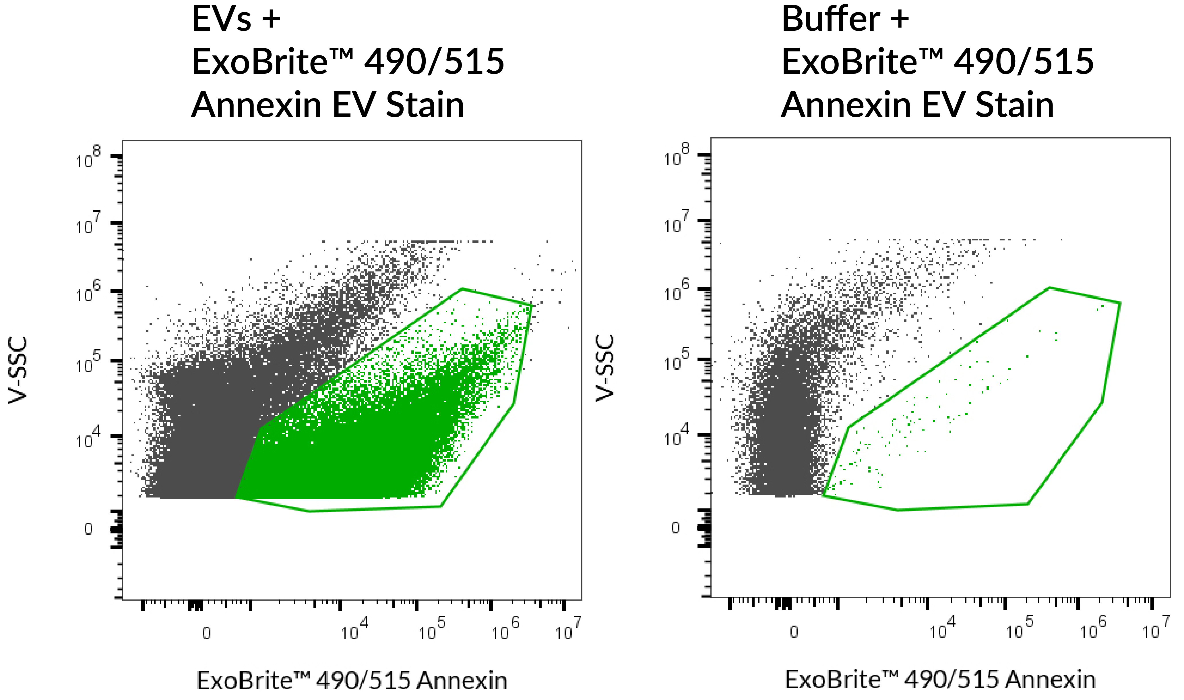
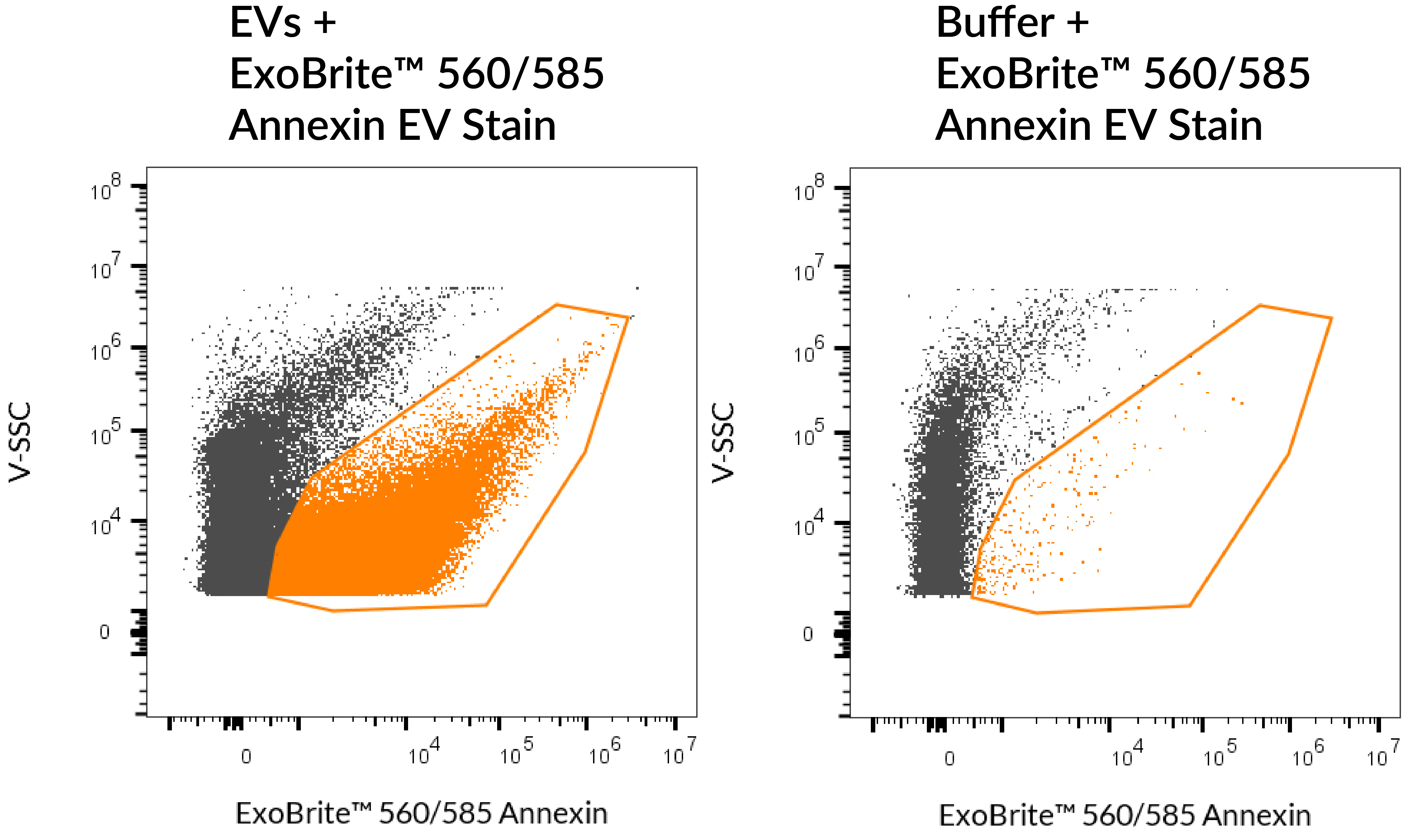
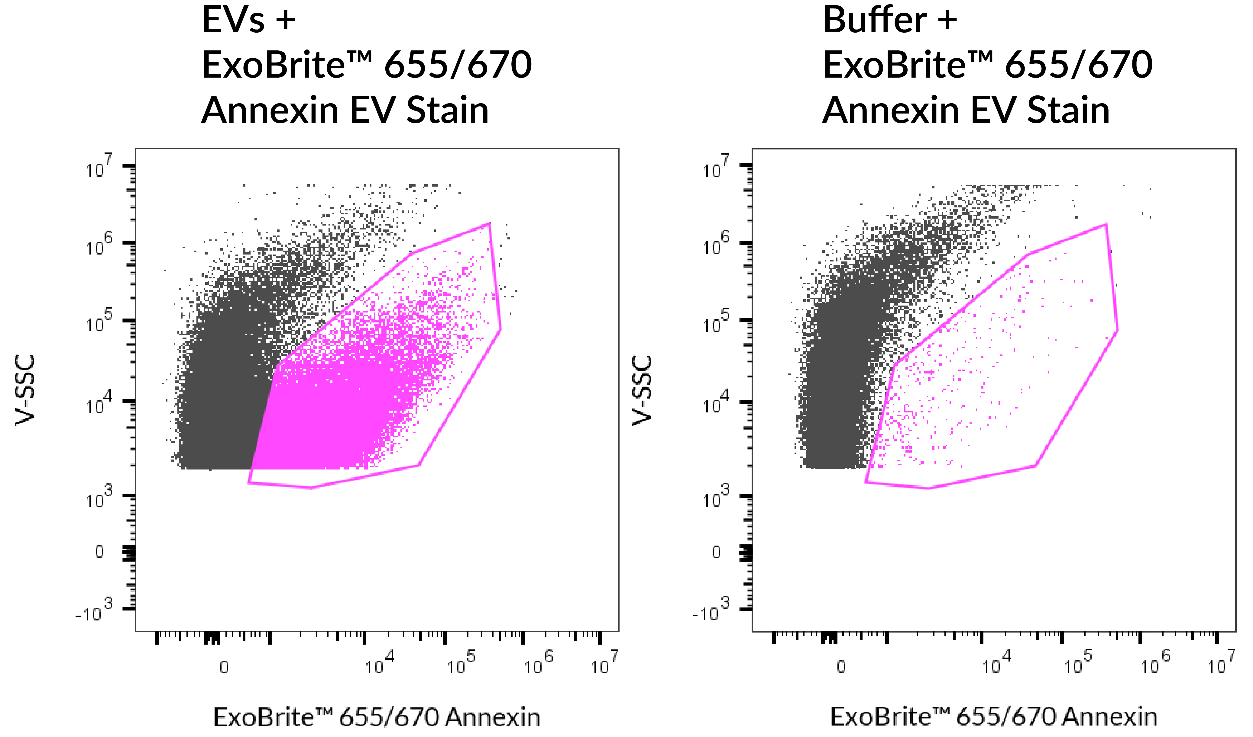
ExoBrite™ Annexin EV Staining Kits were designed to overcome some of the challenges of EV detection, particularly in flow cytometry. ExoBrite™ Annexin EV Stains bind to molecules in the EV membrane for bright, specific staining, with little to no background.
ExoBrite™ Annexin EV Stains are uniquely formulated conjugates of Annexin V, a 35-36 kDA calcium-dependent phospholipid-binding protein with high affinity for phosphatidyleserine (PS). Annexin V conjugates have been used to detect EVs due to the presence of PS on most EV membranes.
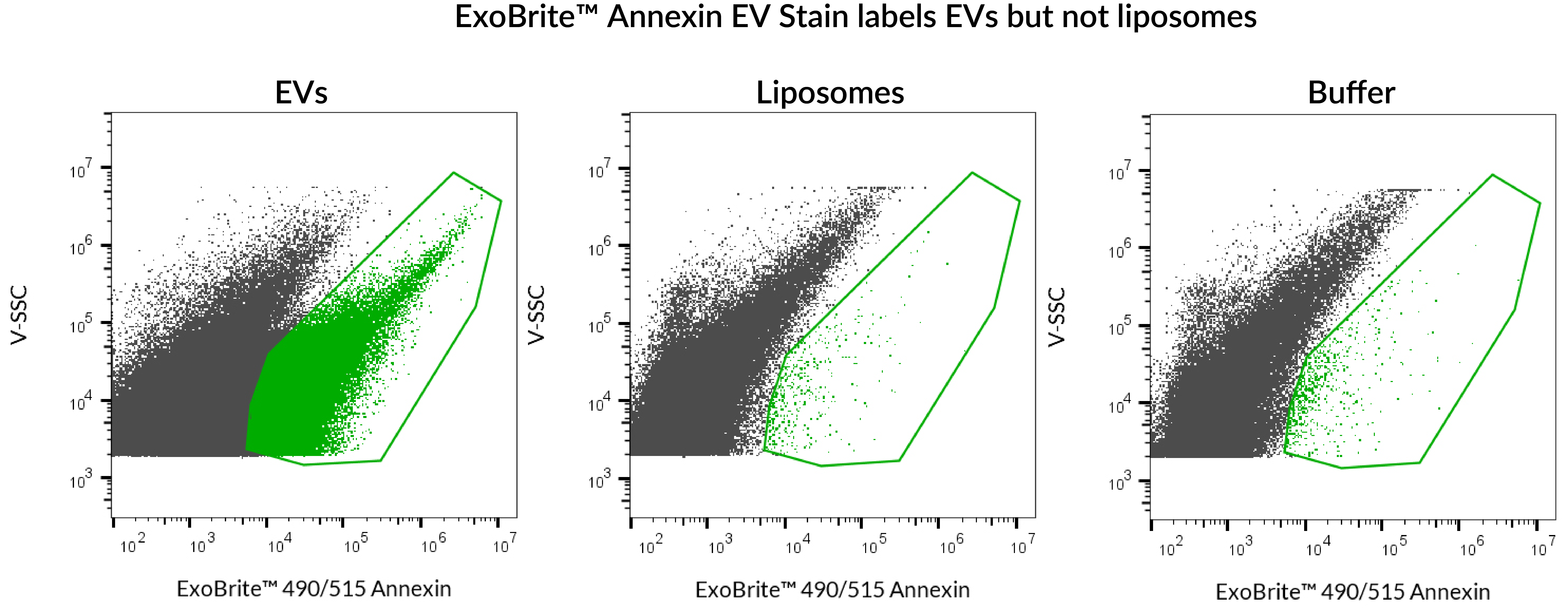
ExoBrite™ Annexin EV Stains were designed to overcome some of the challenges of EV detection, particularly in flow cytometry. For example, lipophilic membrane dyes commonly used to stain EVs can form aggregates of a similar size as exosomes or EVs, thus confounding analysis. Conversely, ExoBrite™ Annexin EV Stains are specially formulated to minimize aggregation in flow cytometry, allowing EVs to be identified with bright staining with minimal background. In addition, ExoBrite™ Annexin EV Stains were designed to offer broad coverage of EVs isolated from different sources. We tested EVs derived from 9 cell lines and ExoBrite™ Annexin EV Stains showed strong staining for all of them.
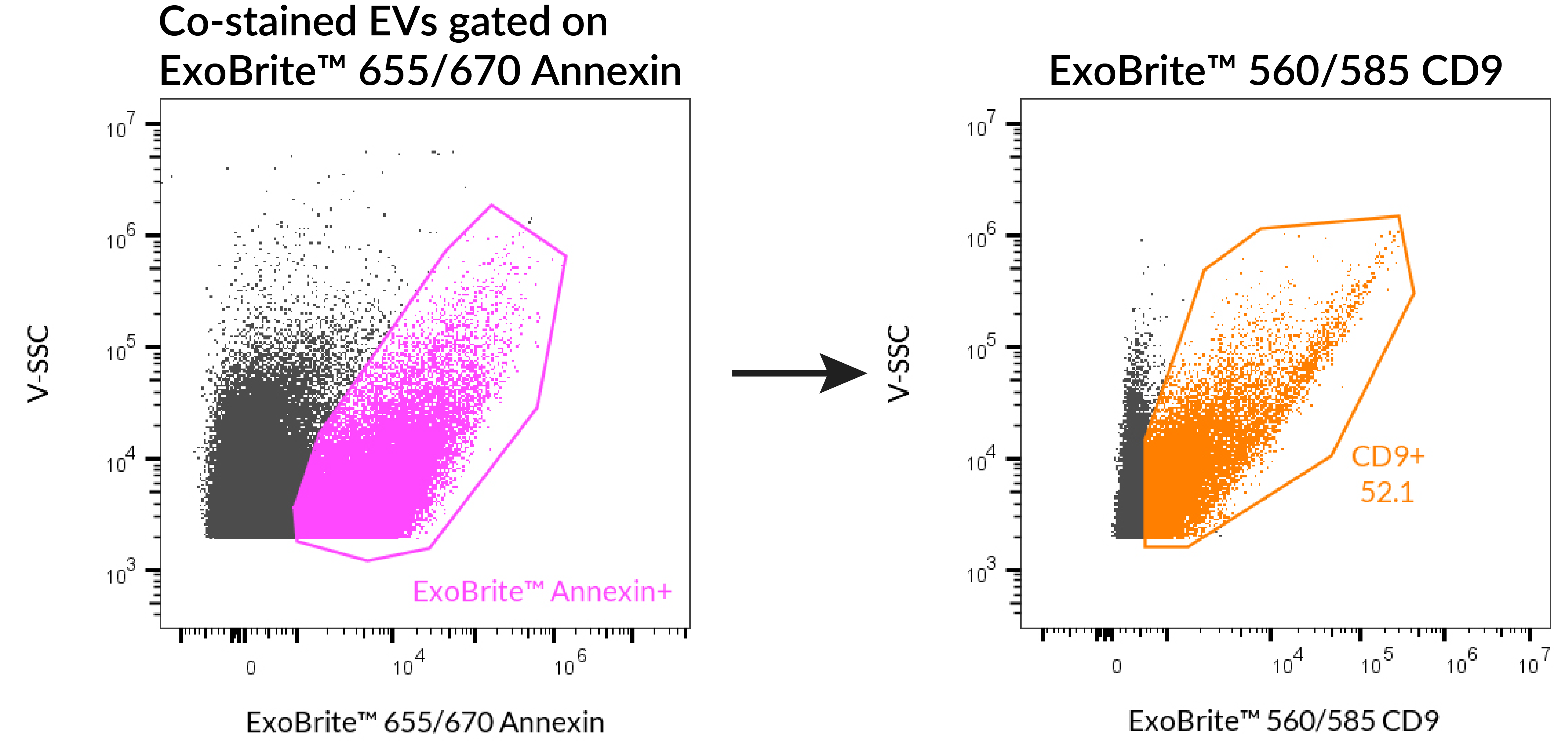
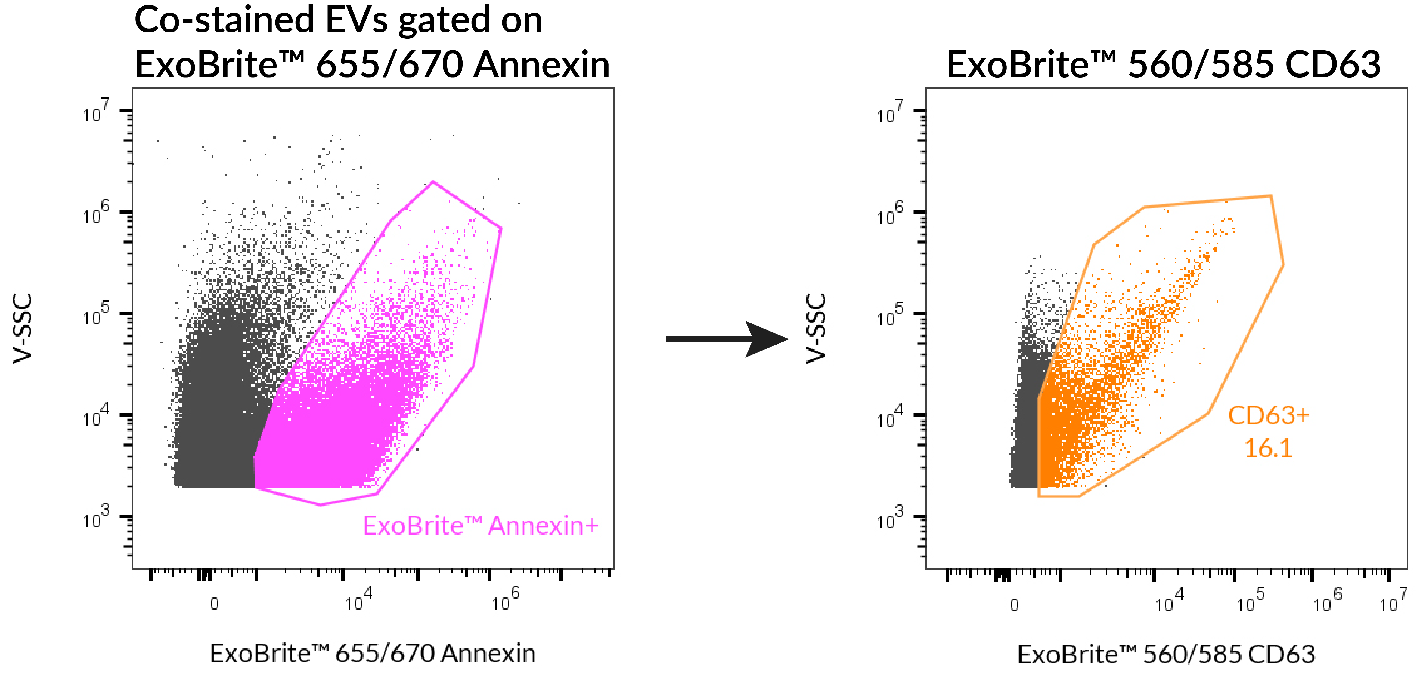
EVs are often labeled with fluorescent antibodies targeting one or more of the tetraspanin proteins CD9, CD63, and CD81. ExoBrite™ Annexin staining can be combined with antibody staining, for multi-parameter analysis.
Notes:
| Product | Ex/Em | Detection channels | Size | Catalog Number |
|---|---|---|---|---|
| ExoBrite™ 410/450 Annexin EV Staining Kit | 416/452 nm | Pacific Blue™ | 100 Labelings | 30119-T |
| 500 Labelings | 30119 | |||
| ExoBrite™ 490/515 Annexin EV Staining Kit | 490/516 nm | FITC | 100 Labelings | 30120-T |
| 500 Labelings | 30120 | |||
| ExoBrite™ 560/585 Annexin EV Staining Kit | 562/584 nm | PE | 100 Labelings | 30121-T |
| 500 Labelings | 30121 | |||
| ExoBrite™ 650/665 Annexin EV Staining Kit | 652/668 nm | APC | 100 Labelings | 30122-T |
| 500 Labelings | 30122 |
| EV Source | ExoBrite™ True EV Membrane Stains | ExoBrite™ CTB Stains | ExoBrite™ WGA Stains | ExoBrite™ Annexin Stains |
|---|---|---|---|---|
| A549 cells | Yes | Yes | Yes | Yes |
| CHO cells | Yes | No | Yes | Yes |
| hASC (human adipose stem cells) | ND | No1 | ND | ND |
| HEK293 cells | Yes | Yes1 | Yes | ND |
| HeLa cells | Yes | No | Yes | Yes |
| HUVEC (human umbilical vein endothelial cells) | ND | No1 | ND | ND |
| J774 cells | Yes | Yes | Yes | Yes |
| Jurkat cells | Yes | Yes | Yes | Yes |
| MCF-7 cells | Yes | Yes | Yes | Yes |
| Plasma | ND | No | ND | Yes |
| Raji cells | ND | Yes | Yes | Yes |
| RAW 264.7 cells | Yes | ND | ND | ND |
| Serum | ND | No | ND | Yes |
| Skeletal myoblasts | ND | Yes1 | ND | ND |
| THP-1 cells | Yes | ND | ND | ND |
| U2OS cells | Yes | No | Yes | Yes |
| U937 cells | Yes | No | Yes | Yes |
| NIH3T3 cells | Yes | ND | ND | ND |
| HepG2 cells | ND | ND | Yes | ND |
| Yeast (S. cerevisiae) | Yes | No | Yes | Yes |
Learn about Biotium's new ExoBrite™ True EV Membrane Stains. These genuine lipophilic membrane dyes are designed for superior pan-EV labeling over other membrane dyes including PKH, DiO, DiI, and DiD. Biotium also offers ExoBrite™ CTB EV Stains (cholera toxin B conjugates) and ExoBrite™ WGA EV Stains (wheat germ agglutinin) optimized for bright and sensitive staining of EVs. The ExoBrite™ EV Surface Stain Sampler Kit contains each of Biotium’s ExoBrite™ EV Surface Stains (CTB, WGA, and Annexin V) for assessing which stain offers the best coverage for the EV samples of interest. Biotium also offers ExoBrite™ Antibody Conjugates for optimal detection of CD9, CD63, and CD81 EV markers by flow cytometry and western blotting. For super-resolution imaging by STORM, learn about our ExoBrite™ STORM CTB EV Staining Kits available in four CF® Dyes validated for STORM.
Note: We do not recommend using ExoBrite™ 410/450 Annexin EV Stain or ExoBrite™ 490/515 Annexin EV Stain to stain bead-bound EVs. For bead-bound EVs we recommend using ExoBrite™ 560/585 Annexin EV Stain, ExoBrite™ 650/665 Annexin EV Stain, as well as ExoBrite™ CTB EV Stains.
While early studies of EVs attempted to use first-generation membrane dyes like DiI or PKH to stain EVs, more recently this class of dyes has been found to be largely unsuitable for EV staining due to their high degree of aggregation. Dye aggregation not only generates nonspecific particles that are indistinguishable from EVs in flow cytometry, but also results in poor EV labeling efficiency. Biotium developed the ExoBrite™ True EV Membrane Stains in response to our customers difficulties with using traditional membrane dyes to stain EVs. See our Literature Digest for more information.
We strongly recommend our ExoBrite™ Flow Antibody Conjugates for staining both purified or bead-bound EVs. The antibodies are validated and optimized to offer bright signal and low background. They are available against human or mouse CD9, CD63, and CD81 tetraspanin proteins.
Yes, EVs can be stained simultaneously with an ExoBrite™ True EV Membrane Stain and a fluorescent antibody.
With purified EVs, we have seen good results when EVs were stained in 500 mL of 1X ExoBrite™ plus 1 ug/mL fluorescent antibody. Please view our Product Information Sheet for detailed protocols.

Content #1
Content #1
Content #1
Content #2
Content #3