 New Products
 Earth-Friendly Products
 Biotium Choice Antibodies
 Special Offers


Content #1
Content #1
Content #1
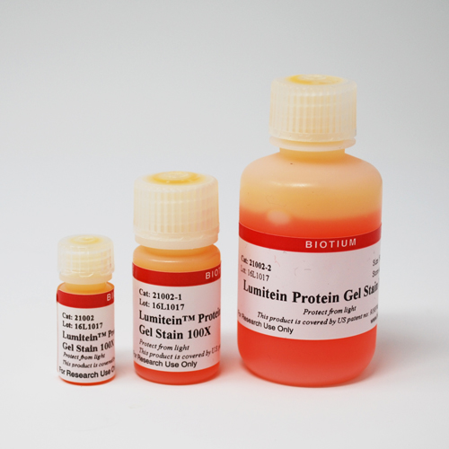
A luminescent dye designed for detecting proteins in SDS polyacrylamide (SDS-PAGE) gels. As sensitive as silver stain.
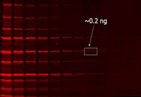
Lumitein™ is a luminescent dye designed for detecting proteins in SDS polyacrylamide (SDS-PAGE) gels, and can also be used to detect proteins in native PAGE gels after an additional SDS incubation step. The stain combines excellent sensitivity, user-friendliness and compatibility with common instruments and downstream analysis. Lumitein™ is as sensitive as silver stain, detecting 1 ng or less protein. For faster, non-toxic alternatives to Lumitein™ for protein gel staining, see our One-Step Lumitein™ and One-Step Blue™ Protein Gel Stains.
Lumitein™ Protein Gel Stain, 100X is an economical concentrated solution. Lumitein™ is also available as a 1X convenient, ready-to-use staining solution (catalog no. 21001).
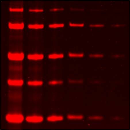
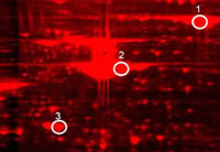
Lumitein™ is as sensitive as the best silver stain by detecting 1 ng or less protein (Figure 1). Unlike silver stain, however, Lumitein™ has a linear detection range of at least 3 orders of magnitude. It is among the simplest protein gel stain by staining protein in gels in 90 minutes or less time without a separate fixation step. Lumitein™ has an excitation spectrum that makes detection possible with either a simple UV box or a high-end laser scanner. Moreover, protein gel staining with Lumitein™ is compatible with downstream protein analyses such as mass spectrometry and Edman-based sequencing (Figure 2).
 |
 |
|
| Figure 1. Two-fold serial dilutions of protein marker were separated via SDS-PAGE and then stained with Lumitein™. Imaged with a GE Typhoon Trio using 532 nm excitation and 610BP30 emission filter. | Figure 2. 2-D gel of human liver protein lysate stained with Lumitein™. The three circled spots were picked for MS analysis by Applied Biomics, Inc. (Hayward, CA), confirming that Lumitein™ staining is fully compatible with MS analysis. | |
|
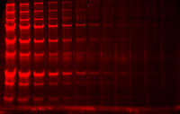
Lumitein™ vs. Coomassie
|
||
 |
 |
|
|
Lumitein |
Coomassie |
|
| Figure 3. PAGE gels containing electrophoretically separated protein marker (loaded in two-fold serial dilution from left to right) were stained with Lumitein™ protein gel stain (left) and Coomassie Blue (right). The Lumitein™-stained gel was imaged using a UV box equipped with EtBr filter (UVP) while the Coomassie-stained gel was imaged using a white light converter (UVP). | ||
1. Meth Mol Biol. 869, 621 (2012).
2. Appl Environ Microbiol (2013) doi:10.1128/AEM.01478-13
3. J Mol Biol (2012) doi:10.1016/j.jmb.2012.10.018
4. Environ Microbiol 12, 2190 (2010).
5. Can J Microbiol 55, 304 (2009).
6. Res Microbiol 160, 401 (2009)
7. Org. Biomol. Chem. (2014) doi:10.1039/C4OB00622D
8. PLoS ONE (2014) doi:10.1371/journal.pone.0112874
9. International Journal of Food Microbiology (2015) doi:10.1016/j.ijfoodmicro.2014.12.012
10. BMC Microbiology (2015) doi: 10.1186/s12866-015-0387-7
11. J Biochem (2015) doi: 10.1093/jb/mvv063
12. Bioscience, Biotechnology, and Biochemistry (2016) doi: 10.1080/09168451.2015.1123609
13. Nature Structural & Molecular Biology (2016) doi:10.1038/nsmb.3237
14. Biophysical Journal (2018) doi:10.1016/j.bpj.2018.03.025
Even though AccuOrange™ buffer does contain SDS, which is required for the dye to bind proteins, the assay is very sensitive to small changes in SDS concentration, and also cannot tolerate non-ionic detergents that form mixed micelles with SDS, like Triton®. Therefore we don't recommend using the kit for cell lysates or other samples with significant amounts of detergents.
Gels stained with One-Step Blue® can be dried just like gels stained with Coomassie. The stain will not interfere with the detection of radiolabeled proteins.
The AccuOrange™ assay is a fluorescent dye-based assay. The dye binds to proteins primarily through hydrophobic interactions. Proteins denature upon heating; the dye binds to the exposed hydrophobic pockets of the protein after cooling. The free AccuOrange™ dye is fluorogenic due to non-radioactive decay but becomes highly fluorescent due to the rigid conformation inside the pocket.
The AccuOrange™ assay more sensitive than traditional protein quantitation assays such as BCA, Bradford and Lowry, and shows superior linearity and reproducibility than the NanoOrange® protein quantitation assay (Thermo Fisher Sci.), but has low tolerance for detergents like SDS and Triton® X-100.
