 New Products
 Earth-Friendly Products
 Biotium Choice Antibodies
 Special Offers


 Powered by Bioz


Content #1
Content #1
Content #1
A visible three-color protein marker for SDS-PAGE or western blotting, with 12 bands ranging from 8 to 245 kDa.
Peacock™ Plus Prestained Protein Marker is a three-color protein ladder that allows you to visually monitor protein separation during SDS-PAGE or protein transfer to membranes for western blotting. We also offer Peacock™ Prestained Protein Marker, a three-color marker with 10 bands ranging from 10 kDa to 180 kDa.
The Peacock™ Plus Prestained Marker contains a total of 12 visible bands. This includes 10 blue bands ranging from 8 kDa to 245 kDa, plus a red band at 75 kDa and green band at 25 kDa for easy band identification. Peacock™ Plus Prestained Protein Marker is ready to load with no heating or other preparation needed. Recommended loading is 3-5 uL per well for mini-gels.
Also visit our Protein Detection, Quantitation, & Analysis technology page to learn about our safe and sensitive protein gel stains and western blotting normalization reagents.
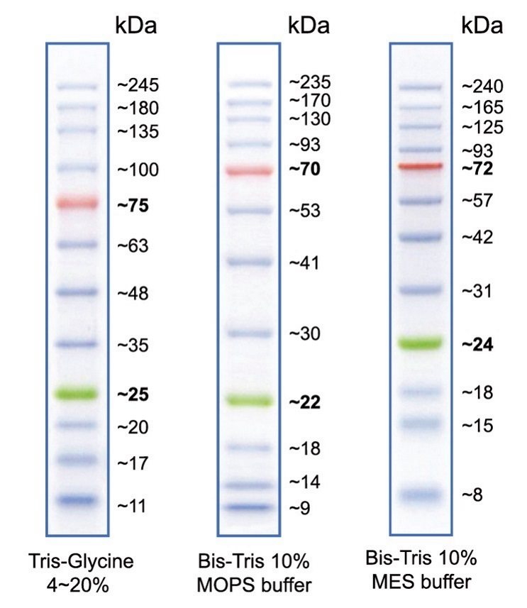
Apparent molecular weights of bands in Peacock Plus Prestained Protein Marker on various SDS-PAGE gels.
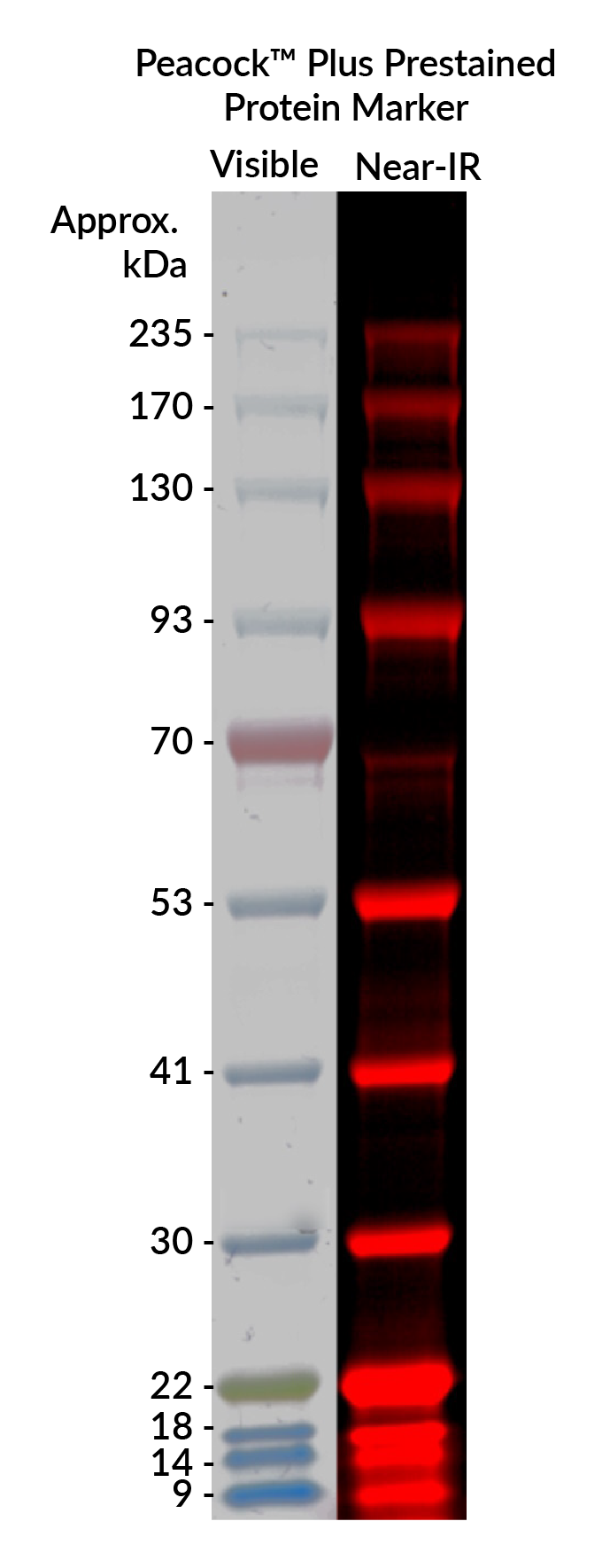
Comparison of Peacock™ Plus Prestained Protein Marker visible and fluorescent bands on a 10% MOPS PAGE gel. The The blue and green marker bands, but not the orange marker band, can be detected using a near-IR fluorescence imager. Fluorescence was imaged in the 700 channel on a LICORbio Odyssey® M imaging system. Approximate band molecular weights are shown next to each marker.
Even though AccuOrange™ buffer does contain SDS, which is required for the dye to bind proteins, the assay is very sensitive to small changes in SDS concentration, and also cannot tolerate non-ionic detergents that form mixed micelles with SDS, like Triton®. Therefore we don't recommend using the kit for cell lysates or other samples with significant amounts of detergents.
Gels stained with One-Step Blue® can be dried just like gels stained with Coomassie. The stain will not interfere with the detection of radiolabeled proteins.
The AccuOrange™ assay is a fluorescent dye-based assay. The dye binds to proteins primarily through hydrophobic interactions. Proteins denature upon heating; the dye binds to the exposed hydrophobic pockets of the protein after cooling. The free AccuOrange™ dye is fluorogenic due to non-radioactive decay but becomes highly fluorescent due to the rigid conformation inside the pocket.
The AccuOrange™ assay more sensitive than traditional protein quantitation assays such as BCA, Bradford and Lowry, and shows superior linearity and reproducibility than the NanoOrange® protein quantitation assay (Thermo Fisher Sci.), but has low tolerance for detergents like SDS and Triton® X-100.
