 New Products
 Earth-Friendly Products
 Biotium Choice Antibodies
 Special Offers


 Powered by Bioz


Content #1
Content #1
Content #1
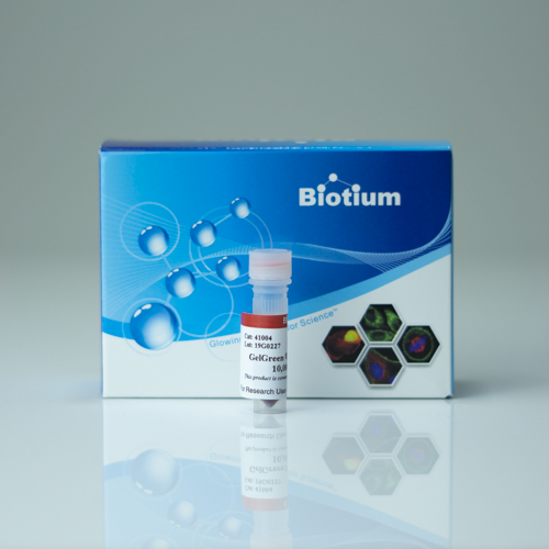
GelGreen® is a sensitive, non-mutagenic and environmentally safer green fluorescent DNA gel stain.
GelGreen® is a highly sensitive, non-toxic green fluorescent nucleic acid dye designed for staining DNA in agarose gels.
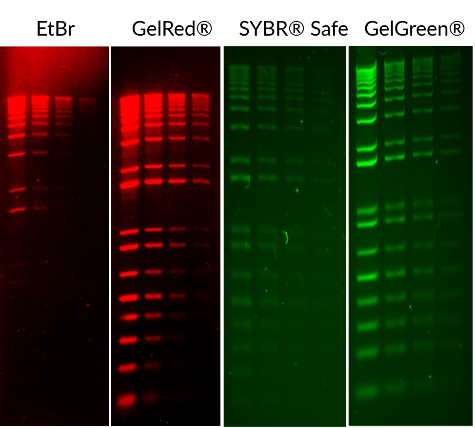
GelGreen® is far more sensitive than SYBR® Safe. Unlike SYBR® dyes, which are known to be unstable, GelGreen® is very stable, both hydrolytically and thermally. GelGreen® is compatible with a 254 nm UV transilluminator, and can be imaged using a SYBR® Green or GelStar® filter. It also can be used with visible blue light excitation imagers (blue LED light box or Dark Reader®). With blue light illuminators, researchers can avoid exposure to UV irradiation for themselves and their DNA samples, for a safer work environment and higher cloning efficiency.
A series of safety tests have confirmed that GelGreen® is noncytotoxic, nonmutagenic and nonhazardous at concentrations above those used for gel staining. As a result, working strength GelGreen® can be safely disposed of down the drain or in regular trash, providing convenience and reducing cost in waste disposal. For detailed test results, you may download a complete GelRed®/GelGreen® Safety Report.
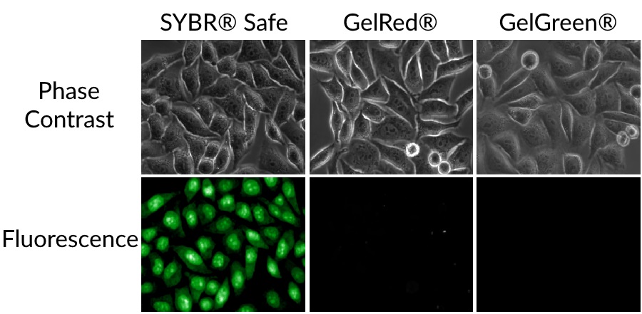
Many so-called “safe” DNA dyes like SYBR® Safe, Midori Green, GreenSafe, SafeView™, and RedSafe™ not only have low sensitivity, but also readily penetrate living cells to bind DNA, and some are cytotoxic. Unlike these dyes, GelGreen® is cell membrane-impermeant, so it cannot enter living cells to interact with their DNA. See our Gel Stains Comparison Flyer or Gel Stains Comparison White Paper for details.
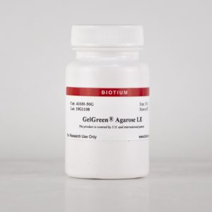
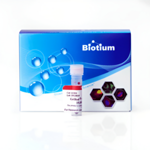
For new users, we recommend GelGreen® 10,000X in water (catalog no. 41005), our latest formulation that eliminates the hazards of handling DMSO for better safety. We continue to offer GelGreen® 10,000X solution in DMSO for established users who do not wish to change their existing laboratory protocols. We also offer GelGreen® Agarose for convenient and safer preparation of precast gels. Also see GelRed® Nucleic Acid Gel Stain, our safer replacement for ethidium bromide. GelGreen® can be used to stain ssDNA and RNA, but we recommend GelRed® for this application because it is five times more sensitive for single stranded nucleic acids than GelGreen®. Also learn about DNAzure® 2.0 Visible Blue DNA Gel Stain Kit for visualizing DNA bands with the naked eye with no need for a UV light source. DNAzure® 2.0 delivers excellent sensitivity, detecting as little as ~1 ng of double-stranded DNA in both agarose and polyacrylamide gels.
| Product / Method | Procedure | Advantages | Disadvantages | Recommended for |
|---|---|---|---|---|
| DNA staining with EMBER™ Ultra DNA Gel Kit | Agarose is supplied pre-coated with EMBER™ Ultra Dye, just dissolve, heat, and pour. | • Safer and more convenient, no need to handle concentrated dye • Superior sensitivity, detect as little as ≤1 ng DNA • No need for post-electrophoresis staining • Optimal for blue LED gel imagers | • Not suitable for PAGE, DGGE, EMSA, or PFGE gels • Dye may cause band migration issues when loading larger amounts of DNA (more than ~200 ng/band), or for some restriction digests | • Routine agarose gels |
| RNA staining with EMBER™ Ultra RNA Gel Kit | Agarose is supplied pre-coated with EMBER™ Ultra Dye, just dissolve, heat, and pour. | • Safer and more convenient stain for RNA, no need to handle concentrated dye • Superior sensitivity, detect as little as ≤5 ng RNA • No need for post-electrophoresis staining • Included loading dye contains formamide for denaturing • Optimal for blue LED gel imagers | • Will stain DNA as well as RNA • Dye may cause band migration issues when loading larger amounts of RNA (more than ~200 ng/band) | • Routine RNA gel electrophoresis • Evaluate total RNA integrity and DNA contamination |
| DNA prestaining with GelRed® Prestain Plus 6X DNA Loading Dye | GelRed® loading buffer is added directly to the DNA sample before loading | • Fast & simple: one-step sample loading & DNA staining • Less concentrated dye for safer handling • Can re-run a gel to use empty lanes | • Not recommended for PAGE, DGGE, EMSA, or PFGE gels • Dye may cause band migration issues when loading larger amounts of DNA (more than ~100 ng/band), or for some restriction digests | • Routine agarose gels • Recommended loading 50-200 ng ladder or 2-5 uL PCR product ( ~100 ng/band or less) |
| Precast staining with GelRed® 10,000X in water or GelGreen® 10,000X in water | GelRed® or GelGreen® is mixed with molten agarose before gel casting | Familiar protocol, rapid results | ||
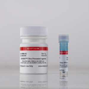
| Precast staining with GelRed® Agarose LE or GelGreen® Agarose LE | Agarose is supplied pre-coated with GelRed® or GelGreen®, just dissolve, heat, and pour | Safer & more convenient, no need to handle concentrated dye | ||
| Post-electrophoresis staining with GelRed® 10,000X in water or GelGreen® 10,000X in water - or - GelRed® 3X in water | No fluorescent dye is added to the gel, it is stained in 3X GelRed® or 3X GelGreen® solution after electrophoresis | • Most accurate sizing/sharpest bands • Staining solution can be re-used • Enhance sensitivity by adding NaCl | Extra staining step (up to 30 minutes) after electrophoresis (some customers report good results after only 5 minutes if dye is not reused) | • Highly accurate band sizing • Gels with more than ~100 ng DNA per band • Analyzing restriction digests |
| Post-electrophoresis staining with DNAzure® 2.0 Visible Blue DNA Gel Stain Kit | No fluorescent dye is added to the gel, it is stained in DNazure® 2.0 solution and then exposed to a bright light source to generate visible blue DNA bands. We recommend the Glo-Plate™ White Photoactivation Device as a light source for developing DNAzure® 2.0-stained gels | • Allows visualization of DNA bands by the naked eye, no need for a UV light source • Detect as little as ~1 ng DNA • Stained bands are stable in gel for weeks • Also emits near-IR fluorescence (~700 nm) for detection on near-IR imaging systems | Extra staining step (up to 30 minutes) followed by a light exposure step (up to 30 minutes) to generate visible blue DNA bands | • Routine DNA agarose gels • Visualizing gels without the UV light or expensive imaging systems • Recommended loading 50-200 ng DNA per lane |
| Post-electrophoresis staining of PAGE gels using PAGE GelRed® 10,000X or 1X in water | No fluorescent dye is added to the gel, it is stained in 1X PAGE GelRed® solution after electrophoresis | • Formulated for efficient penetration and staining of polyacrylamide gels • Like the classic GelRed®, it is safe and environmentally friendly | Extra staining step of approx. 30 minutes after electrophoresis | Staining of nucleic acids in PAGE gels |
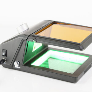
Biotium also offers the Gel-Bright™ Laser Diode Gel Illuminator, a unique laser-diode-based illuminator that offers sensitive staining for both red and green dyes. Also learn about our Go-Go™ Fast DNA Gel Running Buffer for running gels 3X faster than with TAE or TBE buffer.
For more information, view our DNA Stain technology page, and see our GelRed® & GelGreen® FAQs.
Download a list of curated references for GelRed® and GelGreen®.
Download a list of curated references for GelRed® and GelGreen®.
We and other users have often observed that GelGreen® stains ssDNA and RNA orange/ pink and dsDNA green. We have also seen that smaller dsDNA fragments can appear orange-pink, the color ranging from white-pink-orange. We are not sure about the underlying mechanism, possibly the structure of single-stranded nucleic acids favors an altered stacking interaction of GelGreen® monomers leading to the formation of J-aggregates that have red emission.
Yes, use the post-staining protocol for polyacrylamide gels. For polyacrylamide gels containing 3.5-10% acrylamide, typical staining time is 30 minutes to 1 hour with gels of higher acrylamide content requiring longer staining time.,
Biotium also offers PAGE GelRed® a non-toxic, non-mutagenic dye specifically designed for staining DNA in polyacrylamide gels.
We don’t recommend adding GelRed® or GelGreen® directly to loading buffer, because this can result in inaccurate band migration. Biotium offers 6X GelRed® Prestain Loading Buffers designed for this application, although we do not recommended them for applications where precise DNA band sizing is required. For the most accurate determination of DNA band sizes, we recommend using GelRed® post-staining (see the GelRed® protocol for details).
There are a few possibilities:
Many customers use GelRed® or GelGreen® precast gels for convenience. However, because GelRed® and GelGreen® are high affinity dyes designed to be larger dyes to improve their safety, they can affect the migration of DNA in precast gels. Some samples, such as restriction digested DNA may migrate abnormally in GelRed® or GelGreen® precast gels.
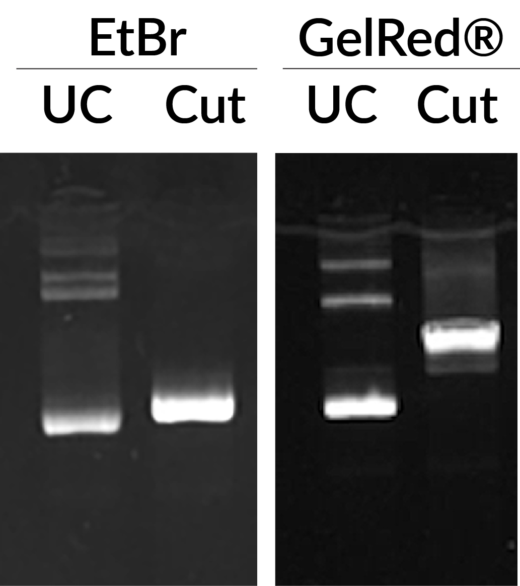
Ethidium bromide (EtBr) and GelRed® in precast staining of uncut pGL3 plasmid (UC), or pGL3 plasmid cut with Mul1 (Cut). GelRed® displays abnormal band migration of cut pGL3 plasmid compared to EtBr. Each lane was loaded with 75 ng DNA.
Tip #1: Load less DNA
Smearing and smiling in GelRed® or GelGreen® precast gels most often caused by overloading of DNA. If you see band migration shifts or smearing and smiling, try reducing the amount of DNA loaded. The recommended loading amount for ladders and samples of known concentration is 50-200 ng/lane. For samples of unknown concentration, try loading one half or one third of the usual amount of DNA. This usually solves band migration problems.
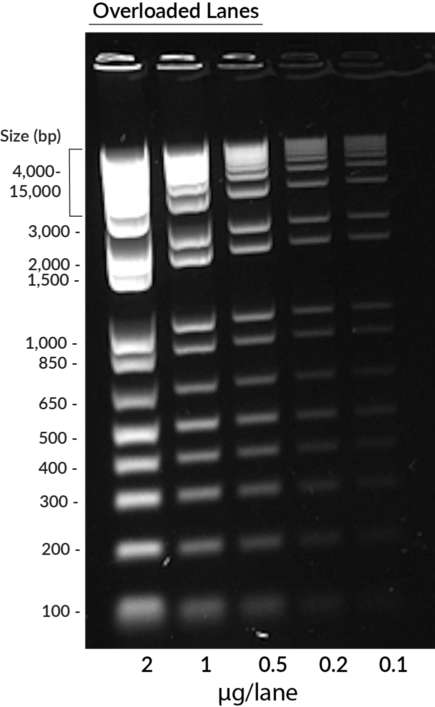
GelRed® in precast staining of 1 kb DNA ladder (Invitrogen) on a 1% agarose TBE gel. Ladder was loaded in the amounts of 2 ug, 1 ug, 0.5 ug, 0.2 ug, and 0.1 ug per lane from left to right. Overloaded lanes with more than 0.2 ug DNA displayed smiling and smearing bands.
Tip #2: Try the post-staining protocol
To avoid any interference the dye may have on DNA migration, we recommend using the post-staining protocol. If your application requires loading more than the recommended amount of DNA, use the post-staining protocol. While we recommend post-staining gels for 30 minutes, you may be able see bands in as little as five minutes, depending on how much DNA is present. Post-staining solutions can be reused. See the GelRed® Product Information Sheet or GelGreen® Product Information Sheet for detailed protocols.
Other tips to improve agarose gel resolution:
