Support & Resources

+1

CD63 Monoclonal Mouse Antibody (MX-49.129.5)

 Request Custom Conjugation

Product Attributes

Antibody number

#0525

Antibody reactivity (target)

CD63

Antibody type

Primary

Host species

Mouse

Clonality

Monoclonal

Clone

MX-49.129.5

Isotype

IgG1, kappa

Molecular weight

26 kDa (core protein); 30-60 kDa (glycosylated)

Synonyms

gp55; granulophysin; Lysosomal-associated membrane protein 3 (LAMP-3); Mast cell antigen AD1; melanoma 1 antigen; Melanoma-associated antigen MLA1; Melanoma-associated antigen ME491; MLA1; NGA; Ocular melanoma-associated antigen; OMA81H; PTLGP40; Tetraspanin-30; TSPAN30

Human gene symbol

CD63

Entrez gene ID

967

SwissProt

P08962

Unigene

445570

Immunogen

Full length CD63 of human origin

Verified antibody applications

Exosome staining (verified), Flow (surface) (verified), IF (verified), IHC (FFPE) (verified), WB (verified)

Antibody target cellular localization

Plasma membrane

Species reactivity

Human, Mouse

Expected antibody applications

IP (published for clone)

Antibody application notes

Higher concentration may be required for direct detection using primary antibody conjugates than for indirect detection with secondary antibody, Immunofluorescence: 0.5-1 ug/mL, Immunohistology formalin-fixed 0.5-1 ug/mL, Staining of formalin-fixed tissues is enhanced by boiling tissue sections in 10 mM citrate buffer, pH 6.0, for 10-20 min followed by cooling at RT for 20 minutes, Flow Cytometry 0.5-1 ug/million cells/0.1 mL, Western blotting 0.5-1 ug/mL, Optimal dilution for a specific application should be determined by user

Positive control

U87MG, SK-MEL-28, HL60, THP-1, NIH/3T3, or MCF-7 cells. Human melanoma, spleen or lymphoma tissue.

Shipping condition

Room temperature

Storage Conditions

Store at 2 to 8 °C, Protect fluorescent conjugates from light, Note: store BSA-free antibodies at -10 to -35 °C

Shelf life

Guaranteed for at least 24 months from date of receipt when stored as recommended

Regulatory status

For research use only (RUO)

Antibody/conjugate formulation

Conjugates: 0.1 mg/mL in PBS/0.1% BSA/0.05% azide, HRP conjugates: 0.1 mg/mL in PBS/0.05% BSA, Purified: 0.2 mg/mL in PBS/0.05% BSA/0.05% azide, Purified, BSA-free: 1 mg/mL in PBS without azide

Cell/tissue expression

Platelets, Granulocytes, Lymphocytes, Monocytes/macrophages

Antibody research areas

Cancer, Exosomes/EVs, Immunology

Conjugation
Don’t see what you’re looking for? Contact us for more conjugation options.
Note: Please be advised that primary antibodies with catalog numbers that begin with “BN” will only be offered conjugated to CF®405S, CF®488A, CF®568, CF®594, CF®640R, CF®647, and biotin as of 5/27/2022. However, previously available primary antibody conjugates that are in stock will remain available until inventory is exhausted. Please view the list of available conjugates on the product page. We also recommend our easy-to-use Mix-n-StainTM Antibody Labeling Kits for labeling antibodies with all CF® Dyes. We apologize for the inconvenience.
Size
Catalog #
price
Qty
50 uL
100 uL
250 uL
500 uL
Clear selection
ADD TO CART
(Please select quantity above )

Wishlist updated! View wishlist

Product Description

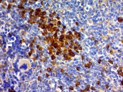
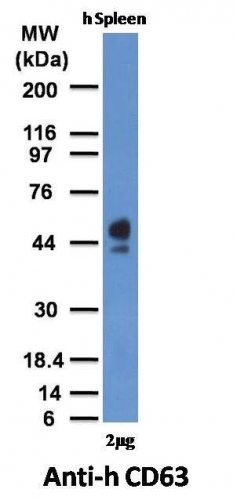
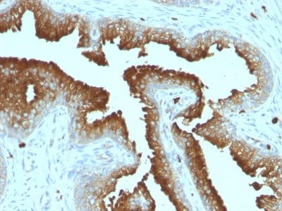
This MAb recognizes protein of 26 kDa-60 kDa, which is identified as CD63. Its epitope is different from that of MAb LAMP3/529. The tetraspanins are integral membrane proteins expressed on cell surface and granular membranes of hematopoietic cells and are components of multi-molecular complexes with specific integrins. The tetraspanin CD63 is a lysosomal membrane glycoprotein that translocates to the plasma membrane after platelet activation. CD63 is expressed on activated platelets, monocytes and macrophages, and is weakly expressed on granulocytes, T cell and B cells. It is located on the basophilic granule membranes and on the plasma membranes of lymphocytes and granulocytes. CD63 is a member of the TM4 superfamily of leukocyte glycoproteins that includes CD9, CD37 and CD53, which contain four transmembrane regions. CD63 may play a role in phagocytic and intracellular lysosome-phagosome fusion events. CD63 deficiency is associated with Hermansky-Pudlak syndrome and is strongly expressed during the early stages of melanoma progression.
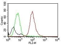
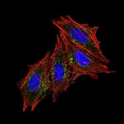
Primary antibodies are available purified, or with a selection of fluorescent CF® dyes and other labels. CF® dyes offer exceptional brightness and photostability. See the CF® Dye Brochure for more information. Note: Conjugates of blue fluorescent dyes like CF®405S and CF®405M are not recommended for detecting low abundance targets, because blue dyes have lower fluorescence and can give higher non-specific background than other dye colors.

Stock status: Because Biotium offers a large number of antibody and conjugation options, primary antibody conjugates may be made to order. Typical lead times are up to one week for CF® dye and biotin conjugates, and up to 2-3 weeks for fluorescent protein and enzyme conjugates. Please email order@biotium.com to inquire about stock status and lead times before placing your order.

Catalog Number Key for Primary Antibody Conjugates

Antibody # prefixConjugationEx/Em (nm)Laser lineDetection channelDye Features
BNC04CF®405S404/431405DAPI (microscopy), AF405CF®405S Features
BNC88CF®488A490/515488GFP, FITCCF®488A Features
BNC68CF®568562/583532, 561RFP, TRITCCF®568 Features
BNC94CF®594593/614561Texas Red®CF®594 Features
BNC40CF®640R642/662633-640Cy®5CF®640R Features
BNC47CF®647650/665633-640Cy®5CF®647 Features
BNCBBiotinN/AN/AN/A
BNUBPurifiedN/AN/AN/A
BNUMPurified,
BSA-free
N/AN/AN/A
Alexa Fluor, Pacific Blue, Pacific Orange, and Texas Red are trademarks or registered trademarks of Thermo Fisher Scientific; Cy is a registered trademark of Cytiva; IRDye, LI-COR, and Odyssey are registered trademarks of LI-COR Bioscience.

References

Note: References for this clone sold by other suppliers may be listed for expected applications.

Mod Pathol (2010) 23: 1418-1428. (immunoprecipitation)


You may also like…