New Products
 Earth-Friendly Products
 Biotium Choice Antibodies
 Special Offers
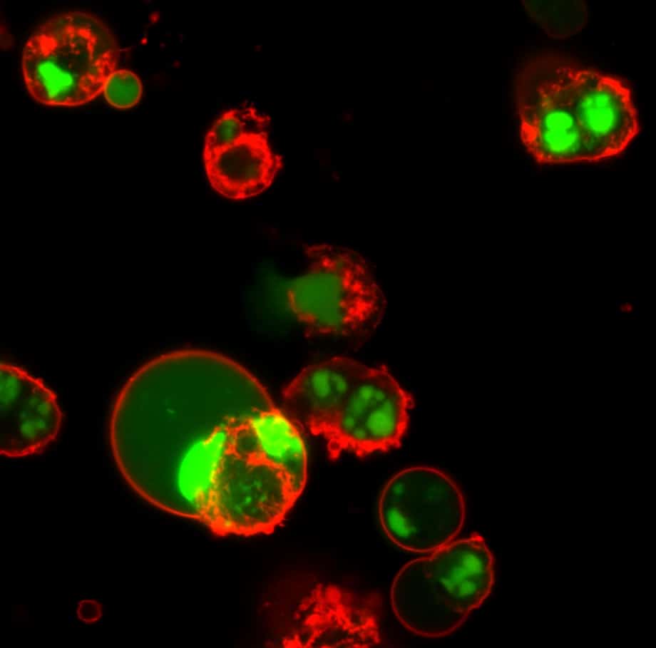
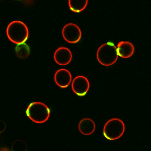
Annexin V Apoptosis Probes
Annexin V is a protein with high affinity for phosphatidylserine (PS). During apoptosis, PS is translocated from the inner to the outer leaflet of the plasma membrane, where it can be stained by Annexin V conjugates. We offer Annexin V labeled with CF® Dyes and other labels, and in Annexin V kits with other apoptotic or necrotic cell stains.
Annexin staining is calcium-dependent, and is traditionally performed in an optimized Annexin binding buffer. Annexin conjugates also can be used to stain cells in culture medium for real-time apoptosis monitoring. Annexin can be fixed with formaldehyde after staining as long as calcium is included in all buffers. However, Annexin staining cannot be performed on fixed cells or tissues. For fixed cell apoptosis stains, see our TUNEL Assays.
Annexin V Conjugates
| Product | Features |
|---|---|
| Annexin V Conjugates | • Wide selection of CF® dyes, R-PE, biotin, and other labels • 50 ug/mL in 10 mM Tris, 1 mM EDTA, 30 mM NaCl, 1 mg/mL bovine serum albumin, 0.1% sodium azide, pH 7.5 |
| CF® Dye Annexin V Conjugates, Azide Free, Lyophilized | • Azide-free for no-wash, real-time live cell imaging in culture medium • Choice of twelve CF® dyes |
| Near-IR CF® Dye Annexin V | • Lyophilized, preservative-free • Choice of five near-IR CF® dyes |
| Annexin V (His Tag) | • Recombinant Annexin V with or without His Tag expressed from E.coli |
| 5X Annexin Binding Buffer | • Concentrated buffer optimized for Annexin V binding |
Alpha-Bungarotoxin Neuromuscular Junction Probes
Alpha-bungarotoxin is a polypeptide snake toxin that binds to the nicotinic acetylcholine receptor found at the neuromuscular junction with high affinity. Fluorescent alpha-bungarotoxin can be used for labeling of nicotinic acetylcholine receptors at neuromuscular junctions in tissue sections. Alpha-bungarotoxin may also be used for detection of GABA A receptor subsets in cells, or for labeling recombinant proteins that express the alpha-bungarotoxin binding site (BBS) epitope tag.
We offer α-bungarotoxin conjugated to a wide selection of CF® Dyes and other labels, as well as unconjugated α-bungarotoxin.
- Choose from 9 bright and stable CF® Dye colors or other labels
- Dye options for super-resolution and 2-photon imaging


α-Bungarotoxin Conjugates
| Product | Features |
|---|---|
| CF® Dye & Other α-Bungarotoxin Conjugates | • Choice of popular CF® dye colors or a selection of classic fluorophores |
| Biotin-XX-α-Bungarotoxin | • Biotinylated α bungartoxin |
| α-Bungarotoxin | • Unconjugated α-bungarotoxin, potent inhibitor of the motor endplate acetylcholine receptor |
Cholera Toxin & Transferrin Conjugates
Cholera Toxin Subunit B Conjugates
Cholera toxin subunit B binds to ganglioside GM1 in lipid rafts on the plasma membrane. Cholera toxin subunit B is reported to be internalized by clathrin-dependent and independent pathways depending on cell type. We offer cholera toxin subunit B conjugated to a selection of CF® Dyes. The conjugates can be used to label lipid rafts on the cell surface, track endocytic vesicles, and as a retrograde neuronal tracer.
Transferrin Conjugates
Transferrin delivers iron to vertebrate cells. After binding to its cell surface receptor, transferrin is internalized and the acidic environment in endocytic vesicles favors dissociation of iron from the transferrin-receptor complex. Following iron release, the apotransferrin is recycled to the plasma membrane and released from its receptor to scavenge more iron. We offer human transferrin labeled with CF® Dyes for imaging recycling endosomes.
Cholera Toxin & Transferrin Conjugates
| Product | Features |
|---|---|
| CF® Dye Cholera Toxin | • Binds lipid rafts, can be used as endocytosis probe or neuronal retrograde tracer • Available with a large choice CF® dye colors |
| Cholera Toxin Subunit B (Recombinant) | • Purified from E. coli as a single, non-glycosylated polypeptide chain • Free of stabilizers and ready to conjugate. |
| CF® Dye Human Transferrin | • Trace recycling endosomes • Available with a selection of popular CF® dye colors |
Carbohydrate-Binding Lectins
Biotium’s CF® Dye Lectin Conjugates offer bright, photostable labeling for clear visualization of cell-surface glycans and tissue structures. Concanavalin A (Con A) and wheat germ agglutinin (WGA) can be used to selectively stain the cell surface of live cells, and withstand fixation and permeabilization. When cells are fixed and permeabilized before staining, fluorescent lectins stain both cell surface and organelles in the secretory pathway. WGA also is a live cell Gram stain for bacteria, while Concanavalin A and WGA conjugates can be used to stain cell wall and bud scars in yeast. See our Microbiology Technology Page for more information.
Biotium also offers Peanut Agglutinin (PNA) for β-galactose, Tomato Lectin (LEL/TL) for vasculature staining, Ulex Europaeus Agglutinin I (UEA I) for staining endothelial cells, Datura Stramonium Lectin (DSL) for GlcNAc oligomers, Sambucus Nigra Lectin (SNA) for sialic acid, and Phaseolus Vulgaris Leucoagglutinin (PHA-L) for stimulating lymphocyte and T cell proliferation. These conjugates are ideal for live or fixed cell staining, glycan profiling, microbiology, and labeling blood vessels or microglia in tissue sections.
View Biotium’s full selection of lectin CF® Dye conjugates below.
Lectin Conjugates
| Product | Features |
|---|---|
| CF® Dye Concanavalin A (Con A) | • Cell surface stain for yeast, fungi, and mammalian cells • Selectively binds to a-mannopyranosyl and a-glucopyranosy residues • Available with a wide selection of CF® Dyes |
| CF® Dye Wheat Germ Agglutinin (WGA) | • Cell surface stain for mammalian cells and gram+ bacteria • Also stains yeast bud scars • Has high affinity for sialic acid and N-acetylglucosamine • Choose from a wide selection of CF® Dyes or HRP |
| CF® Dye Peanut Lectin (PNA) from Arachis hypogaea | • Specific for terminal β-galactose and binds preferentially to galactosyl (β-1,3) N-acetylgalactosamine • Choice of 6 CF® dye colors |
| CF® Dye Lycopersicon Esculentum (Tomato) Lectin (LEL, TL) | • Marker for blood vessels and microglial cells • Binds to [GlcNAc] 1,3-N-acetylglucosamine, glycophorin, and Tamm-Horsfall glycoprotein • Used to study tumor angiogenesis or tracing neovascular development in xenograft models • Choice of 7 CF® Dyes or biotin |
| CF® Dye Ulex Europaeus Agglutinin I (UEA I) | • Marker for human endothelial cells and incompletely differentiated gastrin cells • Binds to glycoproteins and glycolipids containing α-linked fucose residues • Choice of 7 CF® Dyes or biotin |
| CF® Dye Phaseolus Vulgaris Leucoagglutinin (PHA-L) | • Used to stimulate lymphocyte and T cell proliferation • Choice of 7 CF® Dyes or biotin |
| CF® Dye Datura Stramonium Lectin (DSL) | • Binds to (beta-1,4) linked N-acetylglucosamine oligomers • Choice of 7 CF® Dyes or biotin |
| CF® Dye Sambucus Nigra Lectin (SNA, EBL)) | • Binds to sialic acid attached to terminal galactose • Choice of 7 CF® Dyes or biotin |
Dextran, BSA, & Hydrazide Fluid Phase Tracers
Dextrans are water soluble branched-chain polysaccharides. Fluorescently labeled dextrans are used as markers for trafficking of fluid phase endocytic cargo to lysosomes, tracers for epithelial and endothelial permeability, and tracers for neuronal morphology. Biotium offers CF® Dye Dextrans in a variety of molecular weights from 10,000 to 250,000.
Note: Formaldehyde fixation preserves the localization of dextrans in endosomes, but the labeling generally cannot tolerate detergent permeabilization.
CF® Dye Bovine Serum Albumin (BSA) also can be used as a tracer for protein uptake and permeability.
CF® Dye Hydrazides are water soluble dyes that can be used as microinjected cytoplasmic tracers or fluid-phase markers. Hydrazides also can be used for labeling aldehyde or ketone groups, such as carbohydrate molecules after peroxidation with periodate. However, for this application we recommend using CF® Dye aminooxy forms, which are more reactive toward these groups than hydrazide forms. For 2D-DIGE platform, we recommend CFDI hydrazides. Also see our full selection of polar tracer dyes.
CF® Dye Dextrans
| Product | Conjugates |
|---|---|
| CF® Dye Dextran, 3500 MW | UV-excitable CF®350 |
| CF® Dye Dextran, 10,000 MW | Available with a wide selection of CF® Dye colors |
| CF® Dye Dextran, 40,000 MW | Green fluorescent CF®488A, near-IR CF®680, CF®750 or CF®770 |
| CF® Dye Dextran, 70,000 MW | Blue fluorescent CF®350, Green fluorescent CF®488A, Far-red fluorescent CF®633, near-IR CF®680, CF®750 or CF®770 |
| CF® Dye Dextran, 150,000 MW | Green fluorescent CF®488A, near-IR CF®680, CF®750 or CF®770 |
| CF® Dye Dextran, 250,000 MW | Green fluorescent CF®488A, near-IR CF®680, CF®750 or CF®770 |
CF® Dye BSA
| Product | Conjugates |
|---|---|
| CF® Dye BSA | Green fluorescent CF®488A, red fluorescent CF®594, far-red fluorescent CF®640R, or near-IR fluorescent CF®680 |
CF® Dye Hydrazides
| Product | Features |
|---|---|
| CF® Dye Hydrazide | • Fixable polar tracers • Wide selection of bright, photostable, and highly soluble CF® dye colors |
dUTP, cAMP, & Other Labeled Nucleotides
Labeled Nucleotides
Labeled nucleotides can be used to synthesize labeled DNA probes that are commonly used for in situ hybridization and nucleic acid blotting applications. Biotium offers a large selection of labeled nucleotides, including nucleotides conjugated to CF® Dyes, other fluorophores, and biotin, and our biotinylated nucleotides are available with a variety of linker lengths.
Common applications of specific labeled nucleotides include:
- Labeled dUTP: TUNEL assays (also see our CF® Dye TUNEL Assay Kits)
- Aminoallyl nucleotides: introduce free amine groups into probes for subsequent labeling with reactive dyes or biotin
- Bromine and digoxygenin (DIG) labeled nucleotides: detected using specific antibodies
- Bromo-deoxyuridine (BrdU): monitor DNA synthesis in cells (see our anti-BrdU antibodies)
- Fluorescently-labeled cAMP analogs: image cAMP bound to the cAMP receptor on the surface of Dictyostelium cells
Labeled Nucleotides
| Product | Features |
|---|---|
| CF® Dye Nucleotides | • dUTP, dCTP, UTP, and other nucleotides • For probe labeling, TUNEL assay with labeled dUTP, and other applications • Available with a large choice of CF® dye colors |
| Other Fluorescent Nucleotides | • dUTP and other nucleotides with classic fluorescent dyes |
| Biotinylated Nucleotides | • Biotinylated nucleotides for RNA or DNA probe synthesis with a variety of linker lengths |
| Bromine & DIG Labeled Nucleotides | • BrdU, DIG-dUTP, and other nucleotides for detection with specific antibodies |
| Aminoallyl Nucleotides | • For generating amine-functionalized probes for subsequent labeling with amine-reactive dyes or biotin |
| Labeled cAMP analogs | • cAMP receptor probes • Available with CF® dyes, other fluorophores, or biotin |
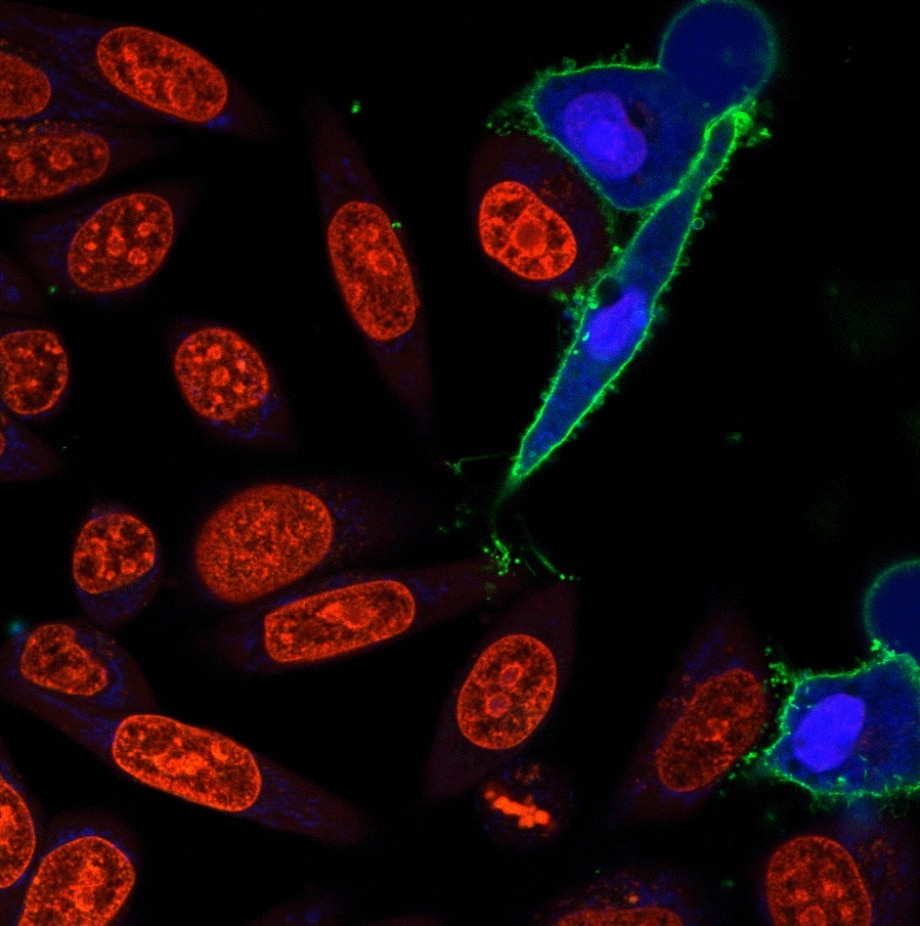
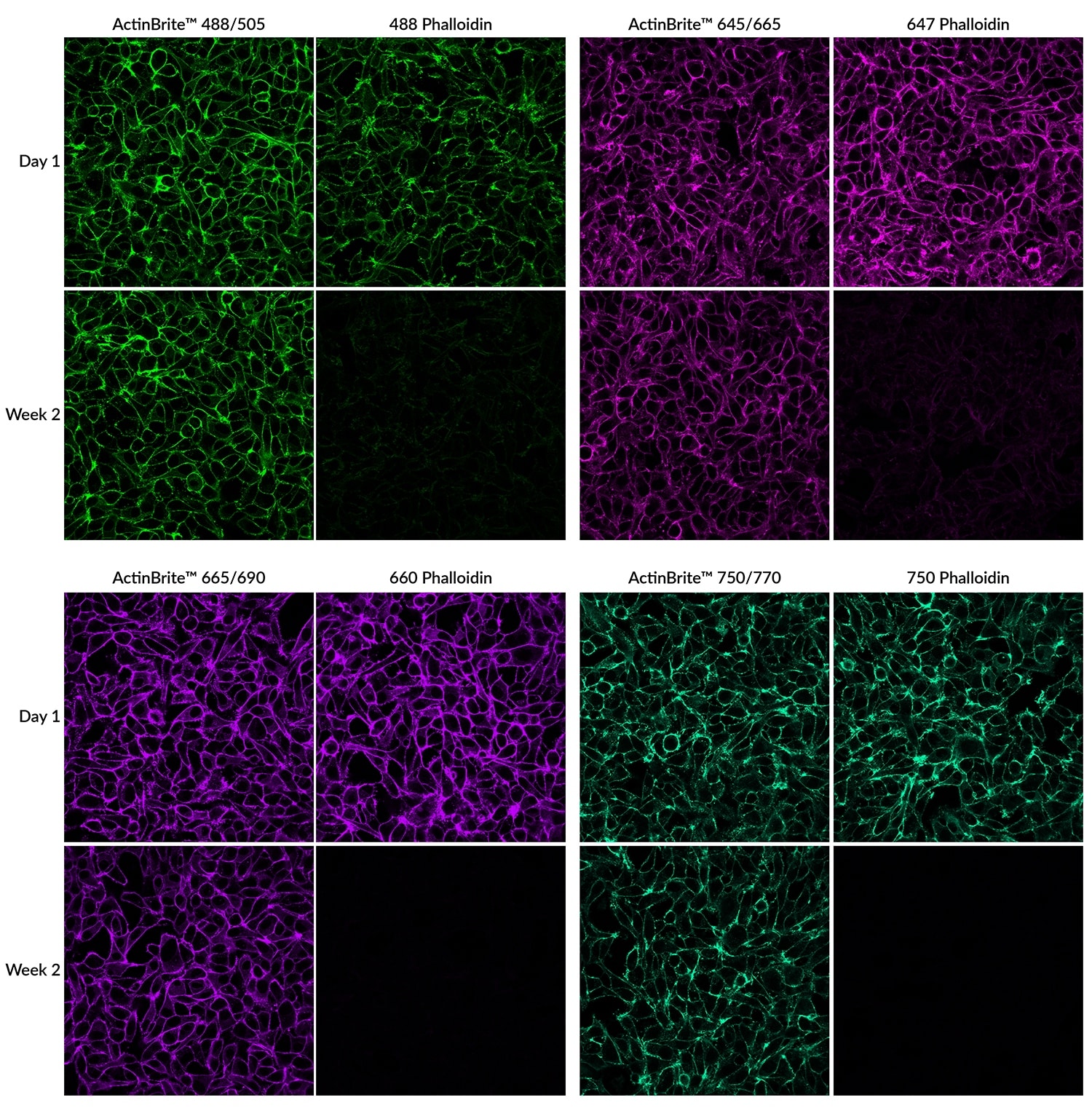
Phalloidin Conjugates for F-Actin Staining
F-actin Staining in Fixed Cells with No Fading
Features
- Novel phalloidin conjugates that preserve strong F-actin binding
- Enables sample storage for 1+ month, depending on conjugate
- Offered in 7 colors from green to near-IR for easy multiplexing
- Direct replacements for any phalloidin conjugate
Biotium also offers phalloidin conjugates with a wide selection of CF® Dyes in addition to traditional dyes and biotin. A number of our CF® Dyes have been validated in super-resolution imaging by STORM, STED, SIM, and other methods.
Phalloidin conjugates are not cell-permeant, and are typically used to stain fixed and permeabilized cells. However, they also can be loaded into live cells via cationic liposomes. For live cell cytoskeletal staining, we also offer ViaFluor® Live Cell Microtubule Stains.
CF® Dye conjugates of Vitamin D-Binding Protein (Vitamin D-BP, also known as GC Globulin) are also available for visualizing monomeric G-actin in fixed and permeabilized cells. They can also be co-stained with phalloidin for comparing G-actin and F-actin distribution.
Phalloidin Conjugates
| Product | Features |
|---|---|
| ActinBrite™ High Affinity Phalloidin Conjugates | • Novel phalloidin conjugates with high F-actin binding affinity • Allows stained samples to be stored for over a month depending on the conjugate • Available in 5 colors from green to near-IR |
| Phalloidin Conjugates | • Wide selection of CF® dyes, classic fluorophores, and biotin • Low cost trial sizes available for CF® dye phalloidins |
Fluorescent Streptavidin Conjugates
Streptavidin is a bacterial protein with four biotin binding sites that binds biotin with extremely high affinity. Streptavidin conjugates are used in many applications to detect biotinylated antibodies or other targets, or to prepare multivalent complexes of biotinylated molecules. Our streptavidin conjugates are high quality streptavidin labeled with a wide selection CF® Dyes and other labels. Also see our Monoclonal Anti-Biotin Conjugates.
| Product | Features |
|---|---|
| Streptavidin Conjugates | • High affinity biotin-binding protein • Wide selection of CF® dyes and other labels |
Biotin Bioconjugates & Fluorescent Biotin
We provide a wide variety of biotin bioconjugates for detection with streptavidin conjugates or anti-biotin antibodies (see above). Also see primary and secondary antibodies available as biotin conjugates. We also offer the Biotin SE Protein Labeling Kit and Mix-n-Stain™ Biotin Antibody Labeling Kit, so you can label your own conjugates.
We also offer fluorescent biotins and biocytin. Potential applications of fluorescent biotin include detection of biotin binding sites and the degree of biotinylation of proteins, and for the measurement of avidin and streptavidin in crude biological samples. Biocytin is biotin with a fixable amine group. Fluorescent biocytin can be used as a polar tracer by microinjection to study the morphology of cells. See our full selection of polar tracers.
Biotin Conjugates
| Product | Features |
|---|---|
| Biotin-XX-α-Bungarotoxin | • Motor endplate acetylcholine receptor probe for neuromuscular junctions |
| Biotin-Annexin V | • Phosphatidylserine-binding protein for labeling apoptotic cells |
| Biotin-XX-Phalloidin | • F-actin probe for fixed cells |
| Biotin Nucleotides | • Selection of nucleotides for probe synthesis or TUNEL assay |
| Biotin-DHPE | • Biotinylated phospholipid |
| Biotin-cAMP | • Biotinylated cAMP receptor probe |
| Biotin-cGMP | • Biotinylated cGMP receptor probe |
| Biotin NTA | • For sensitive detection of His-tagged proteins |
| CF® Dye Biotin | • Fluorescent biotin • Available with red fluorescent CF®568 or far-red fluorescent CF®633 or CF®640R |
| CF® Dye Biocytin | • Fixable biotin polar tracer • Available with green fluorescent CF®488A, red fluorescent CF®568, deep-red fluorescent CF®594, or far-red fluorescent CF®640R |
| Other Fluorescent Biotin | • Biotin & biocytin coupled to FITC, rhodamine, AM3-25 membrane dye, and others |
Methotrexate Conjugates
Methotrexate is a cytotoxic agent used as a chemotherapeutic and immunosuppressant. It is actively taken up by cells and binds to and inhibits dihydrofolate reductase, thereby inhibiting folic acid metabolism and inducing cell cycle arrest. Methotrexate fluorescein is useful for studying antimetabolite resistance and spontaneous gene amplification. Other dye conjugates of methotrexate may not be transported into cells, but may have other applications for studies of drug binding. Biotium offers methotrexate conjugated to a selection of CF® Dyes or fluorescein.