 New Products
 Earth-Friendly Products
 Biotium Choice Antibodies
 Special Offers

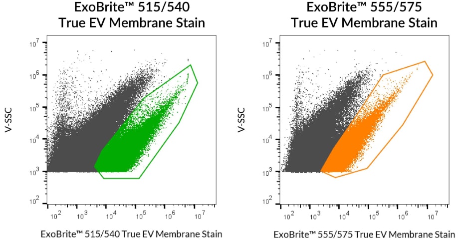
ExoBrite™ True EV Membrane Stains
The Best Choice for Pan-EV Staining
ExoBrite™ True EV Membrane Stains were developed for superior pan-EV labeling, overcoming the aggregation issues seen with dyes like PKH, DiO, and DiI. With unrivaled solubility, near-complete EV coverage, and clearer differentiation from non-specific particles, ExoBrite™ True EV Membrane Stains deliver more reliable results for flow or fluorescence nanoparticle tracking analysis (fNTA).
- Bright lipophilic dyes designed for pan-EV labeling
- Superior to PKH, DiO, DiI, DiD
- High EV coverage, broad sample compatibility
- 4 colors for Pacific Blue®, FITC, PE, and APC
- Compatible with antibody co-staining
- Validated on ZetaView® & Spectradyne ARC™
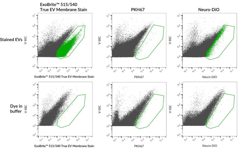
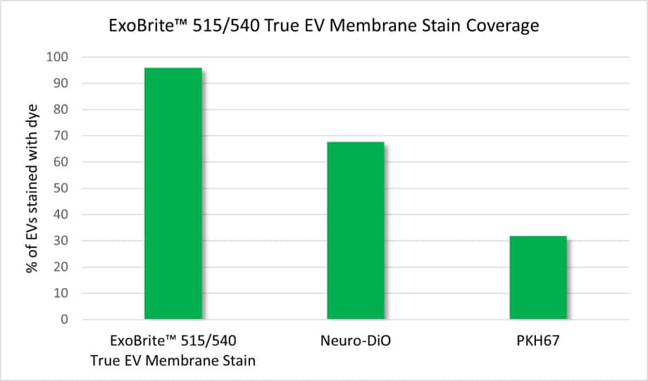
Superior EV Coverage Vs. PKH & DiO
ExoBrite™ True Stains Outperform PKH67 on ARC™
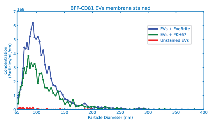
In a collaboration between Biotium and Spectradyne, ExoBrite™ 515/540 True EV Membrane Stain and PKH67 were compared as membrane dyes for extracellular vesicles on the ARC™ Particle Analyzer.
Results demonstrate ExoBrite™ 515/540 True EV Membrane Stain is more effective at staining EVs and is less susceptible to dye aggregation than PKH67 membrane dye.
Download Application Note

Application Note
Spectradyne’s ARC™ Particle Analyzer compares membrane dyes for extracellular vesicles
View Product Page

Test How it Performs With a Free Sample
For a limited time, U.S. customers can request a free 10-labeling sample of any ExoBrite™ True EV Membrane Stain. Click the link below to request a sample.
ExoBrite™ True EV Membrane Stains
| Product | Ex/Em | Detection Channels | Size | Catalog Number |
|---|---|---|---|---|
| ExoBrite™ 400/460 True EV Membrane Stain | 402/460 nm | Pacific Blue® | 100 Labelings | 30136-T |
| 500 Labelings | 30136 | |||
| ExoBrite™ 515/540 True EV Membrane Stain | 515/542 nm | FITC | 100 Labelings | 30129-T |
| 500 Labelings | 30129 | |||
| ExoBrite™ 555/575 True EV Membrane Stain | 556/576 nm | PE | 100 Labelings | 30130-T |
| 500 Labelings | 30130 | |||
| ExoBrite™ 645/675 True EV Membrane Stain | 644/671 nm | APC | 100 Labelings | 30137-T |
| 500 Labelings | 30137 |
ExoBrite™ Flow Antibody Conjugates
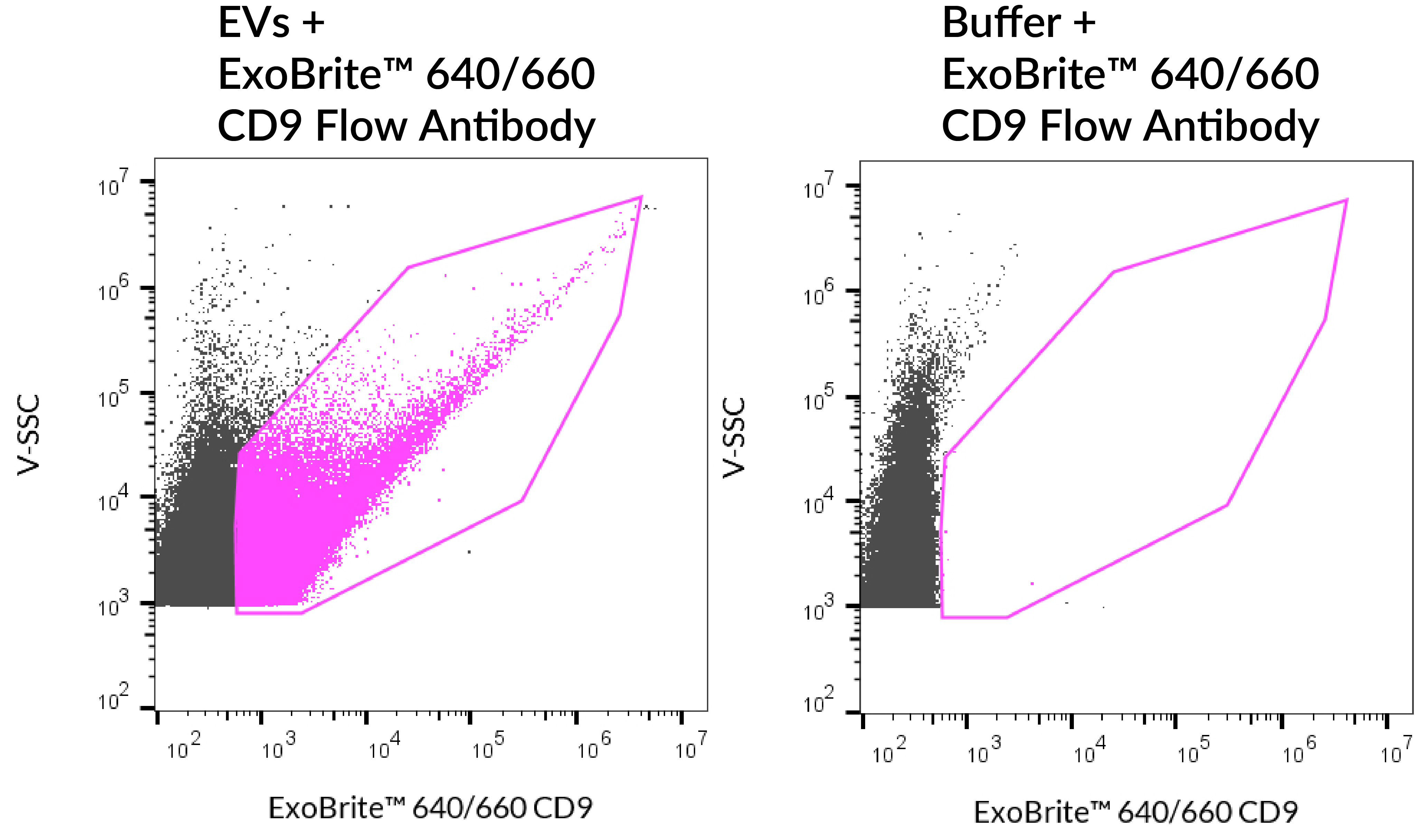
Validated EV Markers Antibodies for Flow
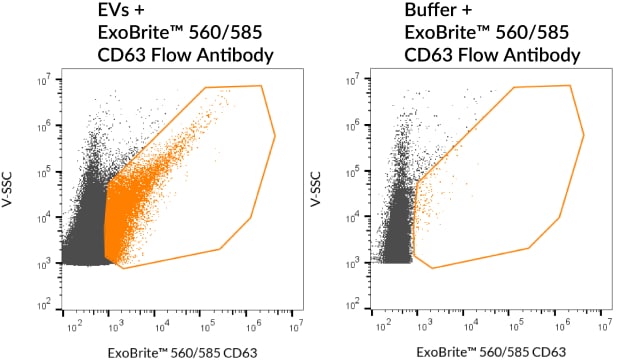
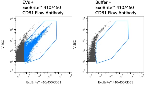
ExoBrite™ Flow Antibody Conjugates target key EV markers with high sensitivity and low background. Carefully selected and validated for robust EV detection, they feature a proprietary buffer to reduce aggregation and enhance staining. Matching isotype controls are also available for accurate flow cytometry analysis of isolated EVs.
Advantages of ExoBrite™ Flow Antibodies:
- Proprietary buffer formulation for enhanced signal-to-noise
- Optimized detection of EV markers by flow and fNTA
- Validated for purified or bead-bound EVs
- Bright signal and low background
- ExoBrite™ Isotype Control Flow Antibody available
- Colors available for Pacific Blue™, FITC, PE, and APC channels
ExoBrite™ Antibody Target Info
ExoBrite™ Flow Antibody Conjugates
| Product | Conjugates | Detection Channels | Sizes | Catalog Number |
|---|---|---|---|---|
| ExoBrite™ CD9 Flow Antibody | ExoBrite™ 410/450 ExoBrite™ 490/515 ExoBrite™ 560/585 ExoBrite™ 640/660 ExoBrite™ R-PE ExoBrite™ APC | Pacific Blue ™ FITC PE APC | 25 tests 100 tests | P003-410... P003-RPE |
| ExoBrite™ CD9 (Mouse) Flow Antibody | ExoBrite™ 410/450 ExoBrite™ 490/515 ExoBrite™ 560/585 ExoBrite™ 640/660 ExoBrite™ APC | Pacific Blue ™ FITC PE APC | 25 tests 100 tests | P018-410... P018-APC |
| ExoBrite™ CD47 Flow Antibody | ExoBrite™ 410/450 ExoBrite™ 490/515 ExoBrite™ 560/585 ExoBrite™ 640/660 | Pacific Blue ™ FITC PE APC | 25 tests 100 tests | P061-410... P061-640 |
| ExoBrite™ CD63 Flow Antibody | ExoBrite™ 410/450 ExoBrite™ 490/515 ExoBrite™ 560/585 ExoBrite™ 640/660 ExoBrite™ R-PE ExoBrite™ APC | Pacific Blue ™ FITC PE APC | 25 tests 100 tests | P004-410... P004-RPE |
| ExoBrite™ CD63 (Mouse) Flow Antibody | ExoBrite™ 410/450 ExoBrite™ 490/515 ExoBrite™ 560/585 ExoBrite™ 640/660 ExoBrite™ APC | Pacific Blue ™ FITC PE APC | 25 tests 100 tests | P022-410... P022-APC |
| ExoBrite™ CD81 Flow Antibody | ExoBrite™ 410/450 ExoBrite™ 490/515 ExoBrite™ 560/585 ExoBrite™ 640/660 ExoBrite™ R-PE ExoBrite™ APC | Pacific Blue ™ FITC PE APC | 25 tests 100 tests | P005-410... P005-RPE |
| ExoBrite™ CD81 (Mouse/Rat) Flow Antibody | ExoBrite™ 410/450 ExoBrite™ 490/515 ExoBrite™ 560/585 ExoBrite™ 640/660 ExoBrite™ APC | Pacific Blue ™ FITC PE APC | 25 tests 100 tests | P019-410... P019-APC |
| ExoBrite™ CD29 Flow Antibody | ExoBrite™ 410/450 ExoBrite™ 490/515 ExoBrite™ 560/585 ExoBrite™ 640/660 | Pacific Blue ™ FITC PE APC | 25 tests 100 tests | P040-410... P040-640 |
| ExoBrite™ IgG1 Isotype Control Flow Antibody | ExoBrite™ 410/450 ExoBrite™ 490/515 ExoBrite™ 560/585 ExoBrite™ 640/660 ExoBrite™ R-PE | Pacific Blue ™ FITC PE APC | 25 tests 100 tests | P008-410... P008-RPE |
| ExoBrite™ Armenian Hamster IgG Isotype Control Flow Antibody | ExoBrite™ 410/450 ExoBrite™ 490/515 ExoBrite™ 560/585 ExoBrite™ 640/660 ExoBrite™ APC | Pacific Blue ™ FITC PE APC | 25 tests 100 tests | P056-410...P056-APC |
| ExoBrite™ Rat IgG2a Isotype Control Flow Antibody | ExoBrite™ 410/450 ExoBrite™ 490/515 ExoBrite™ 560/585 ExoBrite™ 640/660 ExoBrite™ APC | Pacific Blue ™ FITC PE APC | 25 tests 100 tests | P057-410...P057-APC |
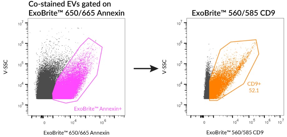
ExoBrite™ Antibody Cocktails
High-Coverage or Robust Phenotype Analysis
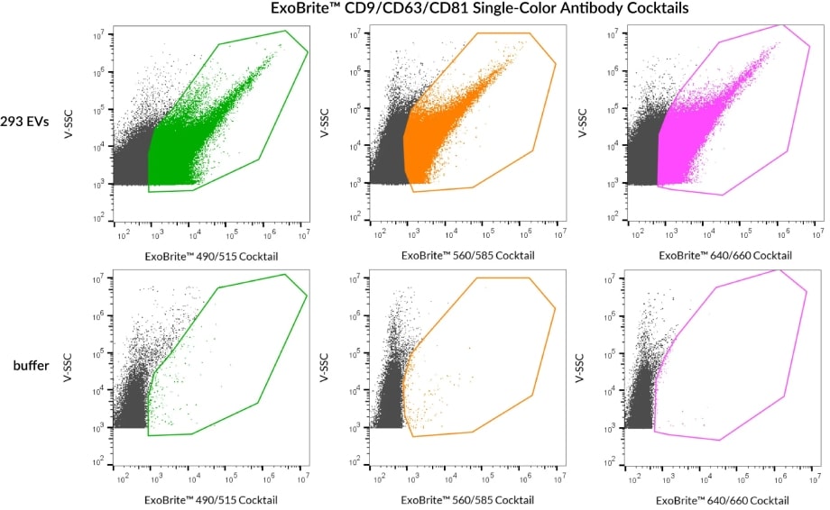
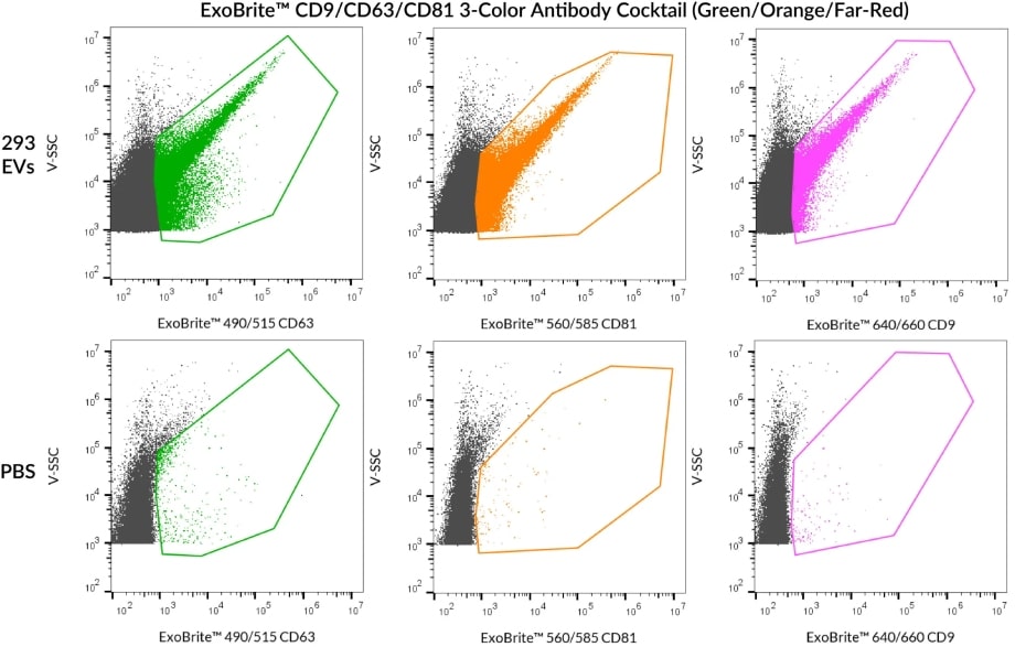
Biotium’s ExoBrite™ CD9/CD63/CD81 Antibody Cocktails offer optimal brightness and low background for EV detection by flow cytometry. Single-color cocktails enable high-coverage EV staining, while 3-color cocktails allow multiplexed detection and robust phenotyping with distinct fluorescent labels. Both formats are validated for low aggregation and high signal-to-noise.
- Single-color cocktails designed for high-coverage EV staining
- 3-color cocktail designed for phenotyping analysis
- Each antibody is optimized to offer the brightest signal
- Formulated for low aggregation, high signal-to-noise
ExoBrite™ Flow Anti-Human CD9/CD63/CD81 Antibody Cocktails
| Cocktail | Ex/Em | Detection Channel | Size | Catalog No. |
|---|---|---|---|---|
| ExoBrite™ CD9/CD63/CD81 Single-Color Antibody Cocktail (Green) | 490/516 nm | FITC | 25 tests | P030-125 |
| 100 tests | P030-500 | |||
| ExoBrite™ CD9/CD63/CD81 Single-Color Antibody Cocktail (Orange) | 562/584 nm | PE | 25 tests | P031-125 |
| 100 tests | P031-500 | |||
| ExoBrite™ CD9/CD63/CD81 Single-Color Antibody Cocktail (Far-Red) | 642/663 nm | APC | 25 tests | P032-125 |
| 100 tests | P032-500 | |||
| ExoBrite™ CD9/CD63/CD81 3-Color Antibody Cocktail (Blue/Green/Far-Red) | 411/452 nm 490/516 nm 642/663 nm | Pacific Blue® FITC APC | 25 tests | P028-125 |
| 100 tests | P028-500 | |||
| ExoBrite™ CD9/CD63/CD81 3-Color Antibody Cocktail (Green/Orange/Far-Red) | 490/516 nm 562/584 nm 642/663 nm | FITC PE APC | 25 tests | P029-125 |
| 100 tests | P029-500 |
ExoBrite™ Western Antibody Conjugates
Validated EV Marker Antibodies for Western Blot
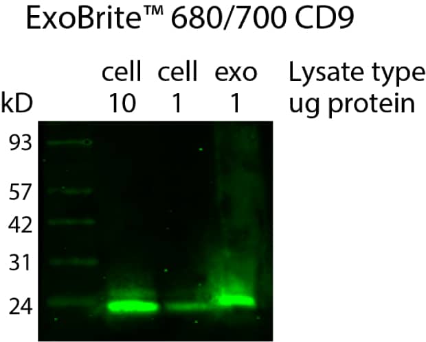
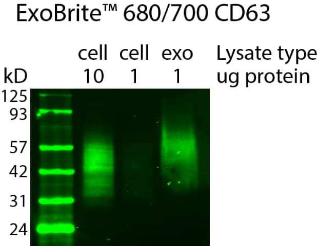
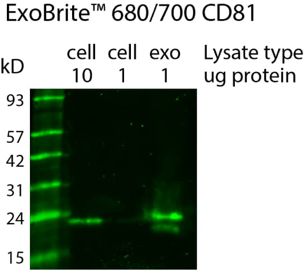
ExoBrite™ Western Antibodies were validated to offer bright signal and low background of EV markers CD9, CD63, and CD81 in EV extracts by chemiluminescence or near-IR fluorescent western blot. The antibodies are offered conjugated to HRP or to the near-infrared fluorescent dyes ExoBrite™ 680/700 and ExoBrite™ 770/800, which provide higher signal-to-noise than dyes with visible light emission for western blotting.
An ExoBrite™ Calnexin Western Antibody detects a protein of the endoplasmic reticulum that is not found in EVs. It is offered as a negative control to assess the purity of isolated EV extracts.
Advantages of ExoBrite™ Western Antibodies:
- For detection of CD9, CD63, and CD81 by fluorescent Western
- Validated for use with EV extracts
- Bright signal and low background
- Choose from two near-IR dyes or HRP
- Negative control ExoBrite™ Calnexin Western Antibody available
| Product | Conjugates | Sizes | Catalog Number |
|---|---|---|---|
| ExoBrite™ CD9 Western Antibody | ExoBrite™ 680/700 ExoBrite™ 770/800 HRP | 25 tests 100 tests 50 tests (HRP only) | P003-680, P003-HRP |
| ExoBrite™ CD63 Western Antibody | P004-680, P004-HRP | ||
| ExoBrite™ CD81 Western Antibody | P006-HRP, P006-770 | ||
| ExoBrite™ Calnexin Western Antibody | ExoBrite™ 770/800 | 25 tests 100 tests | P007-770 |
Custom Antibody Conjugation
Biotium provides custom conjugation of customer-supplied antibodies with ExoBrite™ dyes, formulated in an optimized buffer for sensitive extracellular vesicle detection.
- Customer-supplied antibodies can be conjugated to any ExoBrite™ Dye
- Final conjugate optimized for sensitive EV detection
- Available for whole IgG antibodies only
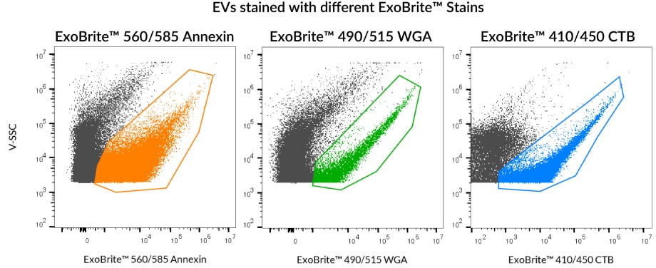
Robust Stains Designed to Detect EVs, Not Dye Aggregates
Membrane dyes, while common tools for labeling EVs, can pose significant challenges when used for EV staining. For example, membrane dyes such as PKH, DiO, and DiI, have poor solubility and thus form aggregates that can be confused with EVs. To overcome these challenges, Biotium developed ExoBrite™ True EV Membrane Stains and ExoBrite™ EV Surface Stains (Annexin V, WGA, and CTB) to offer higher coverage and signal-to-noise over membrane dyes commonly used for EV staining. View our comparison table and reference the validated EV sources below to select the best stain for your next experiment.
ExoBrite™ EV Stains Comparison Guide
| ExoBrite™ EV Surface Stain | Pros | Cons |
|---|---|---|
| ExoBrite™ True EV Membrane Stains | • Near-complete staining of EVs in a sample • Broad compatibility with different EV sources • Validated for flow and fNTA | • Can't be used to stain bead-bound EVs • May have more aggregation than CTB & Annexin |
| ExoBrite™ Annexin EV Staining Kits | • Broad compatibility with different EV sources • Validated for flow and fNTA • Low background aggregates | • May not stain every EV in a sample • Doesn't work well on bead-bound EVs |
| ExoBrite™ WGA EV Staining Kits | • Broad compatibility with different EV sources • Can be used with bead-bound EVs | • May not stain every EV in a sample • Doesn't work well for fNTA |
| ExoBrite™ CTB EV Staining Kits | • Validated for flow and fNTA • Extremely low background • Can be used with bead-bound EVs | • May not stain every EV in a sample • Does not stain EVs from every source |
| ExoBrite™ Antibodies | • Highly specific for human tetraspanins CD9, CD63, CD81, and other EV markers • Validated for EV flow • Broad compatibility for different EV sources • Can be used with bead-bound EVs • Can be used for WB | • Depends on the expression level of the target protein on the EVs |
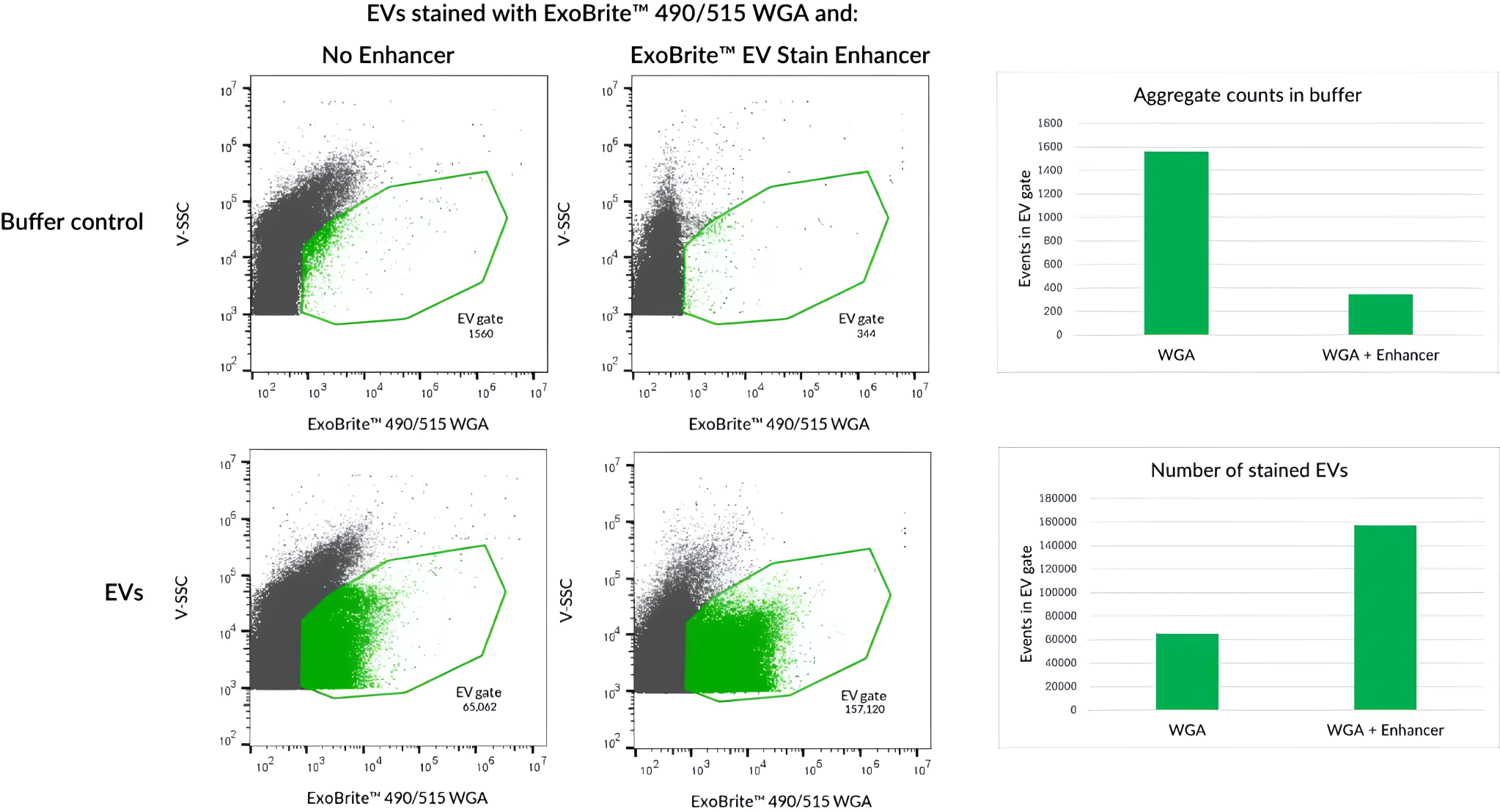
| ExoBrite™ EV Stain Enhancer | • Improves signal-to-noise by reducing or eliminating aggregates of certain EV stains • Validated with several different lectins and Annexin V • Does not interfere with antibody staining of EVs • Easy to use, just add directly to the staining reaction | • Not recommended for use with lipophilic EV stains |
Validated EV Sources for ExoBrite™ EV Surface Stains
| EV Source | ExoBrite™ True EV Membrane Stains | ExoBrite™ CTB Stains | ExoBrite™ WGA Stains | ExoBrite™ Annexin Stains |
|---|---|---|---|---|
| A549 cells | Yes | Yes | Yes | Yes |
| CHO cells | Yes | No | Yes | Yes |
| hASC (human adipose stem cells) | ND | No1 | ND | ND |
| HEK293 cells | Yes | Yes1 | Yes | Yes |
| HeLa cells | Yes | No | Yes | Yes |
| HUVEC (human umbilical vein endothelial cells) | ND | No1 | ND | ND |
| J774 cells | Yes | Yes | Yes | Yes |
| Jurkat cells | Yes | Yes | Yes | Yes |
| MCF-7 cells | Yes | Yes | Yes | Yes |
| Plasma | Yes | No | ND | Yes |
| Raji cells | ND | Yes | Yes | Yes |
| RAW 264.7 cells | Yes | Yes | Yes | Yes |
| Serum | Yes | No | ND | Yes |
| Skeletal myoblasts | ND | Yes1 | ND | ND |
| THP-1 cells | Yes | ND | ND | ND |
| U2OS cells | Yes | No | Yes | Yes |
| U937 cells | Yes | No | Yes | Yes |
| NIH3T3 cells | Yes | Yes | Yes | Yes |
| HepG2 cells | Yes | No | Yes | Yes |
| Yeast (S. cerevisiae) | Yes | No | Yes | Yes |
Value of “Yes” or “No” indicates coverage of EVs based on Biotium’s internal data or customer-reported data. Value of “ND” indicates no data.
ExoBrite™ EV Surface Stains
| Product | Conjugate | Detection channels | Size | Catalog Number |
|---|---|---|---|---|
| ExoBrite™ True EV Membrane Stains | N/A | Pacific Blue™, FITC, PE, APC | 100 Labelings, 500 Labelings | 30129, 30130 |
| ExoBrite™ Annexin EV Staining Kits | Annexin V | Pacific Blue™, FITC, PE, Cy®3, APC | 100 Labelings, 500 Labelings | 30119-30122 |
| ExoBrite™ WGA EV Staining Kits | Wheat Germ Agglutinin (WGA) | 30123-30126 | ||
| ExoBrite™ CTB EV Staining Kits | Cholera Toxin Subunit B (CTB) | 30111-30114 | ||
| ExoBrite™ EV Surface Stain Sampler Kit, Green | Annexin V, Wheat Germ Agglutinin (WGA), Cholera Toxin Subunit B (CTB) | FITC | 100 Labelings | 30127 |
| ExoBrite™ STORM CTB EV Staining Kits | Cholera Toxin Subunit B (CTB) | FITC, Texas Red®, Cy®5, Cy®5.5 | 100 Labelings, 500 Labelings | 30115-30118 |
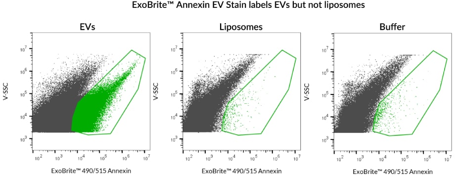
ExoBrite™ Annexin EV Staining Kits
Bright EV Stains with Broad Compatibility
ExoBrite™ Annexin EV Staining Kits are optimized for bright, sensitive EV detection across diverse sources. Like ExoBrite™ CTB stains, they offer high signal with little to no background aggregation, ideal for flow cytometry. They deliver broad EV coverage and strong performance across EVs from 9 tested cell lines—making them a powerful tool for consistent and reliable EV detection.
- Optimally formulated Annexin V conjugates for staining purified EVs
- Broad compatibility, stained EVs isolated from all 9 sources tested
- Bright signal and low background staining for flow cytometry
- Compatible with antibody co-staining
- Available in 4 colors for flexible experimental design
ExoBrite™ Annexin EV Staining Kits
| Product | Ex/Em | Detection channels | Size | Catalog Number |
|---|---|---|---|---|
| ExoBrite™ 410/450 Annexin EV Staining Kit | 416/452 nm | Pacific Blue™ | 100 Labelings | 30119-T |
| 500 Labelings | 30119 | |||
| ExoBrite™ 490/515 Annexin EV Staining Kit | 490/516 nm | FITC | 100 Labelings | 30120-T |
| 500 Labelings | 30120 | |||
| ExoBrite™ 560/585 Annexin EV Staining Kit | 562/584 nm | PE | 100 Labelings | 30121-T |
| 500 Labelings | 30121 | |||
| ExoBrite™ 650/665 Annexin EV Staining Kit | 652/668 nm | APC | 100 Labelings | 30122-T |
| 500 Labelings | 30122 |
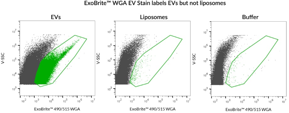
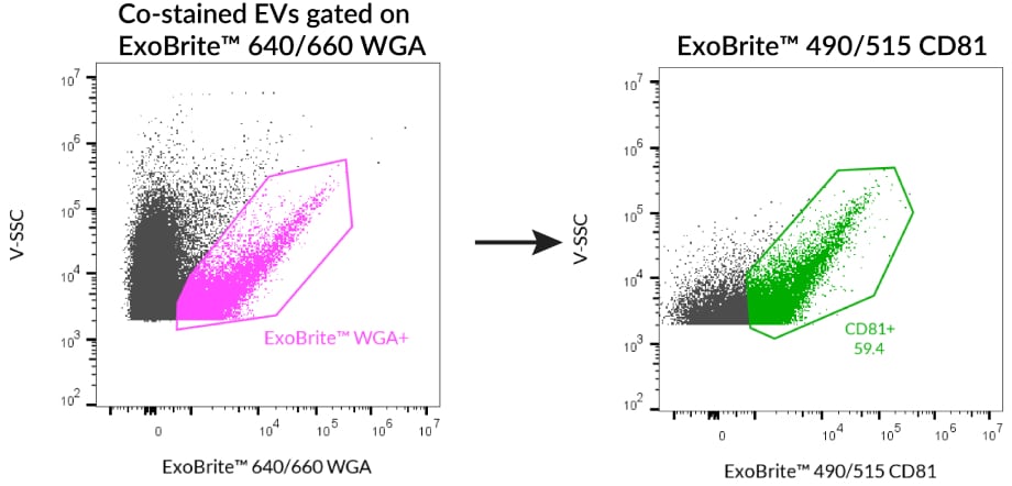
ExoBrite™ WGA EV Staining Kits
Sensitive Stains for Purified or Bead-Bound EVs
ExoBrite™ WGA EV Stains are fluorescent conjugates of wheat germ agglutinin (WGA) for bright and sensitive staining of EVs. Similar to ExoBrite™ Annexin EV Stains, these WGA conjugates offer broad compatibility with EVs. The stains were validated for EVs derived from all 9 cell lines tested. In addition, ExoBrite™ WGA EV Stains are less prone to aggregation than hydrophobic membrane dyes and may be used to stain purified and bead-bound EVs.
- Optimally formulated WGA conjugates for staining purified or bead-bound EVs
- Broad compatibility, stained EVs isolated from all 9 sources tested
- Bright signal and low background staining for flow cytometry
- Compatible with antibody co-staining
- Available in 4 colors for flexible experimental design
ExoBrite™ WGA EV Staining Kits
| Product | Ex/Em | Detection channels | Size | Catalog Number |
|---|---|---|---|---|
| ExoBrite™ 410/450 WGA EV Staining Kit | 416/452 nm | Pacific Blue™ | 100 Labelings | 30123-T |
| 500 Labelings | 30123 | |||
| ExoBrite™ 490/515 WGA EV Staining Kit | 490/516 nm | FITC | 100 Labelings | 30124-T |
| 500 Labelings | 30124 | |||
| ExoBrite™ 560/585 WGA EV Staining Kit | 562/584 nm | PE | 100 Labelings | 30125-T |
| 500 Labelings | 30125 | |||
| ExoBrite™ 640/660 WGA EV Staining Kit | 642/663 nm | APC | 100 Labelings | 30126-T |
| 500 Labelings | 30126 |
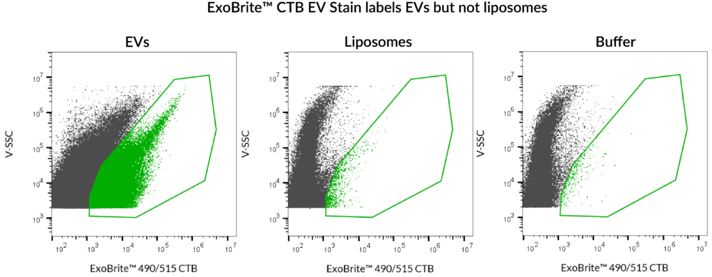
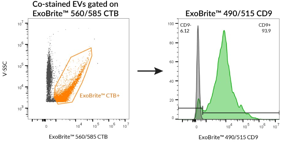
ExoBrite™ CTB EV Staining Kits
Bright EV Stains with Excellent Signal-to-Noise
ExoBrite™ CTB EV Staining Kits are fluorescent conjugates of cholera toxin subunit B (CTB). They offer were extremely low background for detection of EVs by flow cytometry. Also, ExoBrite™ CTB EV stains do not bind non-specifically to polystyrene beads, meaning that they can be used to stain bead-bound EVs.
- Optimally formulated CTB conjugates for staining EVs
- Designed for detection by flow cytometry
- Bright fluorescence and low background for excellent signal-to-noise
- Compatible with antibody co-staining
- Stain purified or bead-bound EVs
- Available in 4 colors for flexible experimental design
ExoBrite™ CTB EV Staining Kits
| Product | Ex/Em | Detection channels | Size | Catalog Number |
|---|---|---|---|---|
| ExoBrite™ 410/450 CTB EV Staining Kit | 416/452 nm | Pacific Blue™ | 100 Labelings | 30111-T |
| 500 Labelings | 30111 | |||
| ExoBrite™ 490/515 CTB EV Staining Kit | 490/516 nm | FITC | 100 Labelings | 30112-T |
| 500 Labelings | 30112 | |||
| ExoBrite™ 560/585 CTB EV Staining Kit | 562/584 nm | PE, Cy®3 | 100 Labelings | 30113-T |
| 500 Labelings | 30113 | |||
| ExoBrite™ 640/660 CTB EV Staining Kit | 642/663 nm | APC | 100 Labelings | 30114-T |
| 500 Labelings | 30114 |
ExoBrite™ EV Stain Enhancer
Boost EV Stain Signal-to-Noise
The ExoBrite™ EV Stain Enhancer is a unique additive that can be added to extracellular vesicle (EV) stain reactions to improve the staining specificity for applications like flow cytometry. The ExoBrite™ Stain Enhancer works by reducing the aggregation of certain EV stains, which allows the conjugate to stain the EVs more efficiently, resulting in a better signal-to-noise ratio and fewer false positives.
- Improves signal-to-noise by reducing or eliminating
- aggregates of certain EV stains
- Validated with several different lectins and Annexin V
- Does not interfere with antibody staining of EVs
- Easy to use, just add directly to the staining reaction
View Product Page
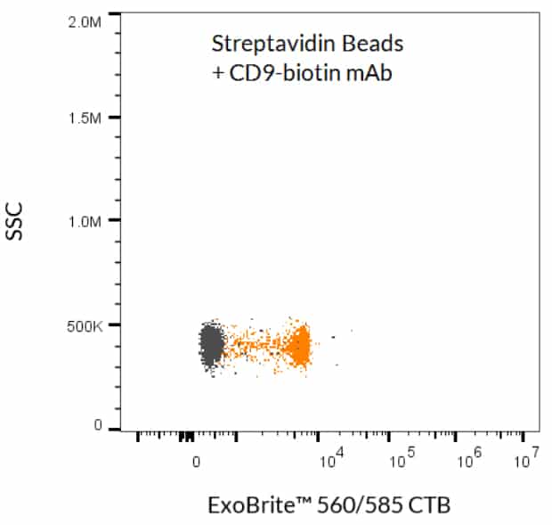
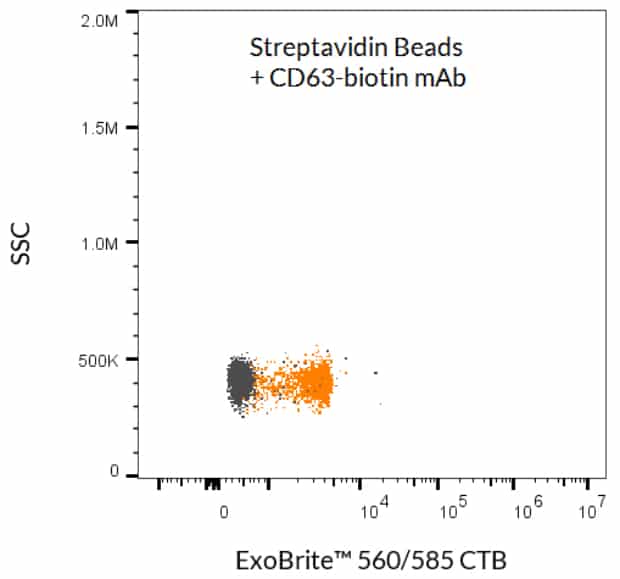
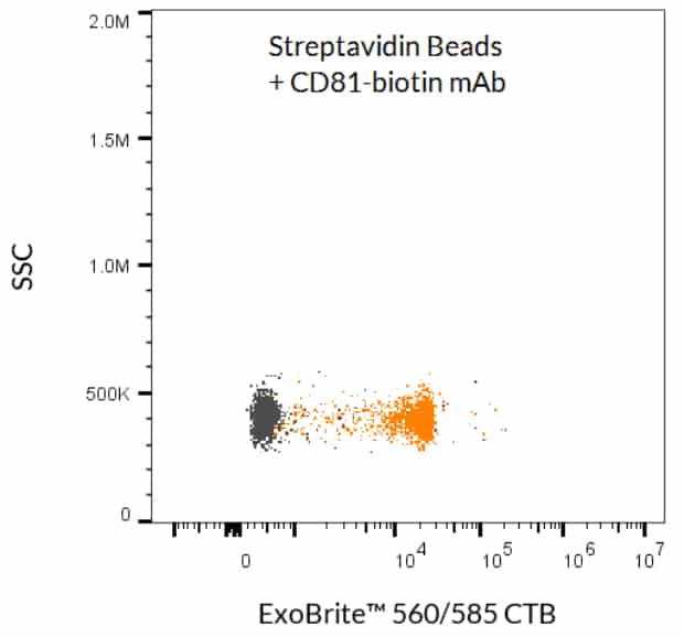
ExoBrite™ Streptavidin Magnetic Beads
Convenient & Optimized Capture of EVs
Streamline your EV and exosome isolation workflows with ExoBrite™ Streptavidin Magnetic Beads, developed to offer lower background and higher sensitivity for EV capture than similar products. These streptavidin-coated and magnetic polystyrene beads (4.5 um diameter) may be used to isolate EVs from cell culture medium or other biological fluids without an overnight precipitation step.
- Combine with a biotinylated antibody (not included) for isolation of EVs
- Isolate EVs from biofluids without an overnight precipitation step
- Lower background and higher sensitivity than competitors
- Convenient magnetic-based separation
- Detect downstream with microscopy, flow cytometry, or western blot
View Product Page
Texas Red and Pacific Blue are registered trademarks of Thermo Fisher Scientific; Cy Dye is a registered trademark of Cytiva.
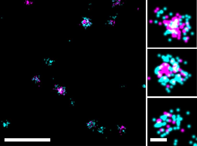
EV Stains for Super-Resolution Imaging
Featuring STORM-Optimized CF® Dyes
Characterizing exosomes and EVs by imaging is difficult due to their small size and light microscopy limits. Super-resolution methods like dSTORM overcome these barriers, revealing EV structure at the single-molecule level. ExoBrite™ STORM Antibodies and CTB EV Stains use CF® Dyes optimized for STORM, offering bright, high-resolution EV imaging with excellent clarity and signal-to-noise.
Advantages of ExoBrite™ STORM Antibodies
- Validated antibodies for detecting EV markers CD9, CD63, and CD81
- Available with excellent STORM dyes CF®498, CF®568, CF®583R, and CF®647Plus
- Proprietary buffer for enhanced signal-to-noise
- Available in 4 colors for FITC, Cy®3, Texas Red®, and Cy®5 channels
Advantages of ExoBrite™ STORM CTB EV Stains:
- Optimally formulated CTB conjugates for imaging EVs by STORM
- Available with excellent STORM dyes CF®505, CF®583R, CF®647, and CF®680
- Allows antibody co-staining and localization studies with EV biomarkers
Note: ExoBrite™ CTB stains have been found to label EVs derived from several tested cell lines, but do not stain EVs from every source. Please visit the product page to view a full list of validated EV sources.
ExoBrite™ STORM Antibodies
| Antibody | Conjugates | Detection Channels | Catalog Number |
|---|---|---|---|
| ExoBrite™ STORM CD9 Antibody | CF®498 CF®568 CF®583R CF®647Plus | FITC Cy®3 Texas Red® Cy®5 | P003-498ST-500 P003-568ST-500 P003-583RST-500 P003-647PST-500 |
| ExoBrite™ STORM CD63 Antibody | CF®498 CF®568 CF®583R CF®647Plus | FITC Cy®3 Texas Red® Cy®5 | P004-498ST-500 P004-568ST-500 P004-583RST-500 P004-647PST-500 |
| ExoBrite™ STORM CD81 Antibody | CF®498 CF®568 CF®583R CF®647Plus | FITC Cy®3 Texas Red® Cy®5 | P005-498ST-500 P005-568ST-500 P005-583RST-500 P005-647PST-500 |
| ExoBrite™ STORM CD9 (Mouse) Antibody | CF®568 CF®647Plus | Cy®3 Cy®5 | P018-568ST-500 P018-647PST-500 |
| ExoBrite™ STORM CD63 (Mouse) Antibody | CF®568 CF®647Plus | Cy®3 Cy®5 | P022-568ST-500 P022-647PST-500 |
| ExoBrite™ STORM CD81 (Mouse/Rat) Antibody | CF®568 CF®647Plus | Cy®3 Cy®5 | P019-568ST-500 P019-647PST-500 |
ExoBrite™ STORM CTB EV Staining Kits
| Product | Ex/Em (nm) | Laser Line(s) (nm) | Detection Channel | Size | Catalog Number |
|---|---|---|---|---|---|
| ExoBrite™ STORM CF®505 CTB EV Staining Kit | 505/519 | 488 | FITC | 100 Labelings | 30115-T |
| 500 Labelings | 30115 | ||||
| ExoBrite™ STORM CF®583R CTB EV Staining Kit | 583/609 | 555 or 561 | Rhodamine or Texas Red® | 100 Labelings | 30116-T |
| 500 Labelings | 30116 | ||||
| ExoBrite™ STORM CF®647 CTB EV Staining Kit | 652/668 | 633-640 | Cy®5 | 100 Labelings | 30117-T |
| 500 Labelings | 30117 | ||||
| ExoBrite™ STORM CF®680 CTB EV Staining Kit | 681/698 | 633-640 | Cy®5.5 | 100 Labelings | 30118-T |
| 500 Labelings | 30118 |
Total RNA Extraction
Efficient total RNA extraction from purified EVs
The ExoBrite™ EV Total RNA Isolation Kit was designed to address the challenges of EV RNA extraction by offering an optimized and easy-to-use kit for total RNA isolation, including mRNA and miRNAs, from purified EVs. The isolated EV RNA can then be used for downstream analysis such as qPCR or RNAseq.
Advantages of ExoBrite™ EV Total RNA Isolation Kit:
- Optimized for total RNA extraction from purified EVs
- Recover ~10-20 ng of RNA from 1×10¹⁰ SEC-enriched EVs
- Compatible with downstream applications such as qPCR or RNAseq
- Simple column-based purification
- No phenol/chloroform or ethanol precipitation steps
View Product Page
Texas Red and Pacific Blue are registered trademarks of Thermo Fisher Scientific; Cy Dye is a registered trademark of Cytiva.


