New Products
 Earth-Friendly Products
 Biotium Choice Antibodies
 Special Offers
Choose from a Spectrum of Fixable Dead Cell Stains
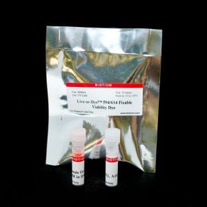
Fixable and Stable Labeling of Dead Cells
Live-or-Dye™ Fixable Viability Stains are cell membrane-impermeant amine-reactive dyes. The dyes enter dead cells that have compromised membrane integrity and covalently label free amines on intracellular proteins. On live cells, the dyes react with surface proteins, but these are much less abundant than intracellular proteins, resulting in low staining levels compared to dead cells.
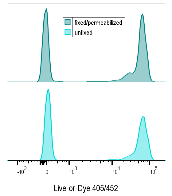
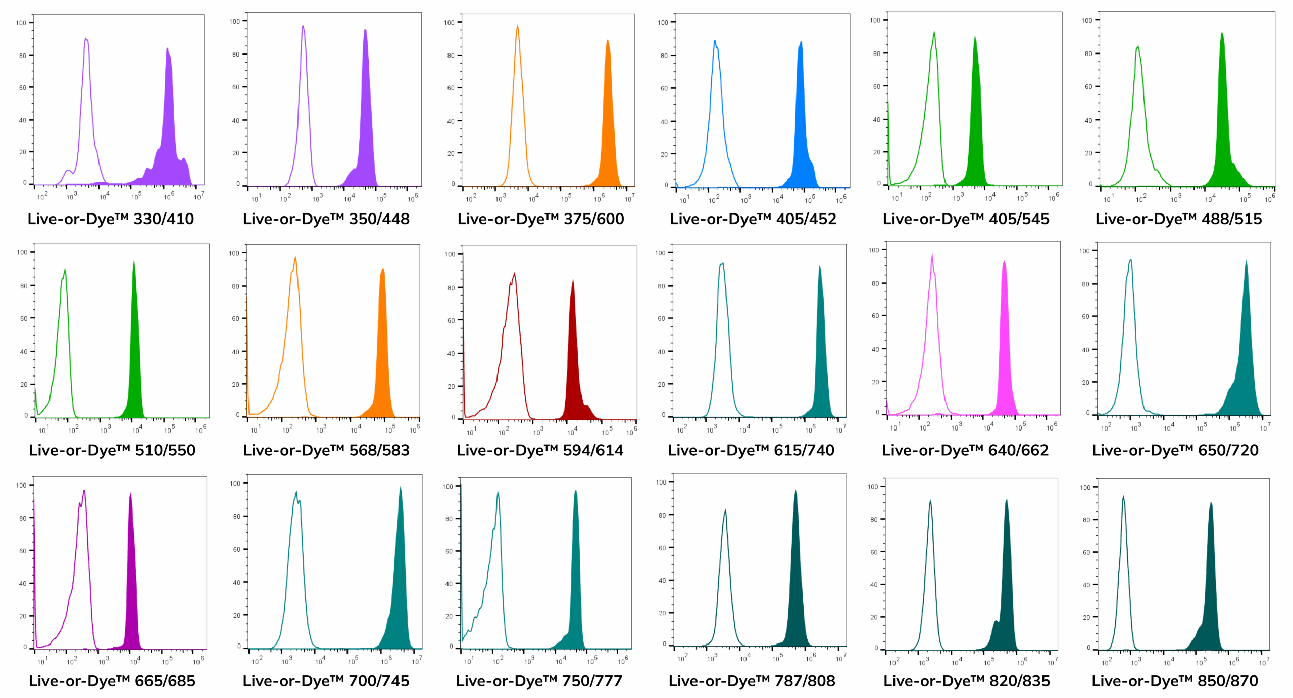
Live-or-Dye™ labeling is extremely stable, allowing the cells to be fixed and permeabilized without loss of fluorescence or dye transfer between cells. Live-or-Dye™ stains are offered in a wide selection of 16 colors for easy panel design. This includes spectrally unique dyes designed for taking advantage of enhanced multiplexing by spectral flow cytometry.

Live-or-Dye™ Advantages
- Wide selection of 16 dye colors for easy panel design
- Exceptionally bright dyes for excellent live/dead separation
- Withstands fixation and permeabilization
- No loss of brightness after fixation
- For flow cytometry or fluorescence microscopy
- Compatible with intracellular staining protocols
- Dyes designed to fit gaps in spectral flow panels
- The most options for 633 nm and 808 nm lasers
- Trial sizes and sample kits available
Live-or-Dye™ Fixable Viability Staining Kits
Cat. No. | Viability Dye | Compatible lasers (nm) | Optimal detection channels | Notes |
|---|---|---|---|---|
| 32018, 32018-T | Live-or-Dye™ 330/410 | 355, 375 | BUV395 | |
| 32002, 32002-T | Live-or-Dye™ 350/448 | 355, 375 | DAPI | |
| 32014,32014-T | Live-or-Dye™ 375/600 | 355, 375, 405 | Spectral scan, BUV615, BV605 | Developed for spectral cytometry |
| 32003, 32003-T | Live-or-Dye™ 405/452 | 405 | Pacific Blue®, BV421, BV450 | |
| 32009, 32009-T | Live-or-Dye™ 405/545 | 405 | AmCyan, BV510 | |
| 32004, 32004-T | Live-or-Dye™ 488/515 | 488 | FITC | Validated for microscopy |
| 32012, 32012-T | Live-or-Dye™ 510/550 | 488, 532 | Spectral scan | Developed for spectral cytometry |
| 32005, 32005-T | Live-or-Dye™ 568/583 | 488, 532, 561 | PE, PI | Validated for microscopy |
| 32006, 32006-T | Live-or-Dye™ 594/614 | 488, 532, 561 | PI, PE-CF®594, PE-Texas Red® | Validated for microscopy |
| 32015, 32015-T | Live-or-Dye™ 615/740 | 633-640 | Spectral scan, APC-Cy®7 | Developed for spectral cytometry |
| 32007, 32007-T | Live-or-Dye™ 640/662 | 633-640 | APC | Validated for microscopy |
| 32023, 32023-T | Live-or-Dye™ 650/720 | 633-640 | APC-700 | |
| 32013, 32013-T | Live-or-Dye™ 665/685 | 633-640 | Spectral scan, AF700 | Developed for spectral cytometry |
| 32024, 32024-T | Live-or-Dye™ 700/745 | 633-640, 785 | APC-Cy®7 | Our best dye for the APC-Cy®7 channel |
| 32008, 32008-T | Live-or-Dye™ 750/777 | 633-640, 785 | APC-Cy®7, IR840 | |
| 32011, 32011-T | Live-or-Dye™ 787/808 | 785, 808 | APC-Cy®7, IR800 | |
| 32021, 32021-T | Live-or-Dye™ 820/835 | 808 | IR840 | |
| 32022, 32022-T | Live-or-Dye™ 850/870 | 808 | IR885 |
Widest Range of Colors for Easy Panel Design
Live/dead stains are useful probes to include in flow cytometry panels because they allow intracellular fluorescence signal from dead cells with permeable plasma membranes to be excluded from analysis. However, the limited number of dye colors from other commercially available live/dead stains can make choosing the right fluorophores difficult when designing highly complex panels. Live-or-Dye™ Fixable Viability Staining Kits are offered in a wide selection of live/dead dye colors, allowing maximal flexibility when designing multi-color flow panels.
Spectrally Unique Dyes for Spectral Flow Cytometry
Spectral flow cytometry is a rapidly growing technology with enhanced multiplexing capabilities over conventional flow cytometry. Where conventional flow cytometer instruments can detect panels with more than a dozen fluorophores, spectral flow cytometers can accommodate panels with upwards of 40 dyes. Biotium offers spectrally unique Live-or-Dye™ stains designed specifically to take advantage of enhanced multiplexing by spectral flow cytometry.
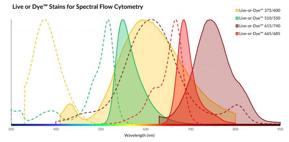
Live-or-Dye™ Stains for Spectral Flow

Learn more about spectral flow cytometry and our high-performance CF® dyes developed to fill emission gaps between available fluorophores.
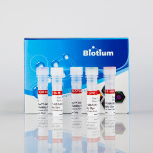
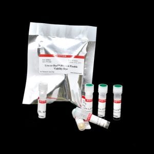
Live-or-Dye™ Sampler Kits for Flow Cytometry
Live-or-Dye™ stains are available in two sampler kits, each containing five different dye colors, for excitation from all of the popular flow cytometry laser lines (see Live-or-Dye™ Sampler Kits table below). The Live-or-Dye™ Sampler Kit, Standard was designed for use on the most common flow cytometer laser and filter configurations, with dyes excitable by UV, Violet, Blue, Yellow-Green, and Red lasers.
The Live-or-Dye™ Sampler Kit, Spectral was designed for use in spectral scanning flow cytometry. It contains dyes excitable by UV, Violet, Blue, and Red lasers, all of which have been validated on the Cytek® Aurora spectral cytometer, and chosen for their ability to fill the gaps in many spectral flow panels.
Live-or-Dye™ Sampler Kits
Kit Name | Catalog No. | Included dyes: Ex/Em (Cat No.) | Application |
|---|---|---|---|
| Live-or-Dye™ Fixable Viability Sampler Kit, Standard | 32016 | • 350/448 (32002A) • 405/545 (32009A) • 488/515 (32004A) • 568/583 (32005A) • 640/662 (32007A) | Designed for use on most standard flow cytometry laser and filter configurations |
| Live-or-Dye™ Fixable Viability Sampler Kit, Spectral | 32017 | • 350/448 (32002A) • 375/600 (32014A) • 510/550 (32012A) • 615/740 (32015A) • 665/685 (32013A) | Designed for use in spectral flow cytometry, to fill in gaps between common fluorophores |
Bright and Photostable Dyes for Microscopy
In microscopy, live/dead stains allow unambiguous visual discrimination of dead cells. Live-or-Dye™ Fixable Viability Staining Kits work just as well for microscopy as they do for flow cytometry, with negligible signal in live cells and strong signal in dead cells.
Four stains have been validated for fluorescence microscopy. The other dyes are expected to work as well, as long as the microscope has the appropriate excitation and emission settings.
Live-or-Dye™ Stains Validated for Microscopy
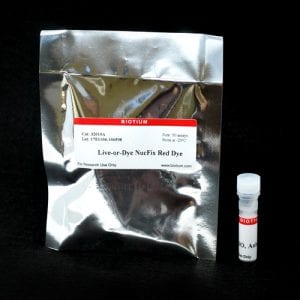
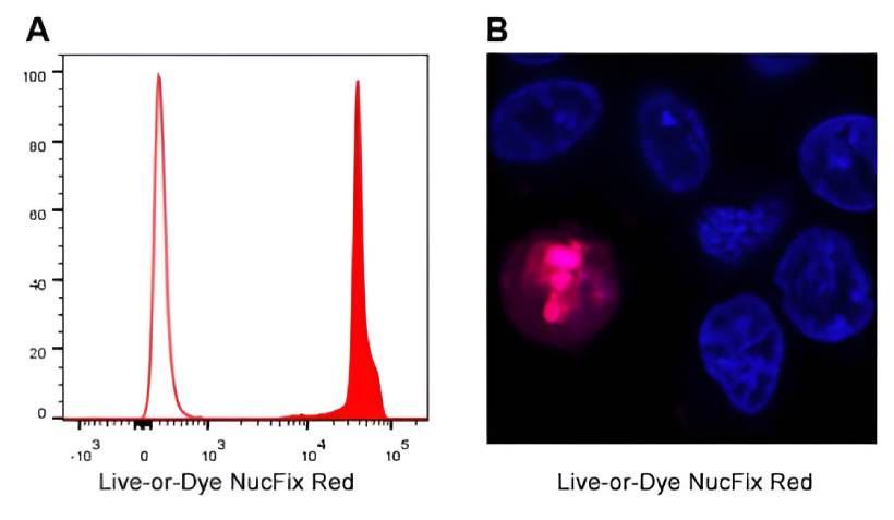
Live-or-Dye NucFix™ Red
Live-or-Dye NucFix™ Red is a unique, cell membrane impermeant dye that specifically stains the nuclei of dead cells.
Unlike other commonly used nuclear stains, such as propidium iodide (PI) or DRAQ7™, Live-or-Dye NucFix™ labeling is covalent, so the dye doesn’t transfer between cells after fixation. Live-or-Dye NucFix™ Red can be used for flow cytometry or fluorescence microscopy.
View Product Page
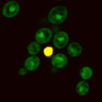
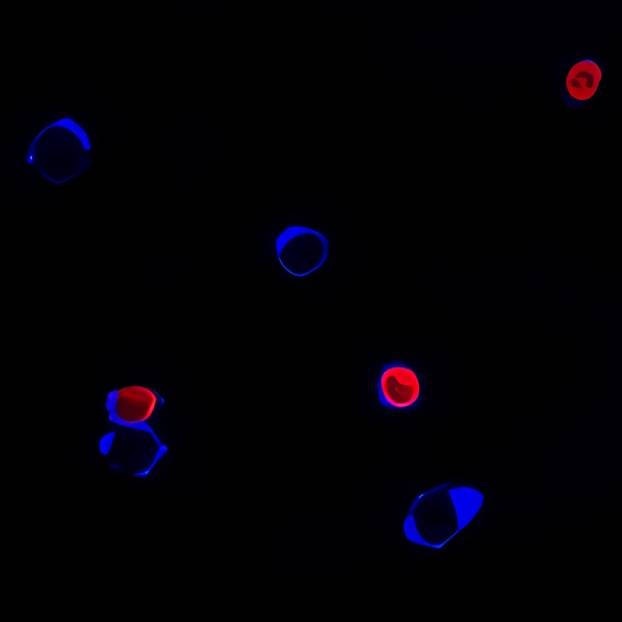
Yeast-Specific Live-or-Dye™ Staining Kits
| Catalog No. | Kit Name | Live-or-Dye™ Color | Other Dye |
|---|---|---|---|
| 31064 | Yeast Live-or-Dye™ Fixable Live/Dead Staining Kit | Live-or-Dye™ 568/583 (stains dead cells red) | Thiazole Orange (stains all cells green) |
| 31063-1 | Yeast Viability Staining Kit (Green/Red) | Live-or-Dye™ 594/614 (stains dead cells red) | CF®488A ConA (stains all cell walls green) |
| 31063-2 | Yeast Viability Staining Kit (Red/Blue) | Live-or-Dye™ 405/452 (stains dead cells blue) | CF®594 ConA (stains all cell walls red) |
| 31063-3 | Yeast Viability Staining Kit (Red/Far-Red) | Live-or-Dye™ 568/583 (stains dead cells red) | CF®640R ConA (stains all cell walls far-red) |
Draq7 is a trademark of Biostatus, Ltd. Texas Red and Pacific Blue are registered trademarks of Thermo Fisher Scientific. Cy Dye is a registered trademark of Cytiva.