 New Products
 Earth-Friendly Products
 Biotium Choice Antibodies
 Special Offers


 Powered by Bioz


Content #1
Content #1
Content #1
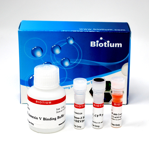
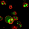
These kits provides two apoptosis markers, our novel NucView® 488 Caspase-3 Substrate and Annexin V conjugate for detecting caspase-3 activation and phosphatidylserine (PS) translocation.
These kits provides two apoptosis markers, our novel NucView® 488 Caspase-3 Substrate and fluorescent CF® dye Annexin V for detecting two important apoptosis events, caspase-3 activation and phosphatidylserine (PS) translocation.
The rate of apoptosis varies from cell to cell even within the same population. As a result, various apoptotic events or markers accompanying the apoptotic process also occur differently among cells. Thus, it is important to be able to detect these apoptotic events on an individual cell basis. Traditionally, caspase activity has been detected either using a membrane-impermeable fluorogenic enzyme substrate such as DEVD-R110, or a fluorescently-labeled inhibitor such as a FLICA reagent. In the former case, cell lysis is required, thus precluding the detection of caspase activity in live cells. In addition, such caspase assays measure only the average caspase activity of a highly heterogeneous cell population at a given time. In the latter case, although a FLICA reagent can enter live cells to detect caspase activity, only the initial fluorescent signal following the application of the reagent can truly reflect the enzyme activity or the state of the apoptotic cells because any detected signal after the initial "snapshot" will need to consider the potential interference of the inhibitor to the enzyme and the apoptotic cell itself.
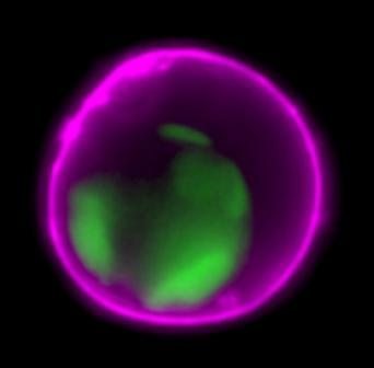
Unlike conventional caspase assays, NucView® 488 Caspase-3 substrate detects caspase-3 activity within individual whole cells in a non-interfering manner. The substrate consists of a fluorogenic DNA dye and a DEVD substrate moiety specific for caspase-3. The substrate, which is both non-fluorescent and nonfunctional as a DNA dye, rapidly crosses cell membranes to enter the cytoplasm, where it is cleaved by caspase-3 to form a high-affinity DNA dye that stains the nucleus bright green. Thus, the NucView® 488 caspase-3 substrate is bi-functional, allowing detection of caspase-3 activity and visualization of apoptotic nuclear morphology.
The kit contains reagents sufficient for 50 flow cytometry assays (200 uL assay volume). The number of fluorescence microscopy assays that can be performed with the kit may vary based on the size of culture vessel and staining volume used.
To learn about the advantages of monitoring apoptosis using NucView® caspase-3 substrates, visit the NucView® Technology Page.
1. Reproductive BioMedicine Online (2008) 16(5), 657-663. www.rbmonline.com/Article/3185
2. Curr Trends Biotech Pharm (2009) 3(3), 278-286. ISSN 0973-8916
3. Malaysian Journal of Chemistry (2009) Vol 11(1), 136-142.
4. Malaysian Journal of Chemistry (2009) 11(1), 143 – 148.
5. Pharmacognosy Res (2010) 2(2), 113-119. doi: 10.4103/0974-8490.62949
6. Mol Cancer Res (2013) 11(4), 405–17. DOI: 10.1158/1541-7786.MCR-12-0551
7. Stem Cells (2013) 31, 1121–1135. doi: 10.1002/stem.1368
8. Stem Cells (2013) 31, 2374–2387. https://doi.org/10.1002/stem.1509
9. Journal of Cell Science (2014) 127, 1738-1750. doi: 10.1242/jcs.138214
10. PLoS Genet (2014) 10(10), e1004594. doi:10.1371/journal.pgen.1004594
11. Cancer Gene Therapy (2016) Cancer Gene Ther 23(6), 178-87. doi:10.1038/cgt.2016.18
12. Current Cancer Drug Targets (2018) 18, 1-9. DOI:10.2174/1568009617666171114144236
13. Sci Rep (2018) 8, 13192. DOI:10.1038/s41598-018-31575-3
Find a list of NucView® references and a list of validated cell lines under Supporting Documents.
Bioscience kits
The guaranteed shelf life from date of receipt for bioscience kits is listed on the product information sheet. Some kits have an expiration date printed on the kit box label, this is the guaranteed shelf life date calculated from the day that the product shipped from our facility. Kits often are functional for significantly longer than the guaranteed shelf life. If you have an older kit in storage that you wish to use, we recommend performing a small scale positive control experiment to confirm that the kit still works for your application before processing a large number of samples or precious samples.
Antibodies and other conjugates
The guaranteed shelf life from date of receipt for antibodies and conjugates is listed on the datasheet sheet which can be downloaded on the product page. Antibodies and other conjugates often are functional for significantly longer than the guaranteed shelf life. If you have an older conjugate in storage that you wish to use, we recommend performing a small scale positive control experiment to confirm that the product still works for your application before processing a large number of samples or precious samples.
For lyophilized antibodies, we recommend reconstituting the antibody with glycerol and antimicrobial preservative like sodium azide for the longest shelf life (note that sodium azide is not compatible with HRP-conjugates).
Chemicals, dyes, and gel stains
Biotium guarantees the stability of chemicals, dyes, and gel stains for at least a year from the date you receive the product. However, the majority of these products are highly stable for many years, as long as they are stored as recommended. Storage conditions can be found on the product information sheet or product safety and data sheet, material safety data sheet, and on the product label. Fluorescent compounds should be protected from light for long term storage.
If you have a Biotium compound that has been in storage for longer than one year that you wish to use, we recommend performing a small scale positive control experiment to confirm that the compound still works for your application before processing a large number of samples or precious samples.
Expiration date based on date of manufacture (DOM)
If your institution requires you to document expiration date based on date of manufacture for reagents, please contact [email protected] for assistance.
Chemical products with special stability considerations:
Esters
Ester compounds include the following:
Ester dyes are stable in solid form as long as they are protected from light and moisture. Esters are not stable in aqueous solution. Concentrated stock solutions should be prepared in anhydrous DMSO (see Biotium catalog no. 90082). Stock solutions in anhydrous DMSO can be stored desiccated at -20°C for one month or longer. Esters should be diluted in aqueous solution immediately before use. Succinimidyl esters (SE) should be dissolved in a solution that is free of amine-containing compounds like Tris, glycine, or protein, which will react with the SE functional group. AM esters and diacetate compounds should be dissolved in a solution that is free of serum, because serum could contain esterases that would hydrolyze the compound.
A note on CF® Dye succinimidyl ester stability
Succinimidyl esters (SE) are generally susceptible to hydrolysis, which can result in lower labeling efficiency. Many commercially available fluorescent dyes used for life science research are heavily sulfonated dyes which makes them particularly hygroscopic, worsening the hydrolysis problem. In addition, for several commercially available SE reactive dyes, the SE group is derived from an aromatic carboxylic acid, while the SE group in all of Biotium’s CF® Dyes is prepared from an aliphatic carboxylic acid. This structural difference reduces the susceptibility of CF® Dye SE reactive groups to hydrolysis, resulting in relatively stable reactive dyes with consistently higher labeling efficiency compared to other SE derivatives of other fluorescent dyes.
Maleimides, MTS and thiosulfate dyes
Like the succinimidyl ester dyes, these dyes are also susceptible to hydrolysis, although generally to a much lower degree. Thus, for long term storage, anhydrous DMSO is recommended for making stock solutions.
Other reactive dyes
Amines, aminooxy (also known as oxylamine), hydrazide, azide, alkyne, BCN, and tyramide reactive dyes, as well as dye free acids, are generally stable in aqueous solution when stored at -20°C for 6-12 months or longer, as long as no compounds are present that may react with the dye’s functional group. See the product information sheets for specific reactive dyes more information.
Coelenterazines and D-luciferin
Coelenterazines are stable in solid form when stored as recommended; they are not stable in aqueous solution. Concentrated coelenterazine stock solutions (typically 1-100 mg/mL) should be prepared in ethanol or methanol; do not use DMSO or DMF to dissolve coelenterazines, because these solvents will oxidize the compounds. Ethanol or methanol stocks of coelenterazine can be stored at -20°C or below for six months or longer; alcohol stocks may evaporate during storage, so use tightly sealing screw cap vials and wrap the vials with Parafilm for long term storage. Propylene glycol also can be used as a solvent to minimize evaporation. If the solvent evaporates, the coelenterazine will still be present in the vial, so note the volume in the vial prior to storage so that you can adjust the solvent volume to correct for evaporation if needed. Prepare working solutions in aqueous buffers immediately before use. Coelenterazines are stable for up to five hours in aqueous solution.
Aquaphile™ coelenterazines are water soluble formulations of coelenterazines. They are stable in solid form when stored as recommended. Aquaphile™ coelenterazines should be dissolved in aqueous solution immediately before use. They are stable for up to five hours in aqueous solution.
Note that coelenterazines are predominantly yellow solids, but may contain dark red or brown flecks. This does not affect product stability or performance. If your coelenterazine is uniformly brown, then it is oxidized and needs to be replaced.
D-luciferin is stable in solid form and as a concentrated stock solution when stored as recommended; it is not stable at dilute working concentrations in aqueous solution. Prepare concentrated D-luciferin stock solutions (typically 1-100 mg/mL) in water, and store in aliquots at -20°C or below for six months or longer. Prepare working solutions immediately before use.
The NucView® caspase-3 substrates are activity-dependent i.e. require active caspase-3 enzyme. In dead, fixed and preserved cells or tissue sections, there are no active caspases and hence these substrates cannot be used. We also offer TUNEL assay kits that are suitable for apoptosis detection in fixed cells and tissues. The kits employ dUTPs conjugated to our exceptionally bright and photostable CF® dyes for single-step fluorescent TUNEL labeling of DNA strand breaks, a hallmark of apoptotic cells, and are suitable for analysis by fluorescence microscopy or flow cytometry.
The NucView® 488 Assay Kit for Live Cells (30029) contains NucView® 488 substrate at 0.2 mM in DMSO and caspase-3 inhibitor Ac-DEVD-CHO. The final DMSO concentration in the kit assay can be fairly high, which is undesirable for sensitive cell types. Because of this, we offer NucView® 488 Caspase-3 Substrate at 1 mM in DMSO (10402) and at 1 mM in PBS (10403), so customers can control the amount of DMSO in their assay. Note that in non-DMSO sensitive cell types, adding DMSO during the substrate incubation can enhance NucView® staining.
We also offer blue fluorogenic NucView® 405 in DMSO (10405) and PBS (10407), and orange fluorogenic NucView® 530 in DMSO (10406) and PBS (10408).
It has been reported in publications that concentrations of serum above 10% in the assay may affect the results.
See the following publications for more information
Our ViaFluor® SE Cell Proliferation assay is a dye dilution assay for cell division, like CFSE and CellTrace™ Violet from Thermo. This type of assay is commonly used to measure lymphocyte proliferative responses in culture and in vivo (if the labeled cells are injected back into mice). It requires flow cytometry to analyze and allows you to count how many cell divisions have occurred in the labeled cells.
For more information and a typical procedure for using fluorescent ViaFluor® SE Dyes with PMBCs, which can easily be adapted for use with other cell types, please see our Tech Tip: Measuring Cell Division in PMBCs by Flow Cytometry
If flow cytometry is not an option, we offer absorbance-based and fluorescence-based microplate assays for quantitating cell numbers. These measure mitochondrial activity (resazurin/MTT/XTT) or intracellular esterase activity (calcein AM) as a readout of viable cell numbers. Please visit the Cell Viability and Apoptosis technology page for more information.
The ATP-Glo™ assay is a luminescence assay for cellular ATP levels, which are proportional to the number of live cells. This assay requires a luminometer.
CellTrace is a trademark of Thermo Fisher Scientific.
Our Resazurin Cell Viability Assay (Cat. No. 30025) has red fluorescence (Ex/Em 530-560/590 nm), and is specifically designed for microplate reader. It is an economical, easy-to-use, and homogeneous (no-wash) assay for quantifying live cells. It is similar to alamarBlue®, PrestoBlue®, and CellTiter-Blue®.
The Calcein AM Cell Viability Assay (Cat. No. 30026) has green fluorescence (Ex/Em 485/530 nm), and also works well for microplate reader. This assay requires culture medium to be removed from cells before adding the viability dye in buffer. We also offer the Viability/Cytotoxicity Assay for Animal Live & Dead Cells, which combines calcein-AM with the fluorescent dead cell stain EthD-III, and is compatible with microplate reader.
