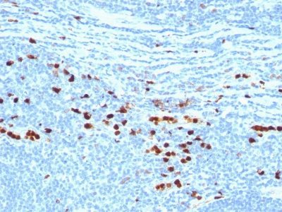
Calgranulin B / MRP-8/-14 / MAC 387 / S100A8/A9 / Macrophage Ag (MAC387)

Please fill in the inquiry form and we will contact you shortly.
Wishlist updated! View wishlist
 Powered by Bioz
Product Description
Calprotectin is a heteroduplex of Protein S100-A8/A9 (also called MRP-8/14, or Calgranulin A/B). This antibody recognizes the 14 kDa B component (Protein S100-A9) of the complex. Protein S100-A9 is a member of the S100 family of proteins containing 2 EF-hand calcium-binding motifs. Altered expression of this protein is associated with the disease cystic fibrosis. This antibody reacts with neutrophils, monocytes, macrophages, and squamous mucosal epithelia and has been shown as an important marker for identifying macrophages in tissue sections.
Primary antibodies are available purified, or with a selection of fluorescent CF® dyes and other labels. CF® dyes offer exceptional brightness and photostability. See the CF® Dye Brochure for more information. Note: Conjugates of blue fluorescent dyes like CF®405S and CF®405M are not recommended for detecting low abundance targets, because blue dyes have lower fluorescence and can give higher non-specific background than other dye colors.
Catalog number key for antibody number 0035, Anti-Calgranulin-B (MAC387)
| Antibody # prefix | Conjugation | Ex/Em (nm) | Laser line | Detection channel | Dye Features |
|---|---|---|---|---|---|
| BNC04 | CF®405S | 404/431 | 405 | DAPI (microscopy), AF405 | CF®405S Features |
| BNC88 | CF®488A | 490/515 | 488 | GFP, FITC | CF®488A Features |
| BNC68 | CF®568 | 562/583 | 532, 561 | RFP, TRITC | CF®568 Features |
| BNC94 | CF®594 | 593/614 | 561 | Texas Red® | CF®594 Features |
| BNC40 | CF®640R | 642/662 | 633-640 | Cy®5 | CF®640R Features |
| BNC47 | CF®647 | 650/665 | 633-640 | Cy®5 | CF®647 Features |
| BNC74 | CF®740 | 742/767 | 633-685 | 775/50 | CF®740 Features |
| BNCB | Biotin | N/A | N/A | N/A | |
| BNUB | Purified | N/A | N/A | N/A | |
| BNUM | Purified, BSA-free | N/A | N/A | N/A |
Note: Listed references are for this antibody clone sold by Biotium and other suppliers.