CD5 Monoclonal Mouse Antibody (C5/473 + CD5/54/F6)

Please fill in the inquiry form and we will contact you shortly.
Wishlist updated! View wishlist
 Powered by Bioz
Product Description
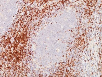
Recognizes a 67 kDa transmembrane protein, which is identified as CD5. The CD5 antigen is found on 95% of thymocytes and 72% of peripheral blood lymphocytes. In lymph nodes, the main reactivity is observed in T cell areas. Anti-CD5 is a pan T-cell marker that also reacts with a range of neoplastic B-cells, e.g. chronic lymphocytic leukemia/small lymphocytic lymphoma (CLL/SLL), mantle cell lymphoma, and a subset (~10%) of diffuse large B-cell lymphoma. CD5 aberrant expression is useful in making a diagnosis of mature T-cell neoplasms. Anti-CD5 detection is diagnostic in CLL/SLL within a panel of other B-cell markers, especially one that includes anti-CD23. Anti-CD5 is also very useful in differentiating among mature small lymphoid cell malignancies. In addition, anti-CD5 can be used in distinguishing thymic carcinoma ( ) from thymoma (-). Anti-CD5 does not react with granulocytes or monocytes.
Primary antibodies are available purified, or with a selection of fluorescent CF® dyes and other labels. CF® dyes offer exceptional brightness and photostability. See the CF® Dye Brochure for more information. Note: Conjugates of blue fluorescent dyes like CF®405S and CF®405M are not recommended for detecting low abundance targets, because blue dyes have lower fluorescence and can give higher non-specific background than other dye colors.
Catalog number key for antibody number 0748, Anti-CD5 (C5/473 CD5/54/F6)
| Antibody # prefix | Conjugation | Ex/Em (nm) | Laser line | Detection channel | Dye Features |
|---|---|---|---|---|---|
| BNC04 | CF®405S | 404/431 | 405 | DAPI (microscopy), AF405 | CF®405S Features |
| BNC88 | CF®488A | 490/515 | 488 | GFP, FITC | CF®488A Features |
| BNC68 | CF®568 | 562/583 | 532, 561 | RFP, TRITC | CF®568 Features |
| BNC94 | CF®594 | 593/614 | 561 | Texas Red® | CF®594 Features |
| BNC40 | CF®640R | 642/662 | 633-640 | Cy®5 | CF®640R Features |
| BNC47 | CF®647 | 650/665 | 633-640 | Cy®5 | CF®647 Features |
| BNC74 | CF®740 | 742/767 | 633-685 | 775/50 | CF®740 Features |
| BNCB | Biotin | N/A | N/A | N/A | |
| BNUB | Purified | N/A | N/A | N/A | |
| BNUM | Purified, BSA-free | N/A | N/A | N/A |
Note: Listed references are for this antibody clone sold by Biotium and other suppliers.