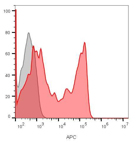
CD8 Monoclonal Mouse Antibody (C8/1035)

Please fill in the inquiry form and we will contact you shortly.
Wishlist updated! View wishlist
Product Description
CD8 is a cell surface receptor expressed either as a heterodimer with the CD8 beta chain (CD8 alpha/beta) or as a homodimer (CD8 alpha/alpha). A majority of thymocytes and a subpopulation of mature T cells and NK cells express CD8a. CD8 binds to MHC class 1 and through its association with protein tyrosine kinase p56lck plays a role in T cell development and activation of mature T cells. For mature T-cells, CD4 and CD8 are mutually exclusive, so anti-CD8, generally used in conjunction with anti-CD4. It is a useful marker for distinguishing helper/inducer T-lymphocytes, and most peripheral T-cell lymphomas are CD4 /CD8-. Anaplastic large cell lymphoma is usually CD4 and CD8-, and in T-lymphoblastic lymphoma/leukemia, CD4 and CD8 are often co-expressed. CD8 is also found in littoral cell angioma of the spleen.
Primary antibodies are available purified, or with a selection of fluorescent CF® dyes and other labels. CF® dyes offer exceptional brightness and photostability. See the CF® Dye Brochure for more information. Note: Conjugates of blue fluorescent dyes like CF®405S and CF®405M are not recommended for detecting low abundance targets, because blue dyes have lower fluorescence and can give higher non-specific background than other dye colors.
Catalog number key for antibody number 1035, Anti-CD8 (C8/1035)
| Antibody # prefix | Conjugation | Ex/Em (nm) | Laser line | Detection channel | Dye Features |
|---|---|---|---|---|---|
| BNC04 | CF®405S | 404/431 | 405 | DAPI (microscopy), AF405 | CF®405S Features |
| BNC88 | CF®488A | 490/515 | 488 | GFP, FITC | CF®488A Features |
| BNC68 | CF®568 | 562/583 | 532, 561 | RFP, TRITC | CF®568 Features |
| BNC94 | CF®594 | 593/614 | 561 | Texas Red® | CF®594 Features |
| BNC40 | CF®640R | 642/662 | 633-640 | Cy®5 | CF®640R Features |
| BNC47 | CF®647 | 650/665 | 633-640 | Cy®5 | CF®647 Features |
| BNC74 | CF®740 | 742/767 | 633-685 | 775/50 | CF®740 Features |
| BNCB | Biotin | N/A | N/A | N/A | |
| BNUB | Purified | N/A | N/A | N/A | |
| BNUM | Purified, BSA-free | N/A | N/A | N/A |
References
Note: References for this clone sold by other suppliers may be listed for expected applications.
Knapp W. et. al. Leukocyte Typing IV, p342-343, Oxford University Press, 1989.