Small Cell Lung Cancer Monoclonal Mouse Antibody (MOC-52)

Please fill in the inquiry form and we will contact you shortly.
Wishlist updated! View wishlist
 Powered by Bioz
Product Description
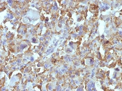
This MAb reacts with a membrane-associated protein present in normal and malignant neuroendocrine tissues including small cell lung cancer (SCLC). It stains neural and a variable number of endocrine tissues and in the lung it reacts preferentially with SCLC and carcinoids. Its epitope is destroyed during formalin fixation. This antibody was categorized during the First International Workshop on Small Cell Lung Cancer Antigens held in London in April 1987. There are two major types of Lung Carcinoma: non-small cell, which accounts for 80% of all cases; and small cell, which accounts for roughly 20% of all lung cancers reported. The lung continues to be a customary place for cancer migration from tumors elsewhere in the body. Treatment depends on the specific cell type of the cancer, level of progression and status of the individual patient.
Primary antibodies are available purified, or with a selection of fluorescent CF® dyes and other labels. CF® dyes offer exceptional brightness and photostability. See the CF® Dye Brochure for more information. Note: Conjugates of blue fluorescent dyes like CF®405S and CF®405M are not recommended for detecting low abundance targets, because blue dyes have lower fluorescence and can give higher non-specific background than other dye colors.
Catalog number key for antibody number 0329, Anti-Small Cell Lung Cancer (MOC-52)
| Antibody # prefix | Conjugation | Ex/Em (nm) | Laser line | Detection channel | Dye Features |
|---|---|---|---|---|---|
| BNC04 | CF®405S | 404/431 | 405 | DAPI (microscopy), AF405 | CF®405S Features |
| BNC88 | CF®488A | 490/515 | 488 | GFP, FITC | CF®488A Features |
| BNC68 | CF®568 | 562/583 | 532, 561 | RFP, TRITC | CF®568 Features |
| BNC94 | CF®594 | 593/614 | 561 | Texas Red® | CF®594 Features |
| BNC40 | CF®640R | 642/662 | 633-640 | Cy®5 | CF®640R Features |
| BNC47 | CF®647 | 650/665 | 633-640 | Cy®5 | CF®647 Features |
| BNC74 | CF®740 | 742/767 | 633-685 | 775/50 | CF®740 Features |
| BNCB | Biotin | N/A | N/A | N/A | |
| BNUB | Purified | N/A | N/A | N/A | |
| BNUM | Purified, BSA-free | N/A | N/A | N/A |
Note: Listed references are for this antibody clone sold by Biotium and other suppliers.