Spectrin Beta III Monoclonal Mouse Antibody (SPTBN2/1778)

Please fill in the inquiry form and we will contact you shortly.
Wishlist updated! View wishlist
Product Description
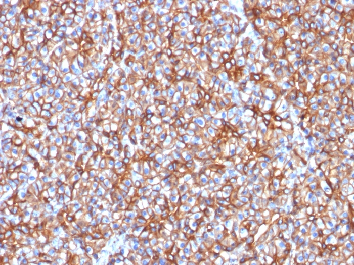
Spectrin is an actin binding protein that is a major component of the plasma membrane skeleton. Spectrins function as membrane organizers and stabilizers by forming dimers, tetramers and higher polymers. Vertebrate spectrins have two alpha-subunits (alpha-I/alpha-II), four beta-subunits (beta-I-beta-IV) and a beta-H subunit creating diversity and specialization of function. Spectrin α and spectrinβ are present in erythrocytes, whereas spectrin α II (also designated fodrin α) and spectrinβ I (also designated fodrinβ) are present in other somatic cells. The spectrin tetramers in erythrocytes act as barriers to lateral diffusion, but spectrin dimers seem to lack this function. Spectrinβ III is highly homologous to both spectrinβ I and spectrinβ II. Spectrinβ III is highly expressed in brain, kidney, pancreas and liver, and at lower levels in lung and placenta. Spectrin beta 3 is primarily expressed in nervous tissues with highest expression levels in the cerebellum, where it is found in Purkinje cell soma and dendrites.
Primary antibodies are available purified, or with a selection of fluorescent CF® dyes and other labels. CF® dyes offer exceptional brightness and photostability. See the CF® Dye Brochure for more information. Note: Conjugates of blue fluorescent dyes like CF®405S and CF®405M are not recommended for detecting low abundance targets, because blue dyes have lower fluorescence and can give higher non-specific background than other dye colors.
Catalog number key for antibody number 1778, Anti-Spectrin Beta III (SPTBN2/1778)
| Antibody # prefix | Conjugation | Ex/Em (nm) | Laser line | Detection channel | Dye Features |
|---|---|---|---|---|---|
| BNC04 | CF®405S | 404/431 | 405 | DAPI (microscopy), AF405 | CF®405S Features |
| BNC88 | CF®488A | 490/515 | 488 | GFP, FITC | CF®488A Features |
| BNC68 | CF®568 | 562/583 | 532, 561 | RFP, TRITC | CF®568 Features |
| BNC94 | CF®594 | 593/614 | 561 | Texas Red® | CF®594 Features |
| BNC40 | CF®640R | 642/662 | 633-640 | Cy®5 | CF®640R Features |
| BNC47 | CF®647 | 650/665 | 633-640 | Cy®5 | CF®647 Features |
| BNC74 | CF®740 | 742/767 | 633-685 | 775/50 | CF®740 Features |
| BNCB | Biotin | N/A | N/A | N/A | |
| BNUB | Purified | N/A | N/A | N/A | |
| BNUM | Purified, BSA-free | N/A | N/A | N/A |