 New Products
 Earth-Friendly Products
 Biotium Choice Antibodies
 Special Offers


Content #1
Content #1
Content #1
Vitamin D-Binding Protein (Vitamin D-BP, also known as GC Globulin) binds monomeric G-actin in fixed and permeabilized cells. Available conjugated to a selection of bright and photostable CF® Dyes for fluorescence microscopy.
Recombinant Human Vitamin D-Binding Protein (Vitamin D-BP, also known as GC Globulin) binds monomeric G-actin (1-3). Fluorescent conjugates of Vitamin D-BP can be used to stain monomeric G-actin in fixed and permeabilized cells.
Vitamin D-BP staining may also be used in combination with fluorescent phalloidin staining of F-actin to visualize the distribution of unpolymerized G-actin relative to actin filaments. Vitamin D-BP is more specific for staining G-actin compared to fluorescent conjugates of DNase-I, which binds to DNA in addition to G-actin. Biotium’s Recombinant Human Vitamin D-BP Conjugates are labeled with a selection of our bright and photostable CF® Dyes for fluorescence microscopy.
Biotium’s next-generation CF® Dyes were designed to be highly water-soluble with advantages in brightness and photostability compared to Alexa Fluor®, DyLight®, and other fluorescent dyes. Learn more about CF® Dyes.
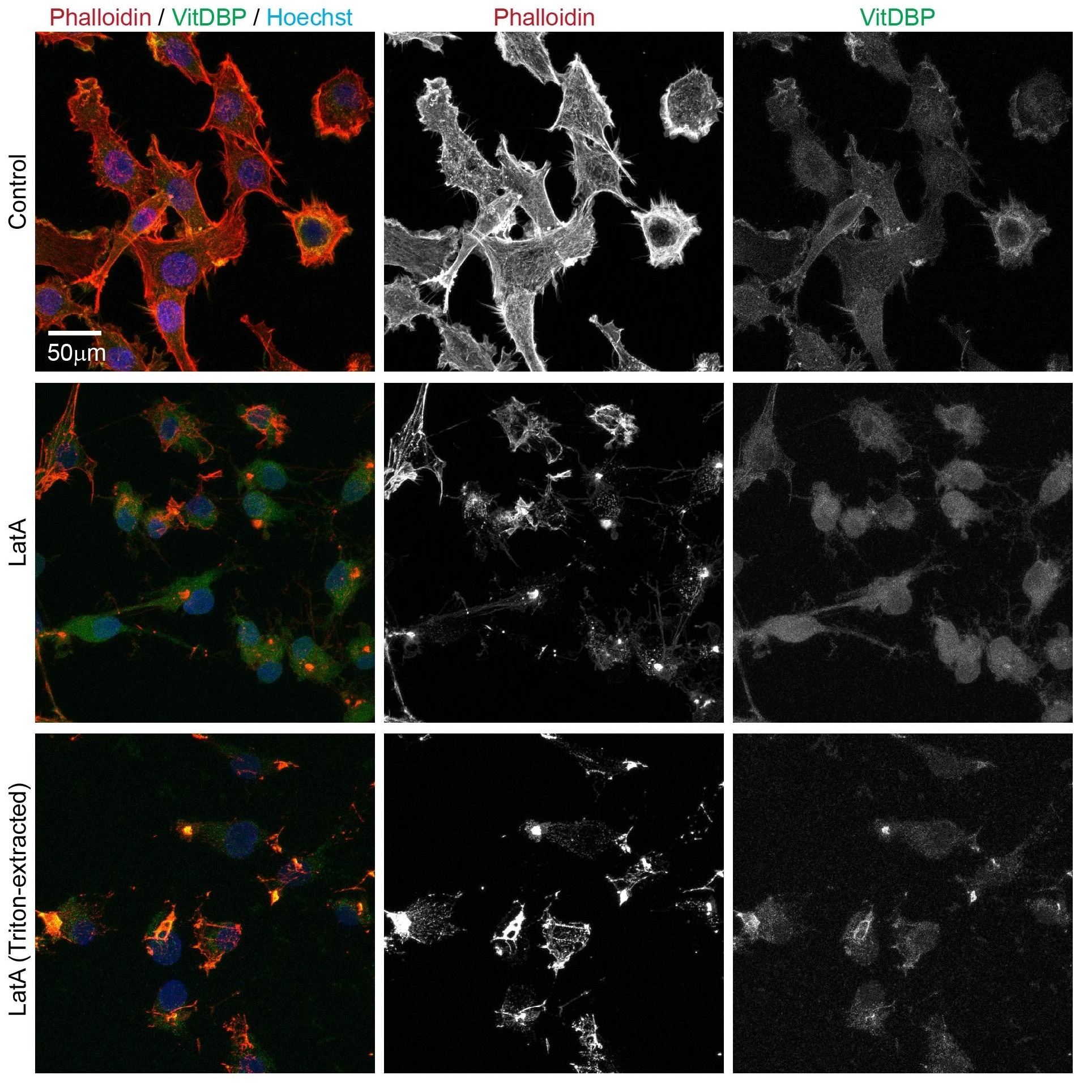
Passaged chondrocytes stained with Rhodamine-Phalloidin and Vitamin D-binding protein CF®488 conjugate to visualize F-actin and G-actin. Passaged chondrocytes were exposed to latrunculin A (1uM) for 30 minutes, and were either fixed immediately in 4%PFA (top and middle row) or pre-solubilized, to remove cytoplasmic G-actin, with 0.1%Triton for 2 minutes prior to fixation in 4%PFA (bottom row). Latrunculin treatment reduced staining for Phalloidin (predominantly F-actin) and increased staining for Vitamin D-binding protein (predominantly G-actin) (see middle row). Pre-treatment with 0.1% Triton prior to PFA fixation reduced staining for Vitamin D-binding protein (see bottom row). Images provided from Parreno Laboratory, Department of Biological Sciences, University of Delaware.
| Conjugation | Ex/Em | Size | Catalog No. | Dye Features |
|---|---|---|---|---|
| CF®488A | 490/516 nm | 1 mL (100 ug) | 70087 | CF®488A Features |
| CF®594 | 593/615 nm | 1 mL (100 ug) | 70088 | CF®594 Features |
| CF®640R | 642/663 nm | 1 mL (100 ug) | 70089 | CF®640R Features |
| CF®647 | 652/668 nm | 1 mL (100 ug) | 70090 | CF®647 Features |
| CF®740 | 742/767 nm | 1 mL (100 ug) | 70091 | CF®740 Features |
For staining F-actin, Biotium recommends ActinBrite™ High Affinity Phalloidin Conjugates which were designed to preserve strong F-actin binding over conventional phalloidin conjugates. With ActinBrite™, samples can be imaged after for a month or more (depending on the conjugate and mounting method)—making delayed imaging easier and more dependable.
Browse Biotium's comprehensive catalog of fluorescent bioconjugates as well novel and classic organelle stains.
Unless specified as isotype-specific, anti-IgG secondary antibodies are raised against whole immunoglobulin, so they are expected to cross-react widely with isotypes other than IgG.
No, you don't always need to use serum from the same species as the secondary antibody for blocking. BSA, fish gelatin, goat serum, or non-fat milk (for western) can be used with secondary antibodies from most host species. However, it is important to avoid using blocking serum from the same host species as your primary antibody that is detected by a secondary antibody against the same species (immunoglobulins in the blocking serum will compete for the secondary binding). For example, avoid using goat serum for blocking if you are using a goat primary antibody with anti-goat secondary antibody.
Our TrueBlack® IF Background Suppressor System and TrueBlack® WB Blocking Buffer Kit contain proteins from non-mammalian sources, and should not interact with secondary antibodies from mammalian or chicken host. Our BSA also is immunoglobulin-free and should not interfere with secondary antibodies.
F(ab) fragment-specific antibodies react with the light chains and not the heavy chains of immunoglobulins from the target species. They can be used to detect antibody fragments, or to avoid cross-reactivity with immunoglobulin heavy chains.
Bioscience kits
The guaranteed shelf life from date of receipt for bioscience kits is listed on the product information sheet. Some kits have an expiration date printed on the kit box label, this is the guaranteed shelf life date calculated from the day that the product shipped from our facility. Kits often are functional for significantly longer than the guaranteed shelf life. If you have an older kit in storage that you wish to use, we recommend performing a small scale positive control experiment to confirm that the kit still works for your application before processing a large number of samples or precious samples.
Antibodies and other conjugates
The guaranteed shelf life from date of receipt for antibodies and conjugates is listed on the product information sheet. Antibodies and other conjugates often are functional for significantly longer than the guaranteed shelf life. If you have an older conjugate in storage that you wish to use, we recommend performing a small scale positive control experiment to confirm that the product still works for your application before processing a large number of samples or precious samples.
For lyophilized antibodies, we recommend reconstituting the antibody with glycerol and antimicrobial preservative like sodium azide for the longest shelf life (note that sodium azide is not compatible with HRP-conjugates).
Chemicals, dyes, and gel stains
Biotium guarantees the stability of chemicals, dyes, and gel stains for at least a year from the date you receive the product. However, the majority of these products are highly stable for many years, as long as they are stored as recommended. Storage conditions can be found on the product information sheet or product safety and data sheet, material safety data sheet, and on the product label. Fluorescent compounds should be protected from light for long term storage.
If you have a Biotium compound that has been in storage for longer than one year that you wish to use, we recommend performing a small scale positive control experiment to confirm that the compound still works for your application before processing a large number of samples or precious samples.
Expiration date based on date of manufacture (DOM)
If your institution requires you to document expiration date based on date of manufacture for reagents, please contact [email protected] for assistance.
Chemical products with special stability considerations:
Esters
Ester compounds include the following:
Ester dyes are stable in solid form as long as they are protected from light and moisture. Esters are not stable in aqueous solution. Concentrated stock solutions should be prepared in anhydrous DMSO (see Biotium catalog no. 90082). Stock solutions in anhydrous DMSO can be stored desiccated at -20°C for one month or longer. Esters should be diluted in aqueous solution immediately before use. Succinimidyl esters (SE) should be dissolved in a solution that is free of amine-containing compounds like Tris, glycine, or protein, which will react with the SE functional group. AM esters and diacetate compounds should be dissolved in a solution that is free of serum, because serum could contain esterases that would hydrolyze the compound.
A note on CF® Dye succinimidyl ester stability
Succinimidyl esters (SE) are generally susceptible to hydrolysis, which can result in lower labeling efficiency. Many commercially available fluorescent dyes used for life science research are heavily sulfonated dyes which makes them particularly hygroscopic, worsening the hydrolysis problem. In addition, for several commercially available SE reactive dyes, the SE group is derived from an aromatic carboxylic acid, while the SE group in all of Biotium’s CF® Dyes is prepared from an aliphatic carboxylic acid. This structural difference reduces the susceptibility of CF® Dye SE reactive groups to hydrolysis, resulting in relatively stable reactive dyes with consistently higher labeling efficiency compared to other SE derivatives of other fluorescent dyes.
Maleimides, MTS and thiosulfate dyes
Like the succinimidyl ester dyes, these dyes are also susceptible to hydrolysis, although generally to a much lower degree. Thus, for long term storage, anhydrous DMSO is recommended for making stock solutions.
Other reactive dyes
Amines, aminooxy (also known as oxylamine), hydrazide, azide, alkyne, BCN, and tyramide reactive dyes, as well as dye free acids, are generally stable in aqueous solution when stored at -20°C for 6-12 months or longer, as long as no compounds are present that may react with the dye’s functional group. See the product information sheets for specific reactive dyes more information.
Coelenterazines and D-luciferin
Coelenterazines are stable in solid form when stored as recommended; they are not stable in aqueous solution. Concentrated coelenterazine stock solutions (typically 1-100 mg/mL) should be prepared in ethanol or methanol; do not use DMSO or DMF to dissolve coelenterazines, because these solvents will oxidize the compounds. Ethanol or methanol stocks of coelenterazine can be stored at -20°C or below for six months or longer; alcohol stocks may evaporate during storage, so use tightly sealing screw cap vials and wrap the vials with Parafilm for long term storage. Propylene glycol also can be used as a solvent to minimize evaporation. If the solvent evaporates, the coelenterazine will still be present in the vial, so note the volume in the vial prior to storage so that you can adjust the solvent volume to correct for evaporation if needed. Prepare working solutions in aqueous buffers immediately before use. Coelenterazines are stable for up to five hours in aqueous solution.
Aquaphile™ coelenterazines are water soluble formulations of coelenterazines. They are stable in solid form when stored as recommended. Aquaphile™ coelenterazines should be dissolved in aqueous solution immediately before use. They are stable for up to five hours in aqueous solution.
Note that coelenterazines are predominantly yellow solids, but may contain dark red or brown flecks. This does not affect product stability or performance. If your coelenterazine is uniformly brown, then it is oxidized and needs to be replaced.
D-luciferin is stable in solid form and as a concentrated stock solution when stored as recommended; it is not stable at dilute working concentrations in aqueous solution. Prepare concentrated D-luciferin stock solutions (typically 1-100 mg/mL) in water, and store in aliquots at -20°C or below for six months or longer. Prepare working solutions immediately before use.
Dyes that carry multiple negative charges can introduce background. Usually, this is more of a concern with labeled antibodies that carry many dyes, as opposed to a small toxin like bungarotoxin. When staining tissues, the endogenous autofluorescence of the tissue itself is often the most significant source of background. Endogenous fluorescence background in tissue is usually highest in the blue wavelengths (DAPI channel) and lowest in the far-red (Cy®5 channel). Our CF®633 bungarotoxin (catalog no. 00009) is a far-red conjugate for the Cy®5 channel with a low negative charge that should have low background from either the dye or autofluorescence.
We test fluorescent bungarotoxin on rat skeletal muscle sections. While the tissue shows autofluorescence, the bungarotoxin staining of motor endplates is usually much brighter than the background for all of the dye colors we've tested. However, if you are staining human tissue (especially brain), lipofuscin autofluorescence may be bright in all channels. This usually shows up as bright, punctate dots around cell nuclei. While we would usually recommend our TrueBlack® lipofuscin quenchers for human brain tissue, they are not compatible with bungarotoxin staining. We have, however, found that EverBrite TrueBlack® Mounting Medium (cat. no. 23017) can be used to mount skeletal muscle sections stained with bungarotoxin.
Cy Dye is a registered trademark of Cytiva.
We do not have firsthand experience with LPS labeling, but according to the literature, LPS has been labeled using amine-reactive dyes, like FITC. Our amine-reactive CF® Dye Succinimidyl Esters should also work for this. There is a publication for enzymatic labeling of LPS using dye hydrazides. Our CF® Dye hydrazides could be used in this method. The paper also describes the traditional amine labeling method and purification of the conjugate.
Bioscience kits
The guaranteed shelf life from date of receipt for bioscience kits is listed on the product information sheet. Some kits have an expiration date printed on the kit box label, this is the guaranteed shelf life date calculated from the day that the product shipped from our facility. Kits often are functional for significantly longer than the guaranteed shelf life. If you have an older kit in storage that you wish to use, we recommend performing a small scale positive control experiment to confirm that the kit still works for your application before processing a large number of samples or precious samples.
Antibodies and other conjugates
The guaranteed shelf life from date of receipt for antibodies and conjugates is listed on the product information sheet. Antibodies and other conjugates often are functional for significantly longer than the guaranteed shelf life. If you have an older conjugate in storage that you wish to use, we recommend performing a small scale positive control experiment to confirm that the product still works for your application before processing a large number of samples or precious samples.
For lyophilized antibodies, we recommend reconstituting the antibody with glycerol and antimicrobial preservative like sodium azide for the longest shelf life (note that sodium azide is not compatible with HRP-conjugates).
Chemicals, dyes, and gel stains
Biotium guarantees the stability of chemicals, dyes, and gel stains for at least a year from the date you receive the product. However, the majority of these products are highly stable for many years, as long as they are stored as recommended. Storage conditions can be found on the product information sheet or product safety and data sheet, material safety data sheet, and on the product label. Fluorescent compounds should be protected from light for long term storage.
If you have a Biotium compound that has been in storage for longer than one year that you wish to use, we recommend performing a small scale positive control experiment to confirm that the compound still works for your application before processing a large number of samples or precious samples.
Expiration date based on date of manufacture (DOM)
If your institution requires you to document expiration date based on date of manufacture for reagents, please contact [email protected] for assistance.
Chemical products with special stability considerations:
Esters
Ester compounds include the following:
Ester dyes are stable in solid form as long as they are protected from light and moisture. Esters are not stable in aqueous solution. Concentrated stock solutions should be prepared in anhydrous DMSO (see Biotium catalog no. 90082). Stock solutions in anhydrous DMSO can be stored desiccated at -20°C for one month or longer. Esters should be diluted in aqueous solution immediately before use. Succinimidyl esters (SE) should be dissolved in a solution that is free of amine-containing compounds like Tris, glycine, or protein, which will react with the SE functional group. AM esters and diacetate compounds should be dissolved in a solution that is free of serum, because serum could contain esterases that would hydrolyze the compound.
A note on CF® Dye succinimidyl ester stability
Succinimidyl esters (SE) are generally susceptible to hydrolysis, which can result in lower labeling efficiency. Many commercially available fluorescent dyes used for life science research are heavily sulfonated dyes which makes them particularly hygroscopic, worsening the hydrolysis problem. In addition, for several commercially available SE reactive dyes, the SE group is derived from an aromatic carboxylic acid, while the SE group in all of Biotium’s CF® Dyes is prepared from an aliphatic carboxylic acid. This structural difference reduces the susceptibility of CF® Dye SE reactive groups to hydrolysis, resulting in relatively stable reactive dyes with consistently higher labeling efficiency compared to other SE derivatives of other fluorescent dyes.
Maleimides, MTS and thiosulfate dyes
Like the succinimidyl ester dyes, these dyes are also susceptible to hydrolysis, although generally to a much lower degree. Thus, for long term storage, anhydrous DMSO is recommended for making stock solutions.
Other reactive dyes
Amines, aminooxy (also known as oxylamine), hydrazide, azide, alkyne, BCN, and tyramide reactive dyes, as well as dye free acids, are generally stable in aqueous solution when stored at -20°C for 6-12 months or longer, as long as no compounds are present that may react with the dye’s functional group. See the product information sheets for specific reactive dyes more information.
Coelenterazines and D-luciferin
Coelenterazines are stable in solid form when stored as recommended; they are not stable in aqueous solution. Concentrated coelenterazine stock solutions (typically 1-100 mg/mL) should be prepared in ethanol or methanol; do not use DMSO or DMF to dissolve coelenterazines, because these solvents will oxidize the compounds. Ethanol or methanol stocks of coelenterazine can be stored at -20°C or below for six months or longer; alcohol stocks may evaporate during storage, so use tightly sealing screw cap vials and wrap the vials with Parafilm for long term storage. Propylene glycol also can be used as a solvent to minimize evaporation. If the solvent evaporates, the coelenterazine will still be present in the vial, so note the volume in the vial prior to storage so that you can adjust the solvent volume to correct for evaporation if needed. Prepare working solutions in aqueous buffers immediately before use. Coelenterazines are stable for up to five hours in aqueous solution.
Aquaphile™ coelenterazines are water soluble formulations of coelenterazines. They are stable in solid form when stored as recommended. Aquaphile™ coelenterazines should be dissolved in aqueous solution immediately before use. They are stable for up to five hours in aqueous solution.
Note that coelenterazines are predominantly yellow solids, but may contain dark red or brown flecks. This does not affect product stability or performance. If your coelenterazine is uniformly brown, then it is oxidized and needs to be replaced.
D-luciferin is stable in solid form and as a concentrated stock solution when stored as recommended; it is not stable at dilute working concentrations in aqueous solution. Prepare concentrated D-luciferin stock solutions (typically 1-100 mg/mL) in water, and store in aliquots at -20°C or below for six months or longer. Prepare working solutions immediately before use.
For dyes or reagents that are supplied lyophilized (as solids), it is hard to compare quantities based on appearance of the dye in the tube, because during the lyophilization process the dye can dry down in different ways, either spread out all over the tube, clumped together, or coating the sides or bottom of the tube. Centrifugation of the tube may not help in collecting the dye solid to the bottom of the tube as this generally works for solutions. However, lyophilized solids are packaged based on highly accurate absorbance measurement of the reagent solution prior to drying, so the vial will contain the correct amount of dye.
Biotium ships all antibodies (primary, secondary and conjugates) at room temperature. We guarantee their quality and performance under these conditions based upon our stability testing. Antibodies were subjected to accelerated stability testing by storing them at various temperatures (4°C, room temperature, or 37°C) for 1 week to mimic simulated shipping conditions and tested in immunostaining experiments. All antibodies showed the expected brightness and specificity, even after storage at sub-optimal temperatures for a week or longer. You can also download our Product Storage Statement here.
In line with our goal to be more environmentally friendly by reducing the use of excess packaging, and lowering shipping costs for our customers, products that have passed our stability testing are shipped at room temperature.
Once you have received the antibody vial, please follow the long-term storage instructions on the product information (PI) sheet.

Content #1
Content #1
Content #1
Content #2
Content #3