New Products
 Earth-Friendly Products
 Biotium Choice Antibodies
 Special Offers
Mix-n-Stain™ CF® Dye Antibody Labeling Technology
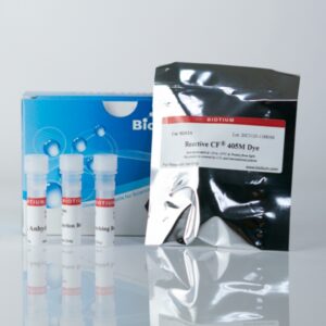
Mix-n-Stain™ Antibody Labeling Kits
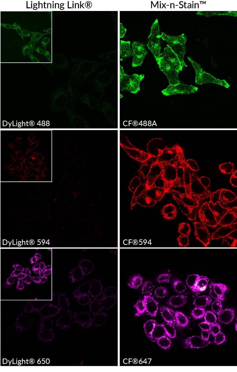
Even though antibody conjugation is dramatically simplified, the conjugates perform comparably to pre-labeled antibodies from leading suppliers. Covalent labeling ensures conjugates are stable for long-term storage, and are ideal for multi-color imaging and multiplex flow cytometry. Each kit includes everything you need, and works with most commercial antibody formulations.
Rapid Antibody Labeling with CF® Dyes & More
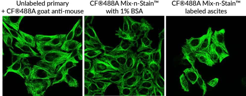
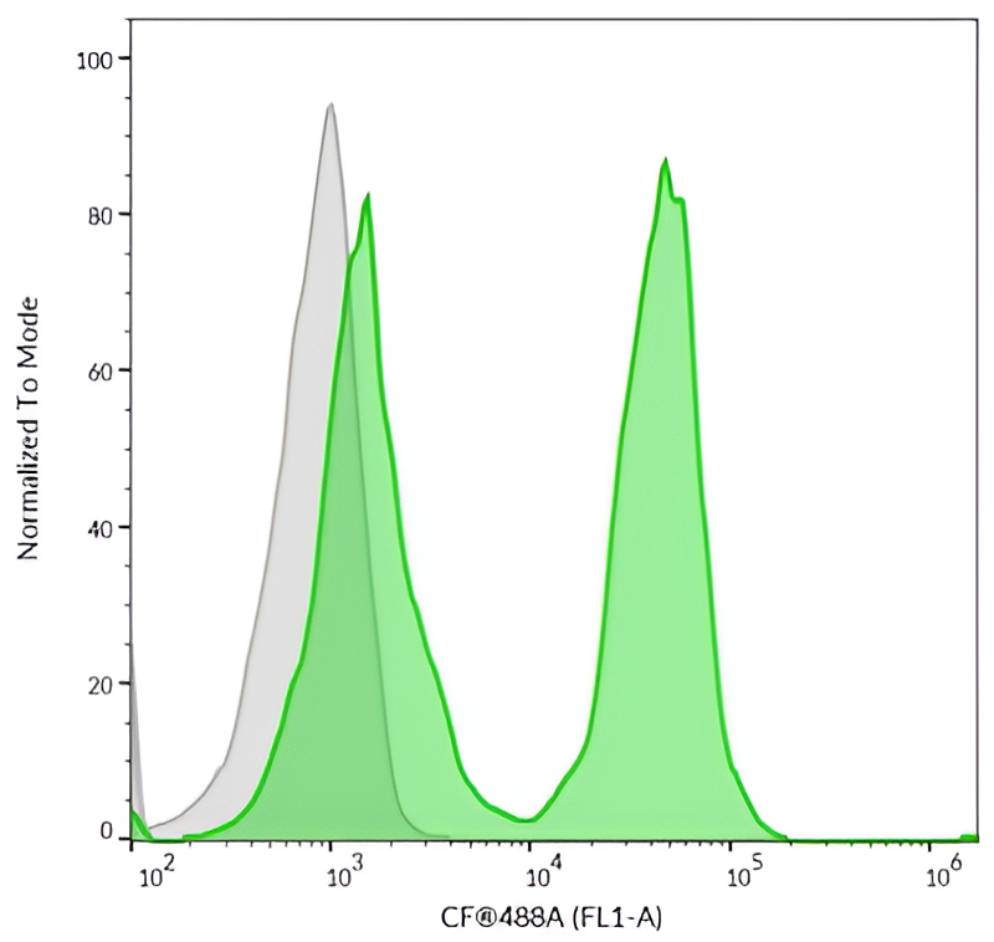
Mix-n-Stain™ CF® Dye Antibody Labeling Kits covalently label antibodies with 100% recovery. CF® Dyes offer superior brightness and photostability versus other fluorescent dyes. High water solubility of the dyes minimizes non-specific background as non-IgG proteins wash away during IF staining.
Mix-n-Stain™ kits allow antibody labeling directly in common buffers containing BSA, gelatin, azide, or low Tris, and with minor protocol adjustments, even in BSA, gelatin, or ascites fluid. This eliminates the need for antibody purification, making the kits compatible with most commercial formulations. See the the Product Protocol for step-by-step compatibility guidance.
Features
- 3 kit sizes: 5–20 µg, 20–50 µg, or 50–100 µg Ab
- Maxi Antibody Labeling Kits for 1 mg scale labeling reactions
- Choice of over 30 CF® Dye colors, biotin, FITC, or haptens
- Less than 30 seconds of hands-on time
- 15 minutes total reaction time
- No purification of the product is required
- Compatible with common antibody stabilizers
Compatible with antibody stabilizers and ascites
Mix-n-Stain™ CF® Dye, Biotin, FITC & Hapten IgG Antibody Labeling Kits
| Product description | Label options | Labeling time |
|---|---|---|
| Mix-n-Stain™ CF® Dye Antibody Labeling Kits | Choice of over 30 bright & photostable CF® Dyes | 15 minutes (~30 seconds hands-on time) Download the product protocol |
| Mix-n-Stain™ Cyanine Dye Antibody Labeling Kits | Cyanine 555 Cyanine 647 |
|
| Mix-n-Stain™ FITC Antibody Labeling Kits | FITC | |
| Mix-n-Stain™ Biotin Antibody Labeling Kits | Biotin | |
| Mix-n-Stain™ DNP Antibody Labeling Kits | DNP | |
| Mix-n-Stain™ Digoxigenin Antibody Labeling Kits | Digoxigenin (DIG) | |
| Mix-n-Stain™ Maxi Antibody Labeling Kits | Choice of 13 bright & photostable CF® Dyes, FITC, Cyanine 555, and Cyanine 647 | 30 minutes (~30 seconds hands-on time) |
| Mix-n-Stain™ STORM CF® Antibody Labeling Kits | Choice of 8 CF® Dyes specialized for STORM super-resolution microscopy |
Mix-n-Stain™ CF® Dye IgM Antibody Labeling Kits
Finally! Streamlined Labeling of IgM Antibodies
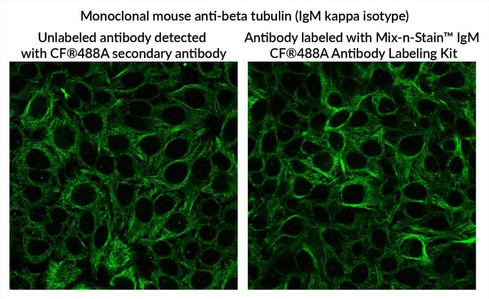
IgM antibodies are difficult to label with fluorescent dyes because the conditions typically used for labeling IgG generally do not result in bright conjugates with good antibody reactivity for IgM. Biotium has resolved this issue by optimizing the reaction conditions for labeling IgM antibodies with Mix-n-Stain™ CF® Dye IgM Antibody Labeling Kits.
Features
- Label 25 µg or 100 µg of IgM antibody
- Choice of 8 CF® Dye colors and FITC
- 15–30 minute total reaction time
- No purification, 100% recovery
View Product Page

Mix-n-Stain™ CF® Dye IgM Antibody Labeling Kits
| Conjugation | Ex/Em | Size | Catalog No. | Dye Features |
|---|---|---|---|---|
| CF®405L | 413/547 nm | 25 ug | 92558 | CF®405L Features |
| 100 ug | 92559 | |||
| CF®405M | 416/452 nm | 25 ug | 92560 | CF®405M Features |
| 100 ug | 92561 | |||
| CF®488A | 490/516 nm | 25 ug | 92562 | CF®488A Features |
| 100 ug | 92563 | |||
| CF®555 | 554/568 nm | 25 ug | 92564 | CF®555 Features |
| 100 ug | 92565 | |||
| CF®568 | 562/583 nm | 25 ug | 92566 | CF®568 Features |
| 100 ug | 92567 | |||
| CF®594 | 593/615 nm | 25 ug | 92568 | CF®594 Features |
| 100 ug | 92569 | |||
| CF®640R | 642/663 nm | 25 ug | 92570 | CF®640R Features |
| 100 ug | 92571 | |||
| CF®647 | 652/668 nm | 25 ug | 92572 | CF®647 Features |
| 100 ug | 92573 | |||
| FITC | 498/517 nm | 25 ug | 92574 | |
| 100 ug | 92575 |
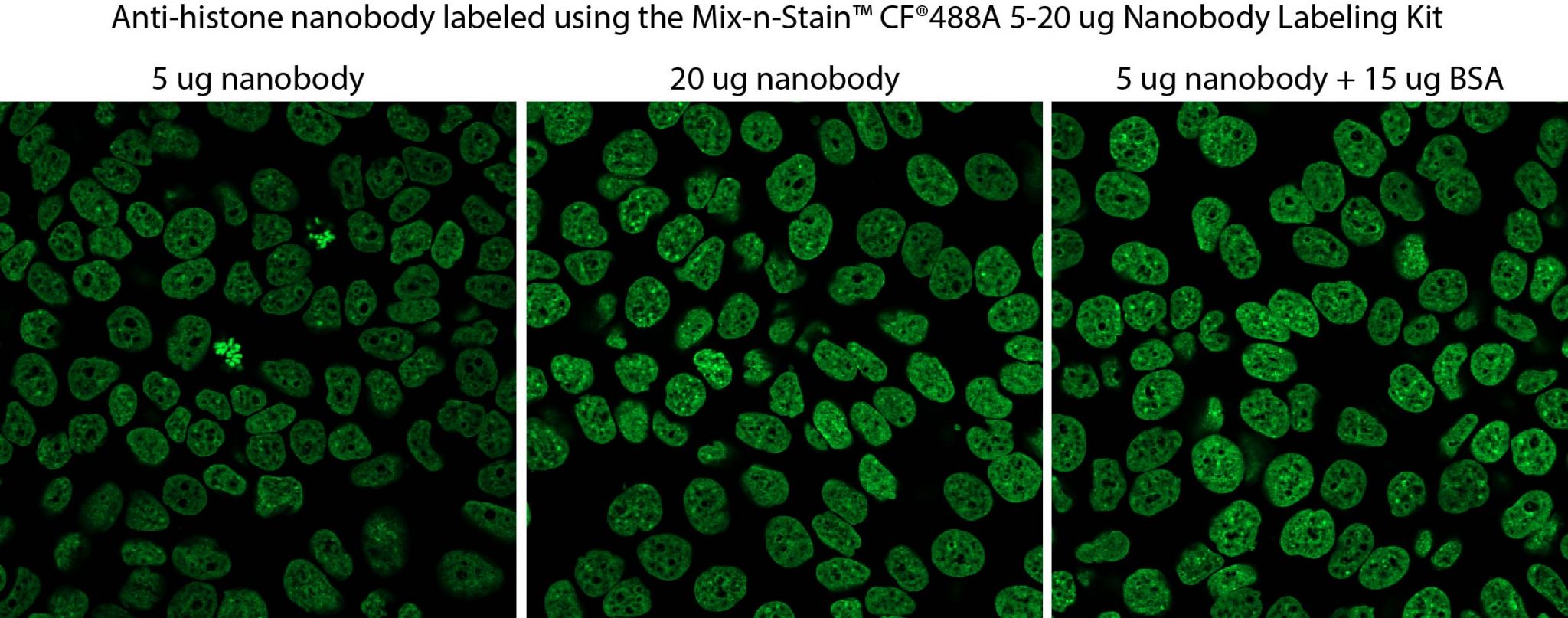
Mix-n-Stain™ Nanobody Labeling Kits
Nanobodies® (also called camelid single variable or VHH domains), are smaller ~15 kDa single-domain antibodies that enable deeper tissue penetration, faster staining times, and improved access to tightly packed or masked epitopes—making them ideal for high-resolution and super-resolution imaging.
Biotium offers Nanobody® labeling kits designed specifically for labeling single-domain Nanobodies® with one of Biotium’s bright and photostable CF® Dyes.
Mix-n-Stain™ Nanobody Labeling Kits are for standard labeling of VHH, while the Mix-n-Stain™ Nanobody Thiol Labeling Kits were designed for labeling VHH containing a single cysteine (1x Cys) residue. The kits allow optimal labeling of between 5-50 ug of Nanobody®, with minimal hands-on time and no purification.
Biotium also offers MiniMab™ single-domain antibodies (SdAbs) labeled with best-in-class CF® Dyes for STORM. MiniMab™ SdAbs are available for popular neuroscience targets or as secondary antibodies.
Mix-n-Stain™ Nanobody Labeling Kit Features
- Ideal for standard labeling of VHH
- Label 5-20 ug or 20-50 ug Nanobody®
- Choice of 7 CF® Dye colors or biotin
- Simple 30 minute labeling, no purification
- Compatible with common stabilizers
- Prevents over-labeling
Mix-n-Stain™ Nanobody Thiol Labeling Kits Features
- Designed for labeling VHH with a single cysteine (1x Cys) residue
- Label 5-20 ug or 20-50 ug Nanobody®
- Choice of 7 CF® Dye colors
- Simple 2 hour labeling, no purification
- No purification, 100% recovery
Mix-n-Stain™ Nanobody Labeling Kits
| Label | Ex/Em | Labeling size (ug Nanobody®) | Catalog number |
|---|---|---|---|
| Biotin | N/A | 5-20 ug | 92500 |
| 20-50 ug | 92501 | ||
| CF®405S | 404/431 nm | 5-20 ug | 92502 |
| 20-50 ug | 92503 | ||
| CF®488A | 490/515 nm | 5-20 ug | 92504 |
| 20-50 ug | 92505 | ||
| CF®568 | 562/583 nm | 5-20 ug | 92506 |
| 20-50 ug | 92507 | ||
| CF®594 | 593/614 nm | 5-20 ug | 92508 |
| 20-50 ug | 92509 | ||
| CF®640R | 642/662 nm | 5-20 ug | 92510 |
| 20-50 ug | 92511 | ||
| CF®647 | 650/665 nm | 5-20 ug | 92512 |
| 20-50 ug | 92513 | ||
| CF®680R | 680/701 nm | 5-20 ug | 92514 |
| 20-50 ug | 92515 |
Mix-n-Stain™ Nanobody Thiol Labeling Kits
| Conjugation | Ex/Em | Labeling size (ug Nanobody®) | Catalog No. | Dye Features |
|---|---|---|---|---|
| CF®488A | 490/516 nm | 5-20 ug | 92585 | CF®488A Features |
| 20-50 ug | 92586 | |||
| CF®568 | 562/584 nm | 5-20 ug | 92587 | CF®568 Features |
| 20-50 ug | 92588 | |||
| CF®583R | 585/609 nm | 5-20 ug | 92589 | CF®583R Features |
| 20-50 ug | 92590 | |||
| CF®594 | 593/615 nm | 5-20 ug | 92591 | CF®594 Features |
| 20-50 ug | 92592 | |||
| CF®647 | 652/668 nm | 5-20 ug | 92593 | CF®647 Features |
| 20-50 ug | 92594 | |||
| CF®680 | 681/698 nm | 5-20 ug | 92595 | CF®680 Features |
| 20-50 ug | 92596 | |||
| CF®740 | 742/767 nm | 5-20 ug | 92597 | CF®740 Features |
| 20-50 ug | 92598 |
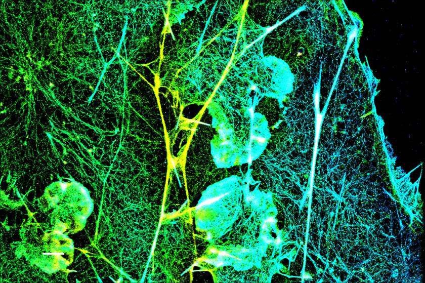
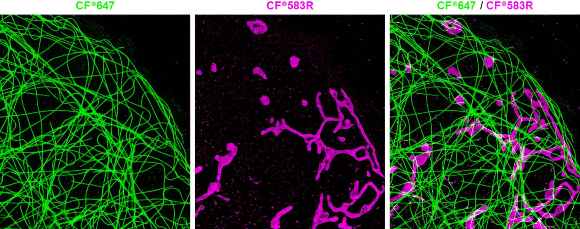
Mix-n-Stain™ STORM CF® Dye Antibody Labeling Kits
Mix-n-Stain™ STORM CF® Dye Antibody Labeling Kits offer a choice of 8 CF® Dyes developed with superior brightness and unique photoswitching properties that are ideal for super-resolution imaging by STORM. In addition, these specialized antibody labeling kits are designed to offer a low degree of labeling (DOL), with 1-2.5 dye molecules per antibody molecule, which is reported to be optimal for STORM (Bittel et al. 2015). Biotium has also developed novel green-excited STORM dyes CF®583R and CF®597R to expand the color palette for multi-color STORM. Learn more about the UC Berkeley and Biotium collaboration paper on CF®583R and CF®597R dyes for STORM, and other fluorescent reagents validated for super-resolution microscopy.
- Choice of 8 CF® Dye colors ideal for STORM
- CF®583R and CF®597R are novel green-excited STORM dyes
- Labeling optimized to provide low 1–2.5 DOL
- Less than 30 seconds of hands-on time
- 30 minutes total reaction time
- No purification, 100% recovery
Two Color (d)STORM Imaging of a Fixed COS-7 Cell
View Product Page
Mix-n-Stain™ STORM CF® Dye Antibody Labeling Kits
| Conjugation | Ex/Em | Size | Catalog No. | Dye Features |
|---|---|---|---|---|
| CF®505 | 505/519 nm | 50 ug | 92549 | |
| CF®568 | 562/584 nm | 50 ug | 92550 | CF®568 Features |
| CF®583R | 585/609 nm | 50 ug | 92551 | CF®583R Features |
| CF®597R | 597/619 nm | 50 ug | 92553 | CF®597R Features |
| CF®647 | 652/668 nm | 50 ug | 92554 | CF®647 Features |
| CF®660C | 667/685 nm | 50 ug | 92555 | CF®660C Features |
| CF®680 | 681/698 nm | 50 ug | 92556 | CF®680 Features |
| CF®750 | 755/779 nm | 50 ug | 92557 | CF®750 Features |
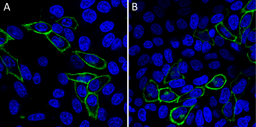
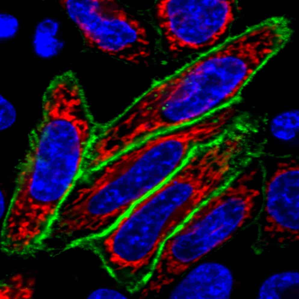
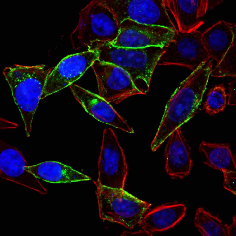
Mix-n-Stain™ CF® Dye Small Ligand Labeling Kits
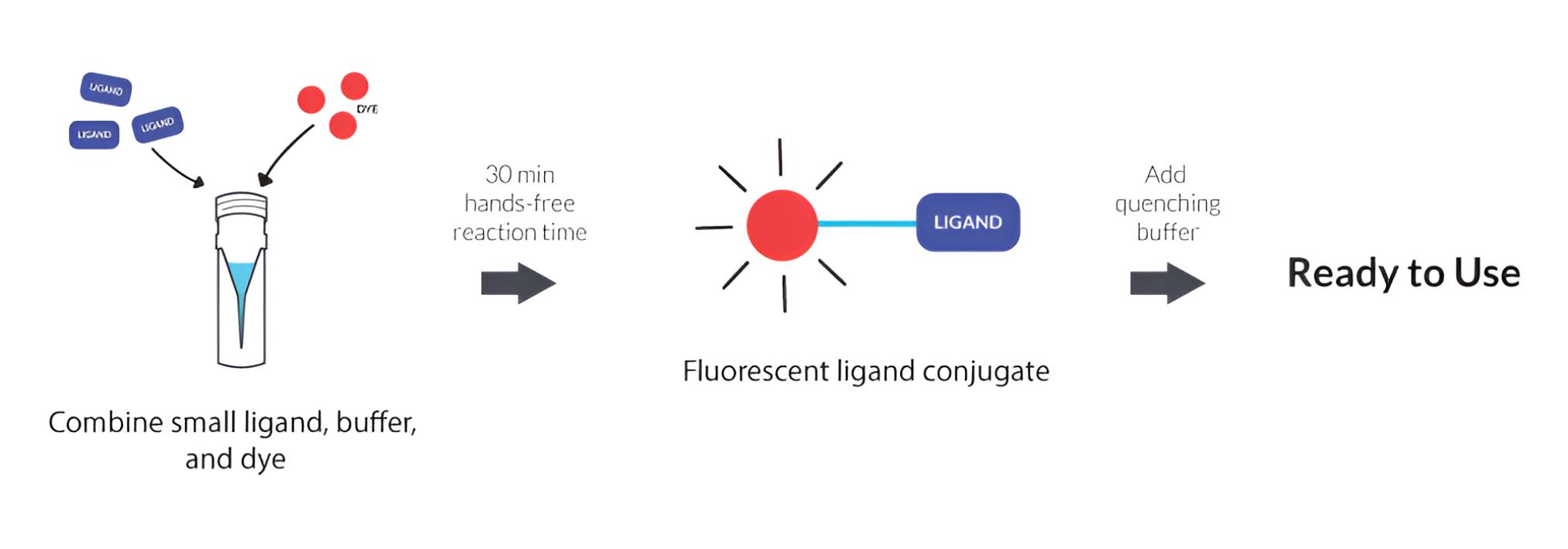
Mix-n-Stain™ CF® Dye small ligand labeling kits are designed for rapid labeling of small (MW ~ 150 – 5,000) and relatively high affinity biological ligands (or substrates). Labeling takes about 30 minutes, without a final purification step. The ligands to be labeled must contain an aliphatic amine group that is not required for biological activity of the ligand. The amine group will form a covalent linkage with the reactive CF® Dye provided in the kit. For example, suitable ligands or substrates include SNAP-tag®, CLIP-tag™ and HaloTag® ligands with an aliphatic amine. Many other small ligands are also possible candidates if they meet the criteria described above.
For live cell imaging, the Mix-n-Stain™ small ligand kits are ideal to generate fluorescent chemical tags that provide smaller, brighter, and more versatile alternatives to fluorescent proteins. Labeling various chemical tags with Mix-n-Stain™ kits yielded highly efficient and specific labeling of the target proteins both on the surface and inside of living cells. The Mix-n-Stain™ small ligand kits have also been proven compatible with fixed cell imaging and gel-based assays.
- Choice of 10 CF® Dye colors for extracellular staining
- Choice of 5 CF® Dye colors for intracellular staining
- For labeling molecules with a MW of 150–5000, containing an inactive aliphatic amine
- Suitable ligands or substrates include SNAP-tag®, CLIP-tag™ and HaloTag® ligands
- 30 minutes total reaction time, no purification necessary
Cell Surface and Intracellular Labeling Options
View Product Page
Mix-n-Stain™ Small Ligand Kits
| Product description | Label options | Labeling time |
|---|---|---|
| Mix-n-Stain™ CF® Dye Small Molecule Labeling Kits | Extracellular Staining: Choice of 10 bright and stable CF® Dyes | ~30 minutes (minimal hands-on time) Download the product protocol |
| Intracellular Staining: Choice of 5 bright and stable CF® Dyes |
CF® Dye SE Protein Labeling Kits
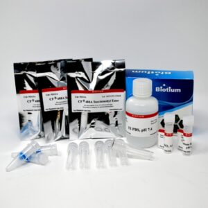
CF® Dye SE Protein Labeling Kits are designed for 3 labelings of up to 1 mg of protein or antibody with bright, photostable CF® Dyes. Unlike Mix-n-Stain™ kits, which are optimized for consistent labeling of small amounts of antibody, the SE Protein Labeling kits allow dye removal after labeling and optimization of dye-to-protein ratio. Ideal for users needing control over labeling conditions and antibody performance.
Features
- Choice of 19 CF® Dye colors or biotin
- Reagents for 3 labeling reactions with purification
- Each reaction can label up to 1 mg of antibody or protein
- Includes instructions for determining degree of labeling
View Product Page
VivoBrite™ Near-IR Antibody Labeling Kits
Label Antibodies for In Vivo Imaging
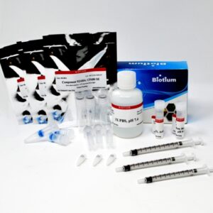
VivoBrite™ Antibody Labeling Kits use the same labeling technology as our SE Protein Labeling Kits. However, the kits include a syringe and syringe filter to sterile filter the labeled protein, allowing it to be used for in vivo small animal studies. Biotium’s near-IR CF® Dyes are best in class for brightness, photostability, and water solubility.
Features
- Choice of 4 near-IR CF® Dye colors optimal for small animal in vivo imaging
- Kit contains reagents for 3 labeling reactions
- Each reaction can label up to 1 mg of antibody
- Kit can also be used to label other proteins
- The resulting labeled protein is purified and sterile
Protein Labeling Kits
| Product description | Label options | Product protocol |
|---|---|---|
| CF® Dye SE Protein Labeling Kits | 19 CF® Dye colors across the spectrum, plus biotin | PI-CF® Dye SE Protein Labeling Kits |
| VivoBrite™ Antibody Labeling Kits for Small Animal In Vivo Imaging | 4 near-IR CF® Dyes | PI-VivoBrite™ Antibody Labeling Kits |
SNAP-tag is a registered trademark of New England Biolabs; CLIP-tag is a trademark of New England Biolabs; HaloTag is a registered trademark of Promega; Nanobody is a registered trademark of Ablynx.