New Products
 Earth-Friendly Products
 Biotium Choice Antibodies
 Special Offers

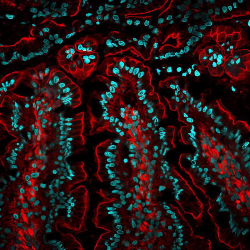
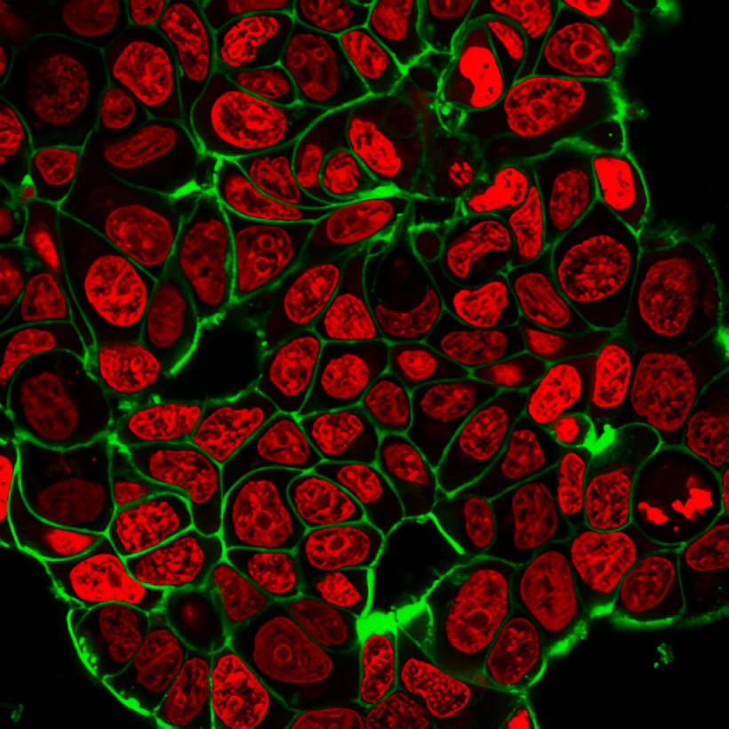
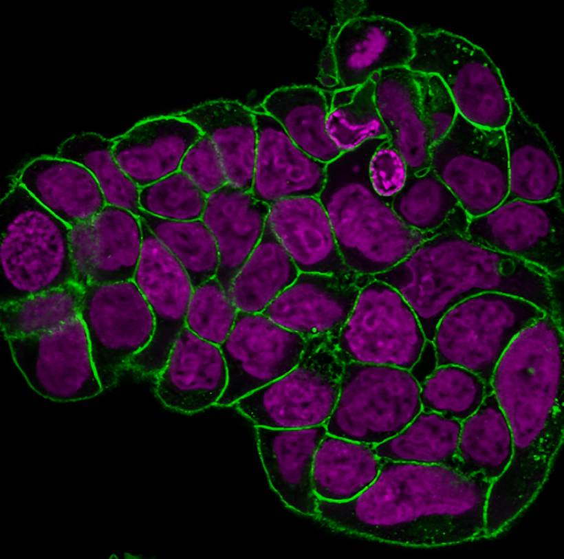
Free Up the DAPI Channel with NucSpot® Counterstains
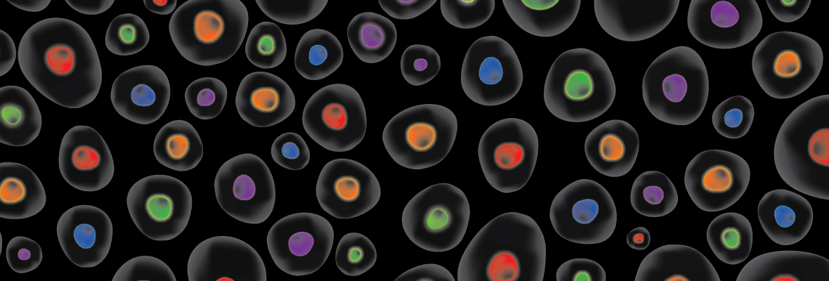
Bright & Specific Nuclear Staining from Green to Near-IR
NucSpot® Nuclear Stains are bright and specific counterstains for fixed cells in a variety of colors from green to near-infrared (near-IR). The stains were developed to address issues of photoconversion that other commonly used nuclear stains, like DAPI and Hoechst, undergo. NucSpot® Nuclear Stains have minimal fluorescence until they bind to DNA and can be used for no-wash nuclear staining. Unlike other nucleic acid dyes that stain both the nucleus and cytoplasm, NucSpot® Nuclear Stains selectively stain the nucleus in fixed and permeabilized cells without the need for RNase treatment.
NucSpot® Nuclear Stains can be used for selective staining of dead cells in unfixed cell cultures for analysis by flow cytometry or fluorescence imaging. Several of the stains can be continuously incubated with cells for multi-day imaging. NucSpot® 470 and NucSpot® Far-Red can be used for DNA content analysis of cell cycle by flow cytometry in fixed and permeabilized cells.
NucSpot® Features
- Bright, specific staining, no RNase treatment needed
- Available in 7 colors from green to near-IR
- Selectively stain dead cells in live culture
- Minimal fluorescence until they bind to DNA
- 10 minute incubation, no wash required
- For microscopy or flow cytometry, with dye options for cell cycle profiling
View Product Page
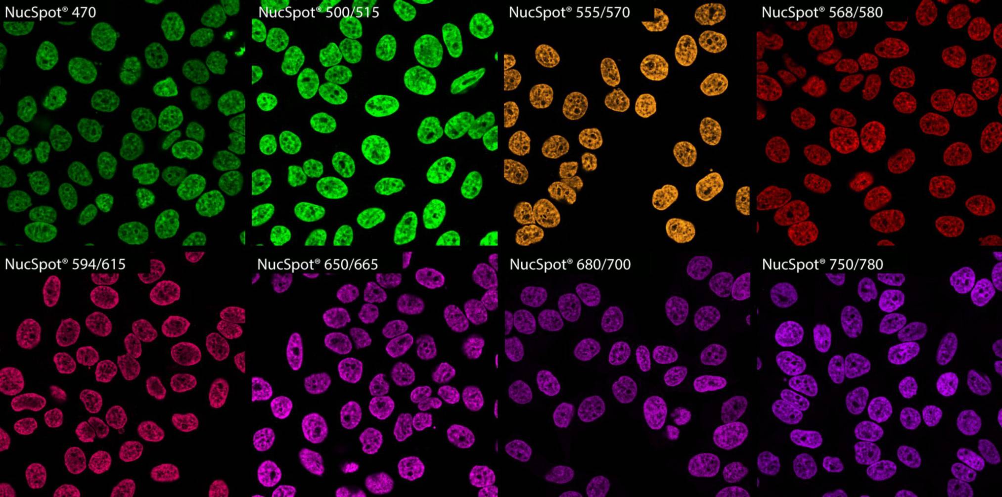
Wide Selection of Colors for Maximum Flexibility
Available in 7 colors from green to near-infrared, NucSpot® Nuclear Stains give you the flexibility to fit your nuclear staining into any channel. In high-plex panels where antibody and conjugate choices are limited, that spectral freedom lets you prioritize the targets that are more difficult to label.
Robust & Specific Nuclear Staining for Macroscopic Imaging
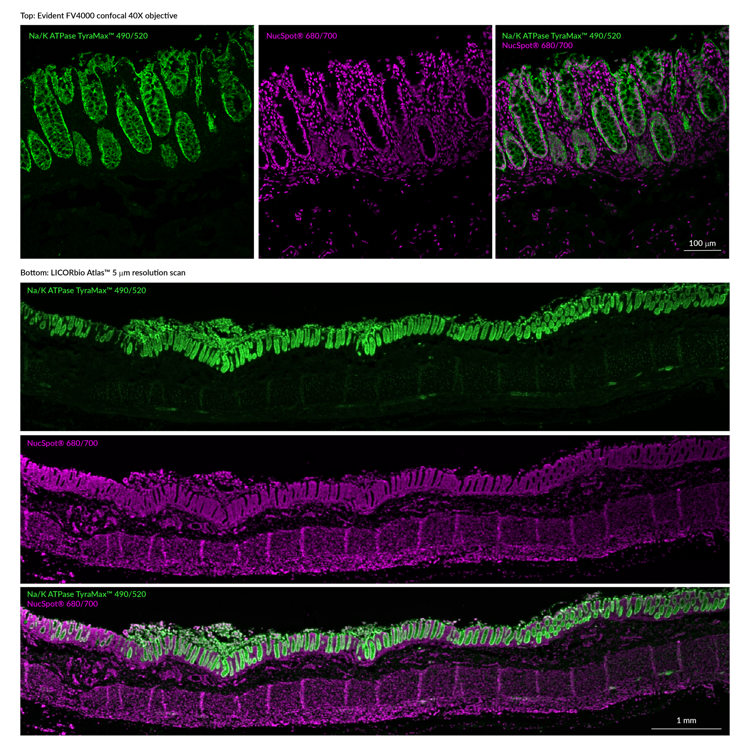
Beware of Artifacts from Photoconversion and Cross-Talk

Literature Digest
Cross-talk and Photoconversion: Assessing the Performance of DAPI and Hoechst Nuclear Stains
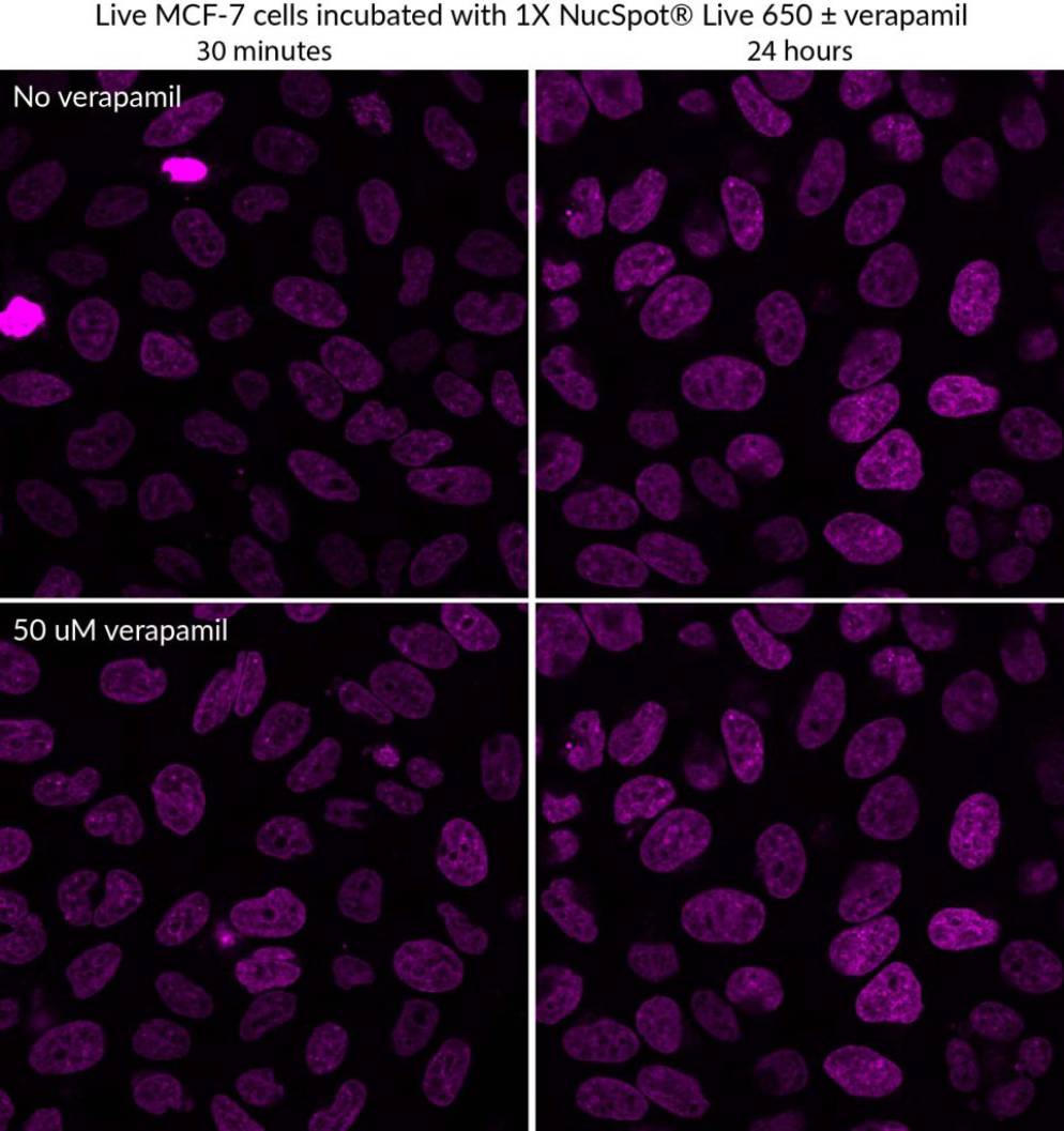
Nuclear Stains Designed for Live Cells
Low-Toxicity Stains for Multi-Day Imaging
NucSpot® Live Nuclear Stains are low-toxicity stains that offer nuclear staining for real-time imaging, available with green or far-red fluorescence. Unlike Draq5™, NucSpot® Live 650 has low cytotoxicity and can be used for longer term imaging.
The dyes are supplied with a vial of the efflux pump inhibitor verapamil for optional use, which may increase probe retention and live cell staining in some cell types.
NucSpot® Live Features
- No wash, nuclear stains
- Low-toxicity for real-time live cell imaging
- Staining is stable for up to 72 hours
- Fix before or after labeling
- NucSpot® Live 650 compatible with SIM, STED, or STORM
- Choose green or far-red fluorescence
Live Cell Staining for Up to 72 Hours
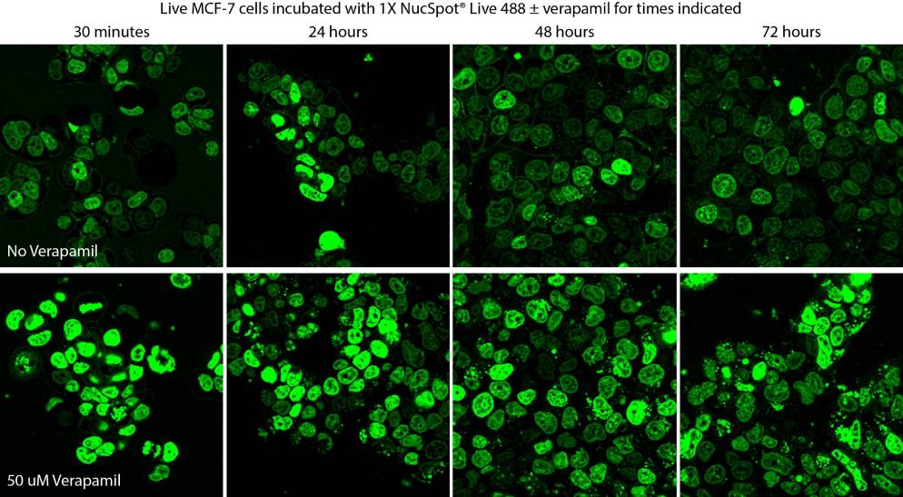
Efflux Pump Inhibitor for Brighter Staining
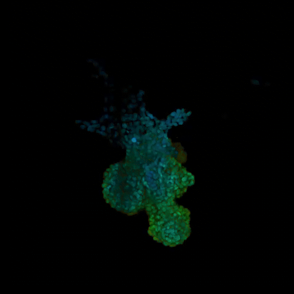
Validated for Organoids & Other 3D Cultures
View Product Page
Mounting Media with Nuclear Counterstains
High performance antifade mounting media with DAPI
Biotium’s EverBrite™ line of mounting media are optimally formulated for preserving fluorescence of our CF® Dyes and other fluorochromes. EverBrite™ Mounting Medium is available in wet-set and hardset formulations with and without DAPI. The EverBrite™ Hardset Mounting Medium hardens completely to form a permanent seal at room temperature in 24 hours and is also available with NucSpot® 640 far-red counterstain. Drop-n-Stain EverBrite™ Mounting Medium is also available with DAPI and provides the wet-set EverBrite™ formulation in a convenient dropper bottle for easy dispensing.
EverBrite™ Mounting Medium Features
- Antifade mounting medium optimally formulated to prevent photobleaching of fluorophores
- Available in wet-set or hardset formulations
- Available with DAPI, hardset formulation available with NucSpot® 640
- Refractive index is well-matched to that of coverslip glass and immersion oil
- Wet-set formulation available as convenient Drop-n-Stain EverBrite™
| Product | Nuclear Counterstain | Cat. No. | Features |
|---|---|---|---|
| EverBrite™ Mounting Medium | None | 23001 | • Wet-set mounting medium • Requires coverslip sealing • Refractive index 1.46 |
| EverBrite™ with DAPI | DAPI | 23002 | |
| Drop-n-Stain EverBrite™ Mounting Medium | None | 23008 | • Wet-set mounting medium • Convenient dropper bottle • Ideal for wells & chambers • Refractive index 1.42 |
| Drop-n-Stain EverBrite™ with DAPI | DAPI | 23009 | |
| EverBrite™ Hardset Mounting Medium | None | 23003 | • Hard-set mounting medium • Forms hard seal after 24 h • No coverslip sealing needed • Refractive index 1.42 after 24 h of curing, and 1.46 four days after curing |
| EverBrite™ Hardset with DAPI | DAPI | 23004 | |
| EverBrite™ Hardset with NucSpot® 640 | NucSpot® 640 | 23016 | |
| EverBrite TrueBlack® Hardset Mounting Medium | None | 23017 | • Unique antifade with lipofuscin quenching • Quenches as it hardens, with low background • Refractive index 1.42 after 24 h of curing, and 1.46 four days after curing |
| EverBrite TrueBlack® Hardset with DAPI | DAPI | 23018 | |
| EverBrite TrueBlack® Hardset with NucSpot® 640 | NucSpot® 640 | 23019 | |
| CoverGrip™ Coverslip Sealant | N/A | 23005 | • For sealing edges of wet-set coverslips |
Unique Alternatives to Draq5™ and Draq7™
RedDot™1
Cell membrane-permeant RedDot™1 stains the nuclei of live cells rapidly and specifically with a spectral profile similar to Draq5™. Validated applications for RedDot™1 include staining nuclei in live organisms and cell cycle analysis by flow cytometry.
Note: For long-term live cell imaging experiments, we recommend our NucSpot® Live Stains.
RedDot™2
Cell membrane-impermeant RedDot™2 is spectrally similar to Draq7™ with selectivity for dead cells. Our NucView® 488 and RedDot™ 2 Apoptosis & Necrosis Kit pairs RedDot™2 with NucView® 488 Caspase-3 Substrate for detection of apoptotic and necrotic cells. Unlike Draq7™, RedDot™2 staining is nuclear specific and provides counterstaining without special blocking steps. RedDot™2 has been validated for tissue clearing protocols such as CUBIC.
View Product Page
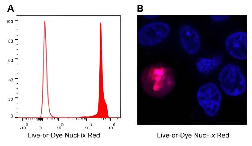
Fixable Dead Cell-Selective Nuclear Staining
Stable Nuclear Labeling of Dead Cells
Live-or-Dye NucFix™ Red is a unique, cell membrane-impermeant dye that stains the nuclei of dead cells. The dye is able to enter into dead cells that have compromised membrane integrity and covalently label the cell nucleus, allowing for clear differentiation of live and dead cells Unlike other commonly used nuclear stains, such as propidium iodide (PI) or DRAQ7™, Live-or-Dye NucFix™ labeling is extremely stable, allowing the cells to be fixed and permeabilized without loss of fluorescence or dye transfer between cells. Live-or-Dye NucFix™ Red can be used to detect dead cells with flow cytometry or fluorescence microscopy.
Live-or-Dye NucFix™ Red Features
- Stains the nucleus in dead cells
- Withstands fixation and permeabilization
- Labeling much more stable than PI and similar stains
- Dead-cell selective in mammalian cells, yeast, or gram-negative bacteria
- Suitable for microscopy or flow cytometry
View Product Page
Dead Cell-Selective Counterstains
Cyanine-based cell membrane-impermeant nucleic acid stains, such as Oxazole Blue (PO-PRO™-1), Oxazole Yellow (YO-PRO®-1), and Thiazole Red (TO-PRO®-3), are dead cell-selective stains suitable for flow cytometry. Using these dyes results in bright and sensitive staining that is localized to the nucleus in dead or dying cells. In addition, staining with these dyes can be done directly in cell culture medium without the need for washing. These dyes are generally known by their Thermo Fisher Scientific brand names and are available from blue to far-red. Biotium offers chemically equivalent forms of these dead cell selective stains at a high purity and lower cost.
Product | Equivalent to | Color (Ex/Em) | Catalog No. |
|---|---|---|---|
| Oxazole Blue, 1 mM in DMSO | PO-PRO™-1 | Blue (434/457 nm) | 40091 |
| Oxazole Blue Homodimer, 1 mM in DMSO | POPO™-1 | Blue (433/457 nm) | 40093 |
| Oxazole Yellow, 1 mM in DMSO | YO-PRO®-1 | Green (491/506 nm) | 40089 |
| Oxazole Yellow Homodimer, 1 mM in DMSO | YOYO®-1 | Green (491/508 nm) | 40090 |
| TO Iodide, 1 mM in DMSO | TO-PRO®-1 | Green (515/531 nm) | 40088 |
| Thiazole Orange Homodimer, 1 mM in DMSO | TOTO®-1 | Green (514/531 nm) | 40079 |
| Oxazole Red, 1 mM in DMSO | YO-PRO®-3 | Far-red (613/629 nm) | 40105 |
| Oxazole Red Homodimer, 1 mM in DMSO | YOYO®-3 | Far-red (612/631 nm) | 40106 |
| Thiazole Red, 1 mM in DMSO | TO-PRO®-3 | Far-red (642/657 nm) | 40087 |
| Thiazole Red Homodimer, 1 mM in DMSO | TOTO®-3 | Far-red (642/661 nm) | 40080 |
Nuclear Stains for Cell Cycle Analysis
Nuclear stains are commonly used for studying the distinct phases of the cell cycle by flow cytometry. The stains bind DNA stoichiometrically and will show fluorescence in proportion to the DNA content of cells in G1, S, or G2 phases of the cell cycle. The stains mentioned below are validated for cell cycle profiling. Biotium also offers classic nuclear dye options for cell cycle analysis, including Propidium Iodide (PI) and 7-AAD. Unlike PI, the dyes mentioned below do not require an RNase step.
RedDot™ 1 Far-Red Nuclear Stain
RedDot™ 1 is a far-red nuclear stain that can be used for cell cycle analysis in live cells and does not require an RNase step, unlike the classic stain Propidium Iodide (PI). RedDot™ 1 can also be used as a far-red nuclear counterstain for live cells in microscopy.
- Far-red cell membrane-permeant nuclear dye similar to Draq5™.
- Can be used for DNA content analysis by flow cytometry like Vybrant™ DyeCycle™ Ruby
- Highly thermostable and photostable
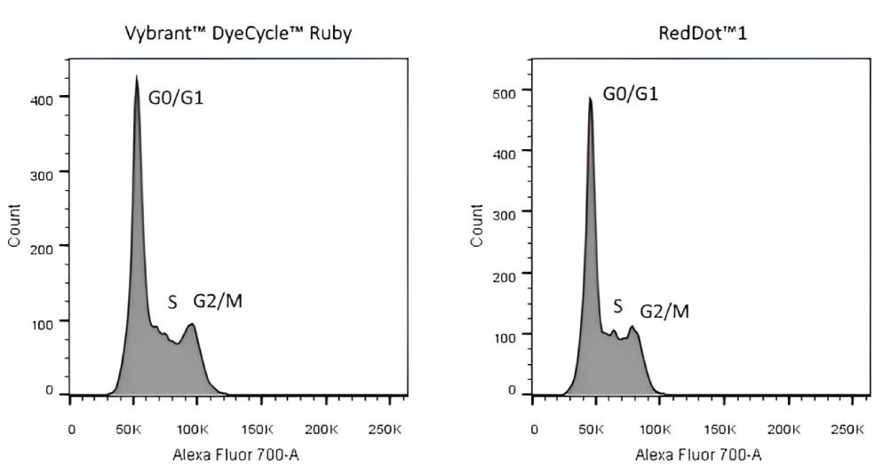
View Product Page
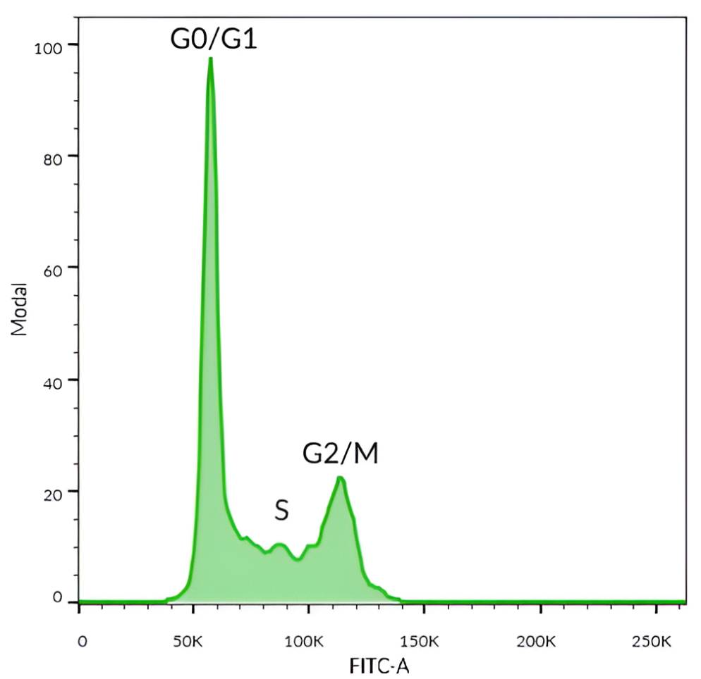
NucSpot® 470 Nuclear Stain
NucSpot® 470 is a cell membrane-impermeant green fluorescent DNA stain for nuclear counterstaining of fixed cells, or selective staining of dead cells.
- Nuclear-specific green counterstain for fixed cells
- Selective detection of dead cells by flow cytometry in the FITC channel
- Perform cell cycle profiling by flow cytometry in fixed/permeabilized cells
- No wash required
View Product Page
NucSpot® Far-Red Nuclear Stain
NucSpot® Far-Red is an improved alternative to the popular flow cytometry dead cell dye 7-AAD. It has red-shifted fluorescence emission compared to 7-AAD, for less bleed-through fluorescence in the PE-Texas Red® channel.
- Improved alternative to 7-AAD
- Selective detection of dead cells by flow cytometry in the PE-Cy®5 or APC channel
- Perform cell cycle profiling by flow cytometry in fixed/permeabilized cells
- Substitute for 7-AAD in any standard protocol
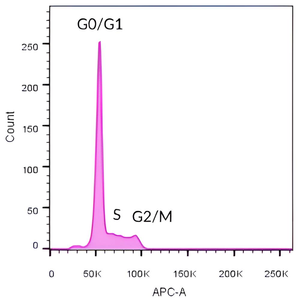
View Product Page
Dyes for Cell Cycle Analysis | Catalog No. | Live or Fixed Cells? | RNase treatment required? | Color (Ex/Em*) | Features |
|---|---|---|---|---|---|
| NucSpot® 470 Nuclear Stain, 1000X in DMSO | 40083 | Fixed | No | Green (460/546 nm) | • Nuclear-specific green counterstain for fixed cells • Selectively stains dead cells in live cultures • Excellent match for blue LED excitation sources |
| Propidium Iodide, 100 mg | 40016 | Fixed | Yes | Red (530/622 nm) | • Widely used dead cell stain • Can be excited by 488 nm laser line for detection in the PE channel |
| Propidium Iodide, 1 mg/mL in Water | 40017 | ||||
| Propidium Iodide Buffer, 50 ug/mL | 40048 | ||||
| 7-AAD, 1 mg | 40037 | Fixed | No | Far-red (546/647 nm) | • Far-red dye for detection in the PE-Cy®5 channel • Can be excited by the 488 nm or 532 nm laser line |
| 7-AAD, 1 mg/mL solution | 40084 | ||||
| NucSpot® Far-Red, 1000X in DMSO | 40085 | Fixed | No | Far-red (597/667 nm) | • Designed as improved replacement for 7-AAD • For the PE-Cy®5 or APC channel • Less bleed into the PE-Texas Red® channel |
| RedDot™1 Far-Red Nuclear Stain, 200X in Water | 40060 | Live | No | Far-red (662/694 nm) | • For short-term live cell staining (≤4 hours) • Useful for cell number normalization for In Cell Western® • Can be excited at wavelengths between 488 and 647 nm • Detect in Cy®5 or APC channel |
Cy Dye is a registered trademark of Cytiva. In Cell Western is a registered trademarks of LI-COR Inc.
Full List of Nuclear Stains
Our complete selection of nuclear stains are listed in the table below and are organized by emission wavelength to help you select the most suitable dye for your specific application. Biotium also carries high-purity and cost-effective versions of classic nuclear stains, including DAPI, Hoechst, Propidium Iodide (PI), and 7-AAD, which are widely used in flow cytometry and fluorescence microscopy.
For detailed guidance on choosing the best stain for your experiment, please see our nuclear stains comparison table.
Nuclear Stains | Catalog | Color (Ex/Em*) | Cell | For live / | Fix after | Features |
|---|---|---|---|---|---|---|
| EverBrite™ Mounting Medium w/DAPI EverBrite™ Hardset w/DAPI Drop-n-Stain EverBrite™ w/DAPI | 23002 23004 23009 | Blue (358/461 nm) | N/A | Mounting fixed samples | N/A | • Hardset and wet-set mounting media • Broad dye compatibility • Available with DAPI for one-step mounting and staining • Drop-n-Stain EverBrite™ comes in convenient dropper bottle |
| DAPI DAPI, 10 mg/mL in H2O DAPI, dilactate 10 mg in H2O | 40011 40043 40009 | Blue (358/461 nm) | Membrane permeant | Live cells Fixed cells | Yes | • Classic nuclear counterstain for fixed cells • Can be used at higher concentrations to stain live cells • Dilactate salt has improved water solubility |
| Hoechst 33258, 10 mg/mL in H2O Hoechst 33258, pentahydrate Hoechst 33342, 10 mg/mL in H2O Hoechst 33342, trihydrochloride trihydrate | 40044 40045 40046 40047 | Blue (358/461 nm) | Membrane permeant | Live cells Fixed cells | Yes | • Classic nuclear counterstain for live cells • Can also be used on fixed cells |
| Oxazole Gold (SYBR® Gold) | 40094 | Green (496/539) | Membrane permeant | Live cells | No 2 | • Aka SYBR® Gold, an ultrasensitive DNA and RNA gel stain • Can be used for live cell staining of nuclei and mitochondrial DNA |
| NucSpot® 470 Nuclear Stain | 40083 | Green (460/546 nm) | Membrane impermeant | Dead cells Fixed cells | No 1 | • Nuclear-specific green counterstain for fixed cells • Selectively stains dead cells in live cultures • Excellent match for blue LED excitation sources |
| Thiazole Green (SYBR® Green I) | 40086 | Green (498/522 nm) | Membrane permeant | Live cells Fixed cells | No 2 | • Aka SYBR® Green I, a well known DNA gel stain and qPCR dye • Can be used as a green nuclear stain for all cells in live cultures • Loses nuclear specificity after fixation • Can be excited by 488 nm laser line |
| NucSpot® Live 488 Nuclear Stain | 40081 | Green (500/515 nm) | Membrane permeant | Live cells Fixed cells | Yes | • Low-toxicity nuclear stain • Fix before or after labeling • Live cell staining may require VRP (included) |
| Live-or-Dye NucFix™ Red | 32010 | Red (520/610 nm) | Membrane impermeant | Dead cells | Yes | • Reactive nuclear stain for dead cells • Specifically stains dead cell nuclei • Fix/permeabilize without dye transfer between cells |
| NucSpot® 555/570 Nuclear Stain | 41033 | Red (559/566 nm) | Membrane impermeant | Dead cells Fixed cells | No 1 | • Nuclear-specific red counterstain for fixed cells • Specifically stains dead cells in live cultures |
| NucSpot® 568/580 Nuclear Stain | 41036 | Red (572/583 nm) | Membrane impermeant | Dead cells Fixed cells | No 1 | • Nuclear-specific red counterstain for fixed cells • Specifically stains dead cells in live cultures • Suitable for multi-day live cell imaging |
| NucSpot® 594/615 Nuclear Stain | 41037 | Red (603/613 nm) | Membrane impermeant | Dead cells Fixed cells | No 1 | • Nuclear-specific red counterstain for fixed cells • Specifically stains dead cells in live cultures • Suitable for multi-day live cell imaging |
| NucSpot® 650/665 Nuclear Stain | 41034 | Far-red (653/671 nm) | Membrane impermeant | Dead cells Fixed cells | No 1 | • Nuclear-specific far-red counterstain for fixed cells • Specifically stains dead cells in live cultures |
| NucSpot® Live 650 Nuclear Stain | 40082 | Far-red (650/675 nm) | Membrane permeant | Live cells Fixed cells | Yes | • Low-toxicity nuclear stain for the Cy®5 channel • Fix before or after labeling • Live cell staining may require VRP (included) • Compatible with SIM, STED, or STORM |
| RedDot™1 Far-Red Nuclear Stain | 40060 | Far-red (662/694 nm) | Membrane permeant | Live cells | No 2 | • For short-term live cell staining (≤4 hours) • Analyze DNA content/cell cycle by flow cytometry • Useful for cell number normalization for In Cell Western® • Can be excited at wavelengths between 488 and 647 nm • Detect in Cy®5 or APC channel |
| RedDot™2 Far-Red Nuclear Stain | 40061 | Far-red (665/695 nm) | Membrane impermeant | Dead cells Fixed cells | No 1 | • Far-red nuclear stain for dead or fixed cells • Selectively stains dead cells • Specific nuclear counterstain for fixed cells • Can be excited at wavelengths between 488 and 647 nm • Detect in Cy®5 or APC channel |
| NucSpot® 680/700 Nuclear Stain | 41035 | Near-IR (683/707 nm) | Membrane impermeant | Dead cells Fixed cells | No 1 | • Nuclear-specific near-IR counterstain for fixed cells • Specifically stains dead cells in live cultures |
| NucSpot® 750/780 Nuclear Stain | 41038 | Near-IR (757/780 nm) | Membrane impermeant | Dead cells Fixed cells | No 1 | • Nuclear-specific near-IR counterstain for fixed cells • Specifically stains dead cells in live cultures • Suitable for multi-day live cell imaging |
1 Dye can transfer from dead to live cells after fixation.
2 Loses nuclear specificity after fixation.
In Cell Western is a registered trademark of LI-COR® Bioscience. Cy Dye is a registered trademark of Cytiva.
Dead Cell Nucleic Acid Stains | Catalog No. | Color (Ex/Em*) | Features |
|---|---|---|---|
| Oxazole Blue, 1 mM in DMSO | 40091 | Blue (434/457 nm) | • Blue cell-impermeant dye • Selectively stains early apoptotic cells • Equivalent to PO-PRO™-1 Iodide |
| Oxazole Blue Homodimer, 1 mM in DMSO | 40093 | Blue (433/457 nm) | • Blue cell-impermeant dye • Equivalent to POPO™-1 Iodide |
| NucSpot® 470, 1000X in DMSO | 40083 | Green (460/546 nm) | • Green cell-impermeant dye • Nuclear-specific counterstain in fixed cells • Useful for cell cycle analysis in fixed cells • Excellent match for blue LED excitation sources |
| Oxazole Yellow, 1 mM in DMSO | 40089 | Green (491/506 nm) | • Green cell-impermeant dye • Selectively stains early apoptotic cells • Equivalent to YO-PRO®-1 Iodide |
| Oxazole Yellow Homodimer, 1 mM in DMSO | 40090 | Green (491/508 nm) | • Green cell-impermeant dye • Equivalent to YOYO®-1 Iodide |
| TO Iodide, 1 mM in DMSO | 40088 | Green (515/531 nm) | • Green cell-impermeant dye • Equivalent to TO-PRO®-1 Iodide |
| Thiazole Orange Homodimer, 1 mM in DMSO | 40079 | Green (514/531 nm) | • High affinity dimeric cyanine dye • Dead cell stain and electrophoresis dye • Equivalent to TOTO®-1 Iodide |
| NucSpot® 555/570 Nuclear Stain | 41033 | Red (559/566 nm) | • Red cell-impermeant dye for the Cy®3 or PE channels • Nuclear-specific counterstain in fixed cells |
| NucSpot® 568/580 Nuclear Stain | 41036 | Red (572/583 nm) | • Red cell-impermeant dye for the Cy®3 or PE channels • Nuclear-specific counterstain in fixed cells • Suitable for multi-day live cell imaging |
| NucSpot® 594/615 Nuclear Stain | 41037 | Red (603/613 nm) | • Red cell-impermeant dye for the Texas Red® or PE-Texas Red® channels • Nuclear-specific counterstain in fixed cells • Suitable for multi-day live cell imaging |
| Propidium Iodide | 40016, 40017, 40048 | Red (530/622 nm) | • Widely used dead cell stain • Can be excited by 488 nm laser line for detection in the PE channel by flow cytometry • Useful for cell cycle analysis in fixed cells (with RNase treatment) |
| Ethidium Homodimer I | 40010, 40014 | Red (527/624 nm) | • High-affinity membrane-impermeant nucleic acid stain • >30-fold fluorescence enhancement upon binding to DNA/RNA • High-purity grade not available from other manufacturers |
| Ethidium Homodimer III | 40050, 40051 | Red (532/625 nm) | • Developed at Biotium as an alternative to Ethidium Homodimer I • 45% brighter than EthDI when bound to DNA |
| Oxazole Red, 1 mM in DMSO | 40105 | Far-red (613/629 nm) | • Far-red cell-impermeant dye for the PE-Cy®5, or APC channel • Useful dead cell stain • Equivalent to YO-PRO®-3 |
| Oxazole Red Homodimer, 1 mM in DMSO | 40106 | Far-red (612/631 nm) | • Far-red cell-impermeant dye for the PE-Cy®5, or APC channel • Useful dead cell stain • Equivalent to YOYO®-3 |
| 7-AAD | 40037, 40084 | Far-red (546/647 nm) | • Far-red dye for flow cytometry detection in the PE-Cy®5 channel • Can be excited by the 488 nm or 532 nm laser line • Useful for cell cycle analysis in fixed cells |
| NucSpot® Far-Red, 1000X in DMSO | 40085 | Far-red (597/667 nm) | • Designed as improved replacement for 7-AAD • For flow cytometry in the PE-Cy®5 or APC channel • Useful for cell cycle analysis in fixed cells • Less bleed into the PE-Texas Red® channel |
| RedDot™2 Far-Red Nuclear Stain | 40061 | Far-red (665/695 nm) | • Far-red cell-impermeant dye for the Cy®5 channel • Nuclear-specific counterstain in fixed cells • Replaces Draq7™ |
| Thiazole Red, 1 mM in DMSO | 40087 | Far-red (642/657 nm) | • Far-red cell-impermeant dye for the Cy®5 channel • Dead cell stain and electrophoresis dye • Equivalent to TO-PRO®-3 Iodide |
| Thiazole Red Homodimer, 1 mM in DMSO | 40080 | Far-red (642/661 nm) | • High affinity dimeric cyanine dye for the Cy®5 channel • Useful dead cell stain • Equivalent to TOTO®-3 Iodide |
| NucSpot® 650/665 Nuclear Stain | 41034 | Far-red (653/671 nm) | • Far-red cell-impermeant dye for the Cy®5 or APC channels • Nuclear-specific counterstain in fixed cells |
| NucSpot® 680/700 Nuclear Stain | 41035 | Near-IR (683/707 nm) | • Near-IR cell-impermeant dye for the Cy®5.5 channel • Nuclear-specific counterstain in fixed cells |
| NucSpot® 750/780 Nuclear Stain | 41038 | Near-IR (757/780 nm) | • Near-IR cell-impermeant dye for the Cy®7 or APC-Cy®7 channels • Nuclear-specific counterstain in fixed cells • Suitable for multi-day live cell imaging |
SYBR, PO-PRO, POPO, Texas Red, TOTO, TO-PRO, YO-PRO, and YOYO are trademarks or registered trademarks of Thermo Fisher Scientific. Cy Dye is a registered trademark of Cytiva. Draq7 is a trademark of Biostatus Ltd.