 New Products
 Earth-Friendly Products
 Biotium Choice Antibodies
 Special Offers


 Powered by Bioz


Content #1
Content #1
Content #1
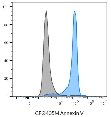
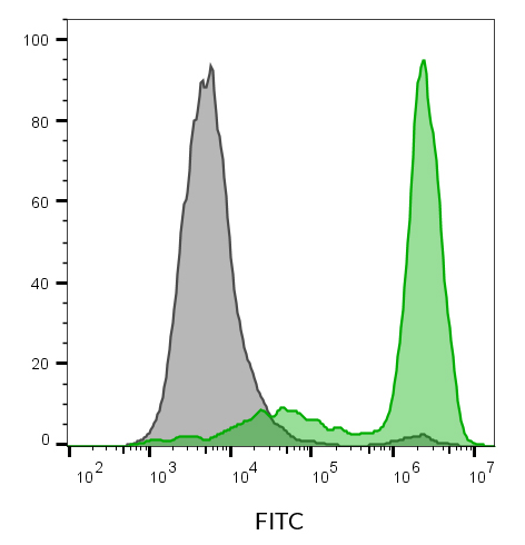
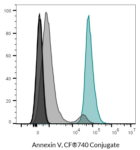
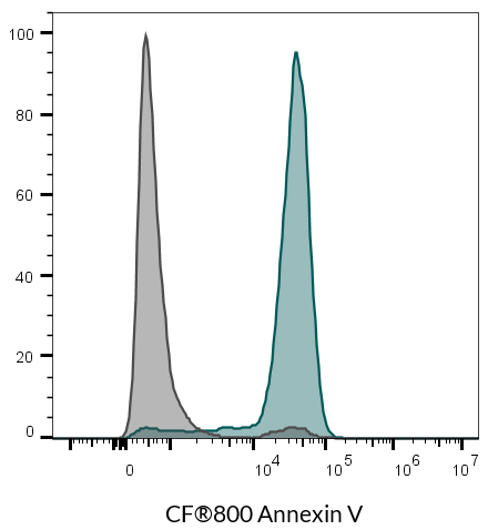
Preservative-free Annexin V conjugates are compatible with real-time staining of apoptotic cells for live cell imaging, fluorescence microscopy, or flow cytometry. Near-IR Annexin V fluorescent conjugates are suitable for in vivo imaging.

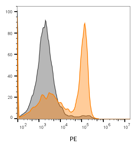
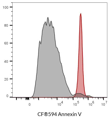
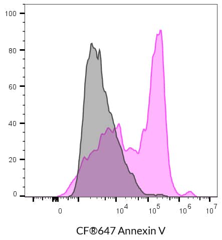
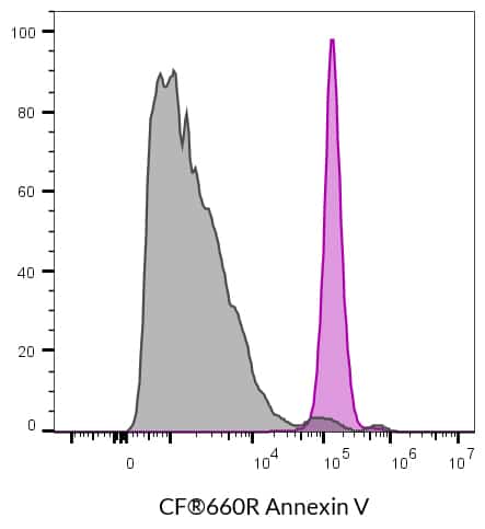
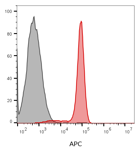
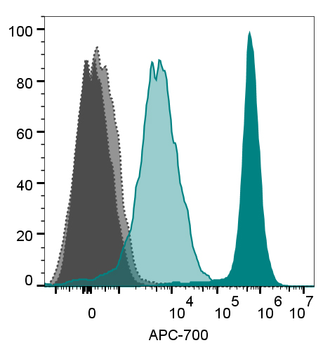
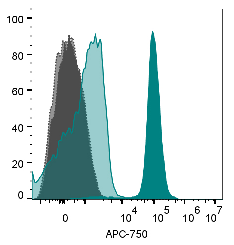
CF® Dye Annexin V Conjugates can be used to stain the surface of apoptotic cells. The human anticoagulant Annexin V is a 35-36 kDa calcium-dependent phospholipid-binding protein with high affinity for phosphatidylserine (PS). In normal viable cells, PS is located on the inner leaflet of the cytoplasmic membrane. In apoptotic cells, however, PS is translocated from the inner to the outer leaflet of the plasma membrane, where it can be detected by fluorescently labeled Annexin V.
Annexin V conjugates typically are supplied as stock solutions with azide as a preservative for end-point staining assays in Annexin V binding buffer. Our azide-free CF® Dye Annexin V Conjugates are supplied as lyophilized solids with no azide or other preservatives that might be incompatible with live cell or in vivo imaging. After reconstitution in buffer, the conjugates can be added to cell culture medium for no-wash, real-time live cell imaging. Our Mini Syringe Filters are convenient for small volume sterile filtration of azide-free Annexin V stock solutions or other aqueous solutions for use in cell culture.
See Annexin V staining in real time:
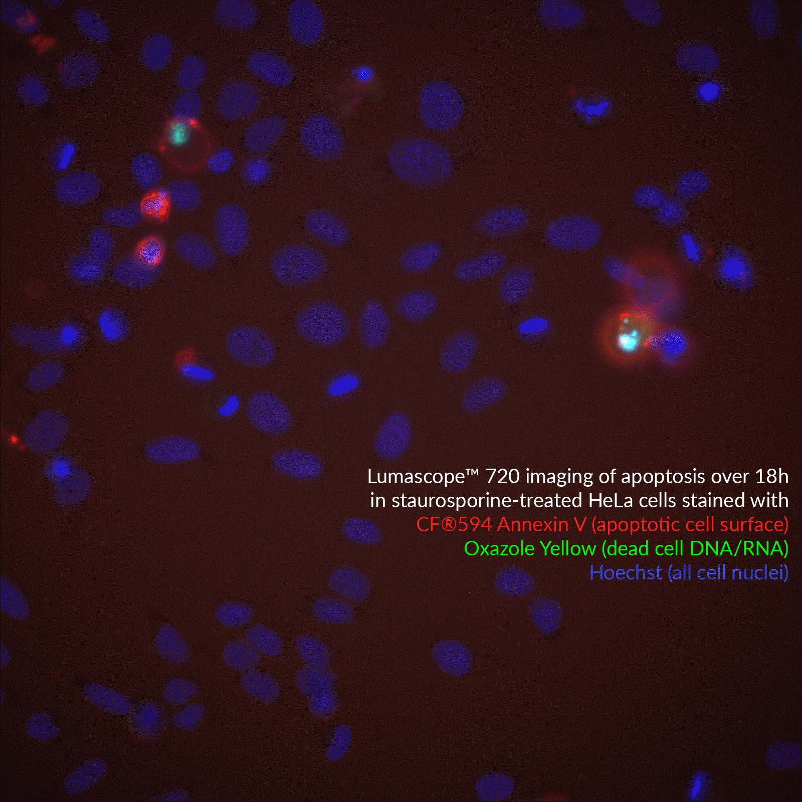
Click to view timelapse.
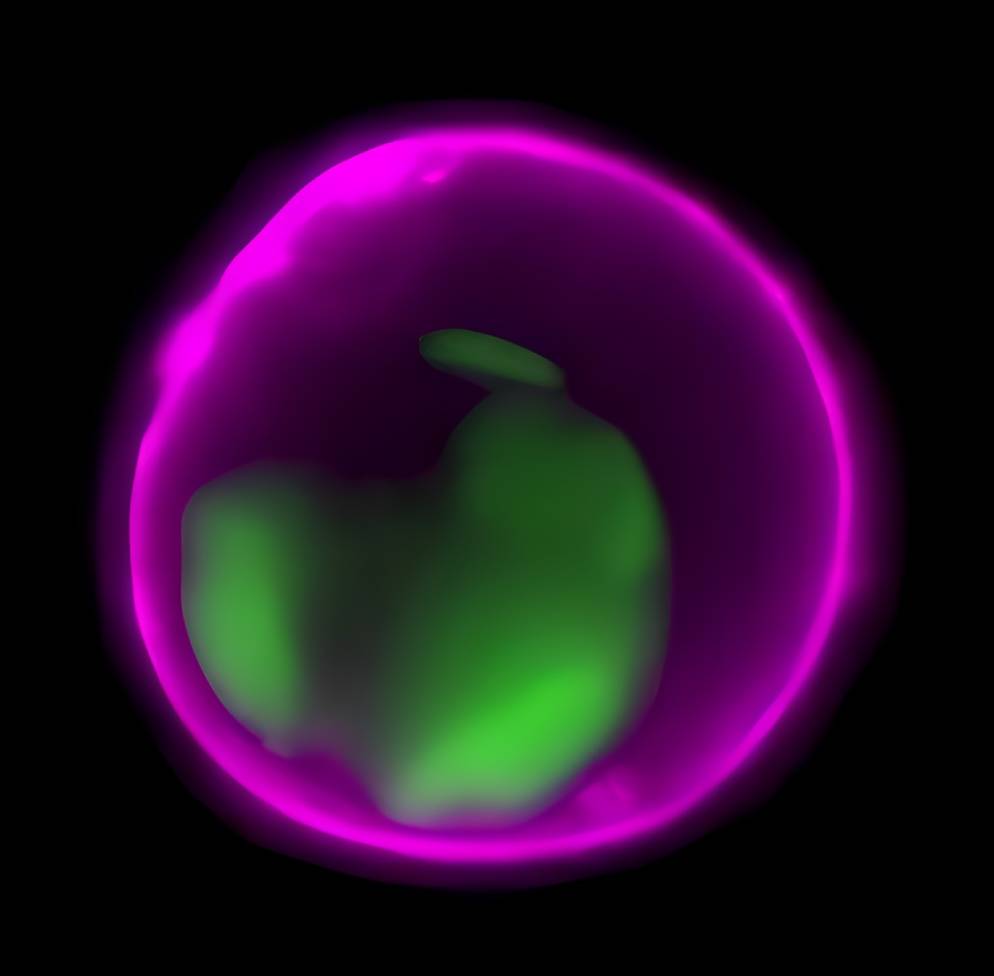
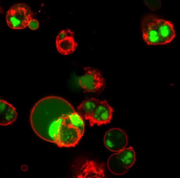
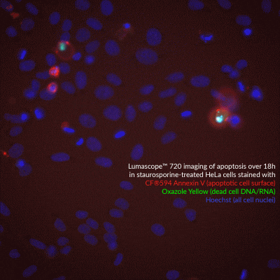
Imaging of apoptosis in staurosporine-treated HeLa cells stained with CF®594 Annexin V (red, apoptotic cell surface probe) and Oxazole Yellow (green, dead cell nuclear stain). All nuclei are stained blue with Hoechst.
Biotium’s next-generation CF® Dyes were designed to be highly water-soluble with advantages in brightness and photostability compared to Alexa Fluor®, DyLight®, and other fluorescent dyes. Learn more about CF® Dyes.
Note: Conjugates of blue-fluorescent dyes like CF®350 and CF®405M are not recommended for detecting low abundance targets and may be challenging to use in tissue specimens. Blue dyes have lower fluorescence and photostability, and cells and tissue have high autofluorescence in blue wavelengths, resulting in lower signal to noise compared to other colors.
We also offer Annexin V Conjugate solutions (with azide) with a large selection of CF® Dyes, biotin, R-PE, APC, and other labels. In addition, we offer Apoptotic and Necrotic Staining Kits containing Annexin V and other probes. See our full selection of Cell Viability and Apoptosis Assays.
| Conjugation | Ex/Em | Size | Catalog No. | Dye Features |
|---|---|---|---|---|
| CF®350 | 347/448 nm | 5 ug | 29012R-5ug | CF®350 Features |
| CF®405M | 408/452 nm | 5 ug | 29009R-5ug | CF®405M Features |
| CF®450 | 450/538 nm | 5 ug | 29083R-5ug | CF®450 Features |
| CF®488A | 490/515 nm | 5 ug | 29005R-5ug | CF®488A Features |
| CF®555 | 555/565 nm | 5 ug | 29004R-5ug | CF®555 Features |
| CF®568 | 562/583 nm | 5 ug | 29010R-5ug | CF®568 Features |
| CF®583R | 586/609 nm | 5 ug | 29085R-5ug | CF®583R Features |
| CF®594 | 593/614 nm | 5 ug | 29011R-5ug | CF®594 Features |
| CF®633 | 630/650 nm | 5 ug | 29008R-5ug | CF®633 Features |
| CF®640R | 642/662 nm | 5 ug | 29014R-5ug | CF®640R Features |
| CF®647 | 650/665 nm | 5 ug | 29003R-5ug | CF®647 Features |
| CF®660R | 663/682 nm | 5 ug | 29069R-5ug | CF®660R Features |
| CF®680 | 681/698 nm | 25 ug | 29007 | CF®680 Features |
| CF®680R | 680/701 nm | 25 ug | 29070 | CF®680R Features |
| CF®700 | 696/721 nm | 25 ug | 29082 | CF®700 Features |
| CF®750 | 755/777 nm | 25 ug | 29006 | CF®750 Features |
| CF®770 | 770/797 nm | 25 ug | 29046 | CF®770 Features |
| CF®790 | 784/806 nm | 25 ug | 29047 | CF®790 Features |
| CF®800 | 797/816 nm | 25 ug | 29078 | CF®800 Features |
Targeted activation of immune checkpoints is an emerging strategy for treating Type 1 Diabetes (T1D), where autoreactive T cells destroy insulin-producing beta cells. Autoimmune diabetes has been linked to disruption of the PD-1/PD-L1 pathway, which normally suppresses T cell activity to maintain immune tolerance. To address the limitations of systemic immunosuppression, an immune modulating monoclonal TCR against autoimmunity (ImmTAAI) molecule was created to selectively activate PD-1 signaling at the beta cell surface. This is a bispecific molecule that combines a T cell receptor targeting a preproinsulin peptide presented by HLA-A2 with a PD-1 agonist domain, enabling localized suppression of autoreactive T cells.
In a 2026 Science Advances publication, Becker et al. evaluated ImmTAAI function in live human pancreas tissue slices through confocal imaging, binding assays, and functional coculture systems using engineered T cell “avatars” designed to mimic the behavior of autoreactive T cells in T1D. Biotium’s CF®647 succinimidyl ester dye was used to label and visualize ImmTAAI molecules in live and fixed human pancreas slices. This labeling enabled precise tracking of ImmTAAI localization and quantification of its binding to beta cells via confocal microscopy without affecting binding affinity, specificity, or functional potency of ImmTAAI.
The researchers found that ImmTAAI binds specifically and dose-dependently to beta cells in a human leukocyte antigen (HLA)-dependent manner, with increased binding under inflammatory conditions. ImmTAAI treatment increased T cell movement, reduced T cell–beta cell interactions, and suppressed cytotoxic activity of target beta cells. Furthermore, ImmTAAI conferred protection of beta cells from immune attack in cell culture and in tissue slices, and helped preserve insulin secretion in live pancreas slices from a donor recently diagnosed with type 1 diabetes.
CF®647 enabled colocalization studies in a PD/PD-L1 therapeutic model and confirmed selective targeting within complex tissue environments. These findings highlight the value of bright, photostable far-red dyes like CF®647 for imaging-driven validation of targeted immunotherapies.
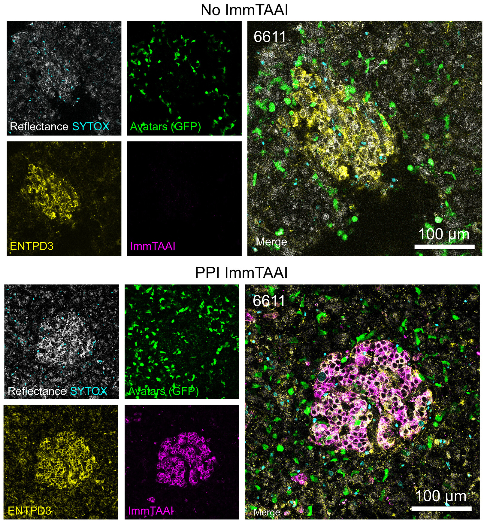
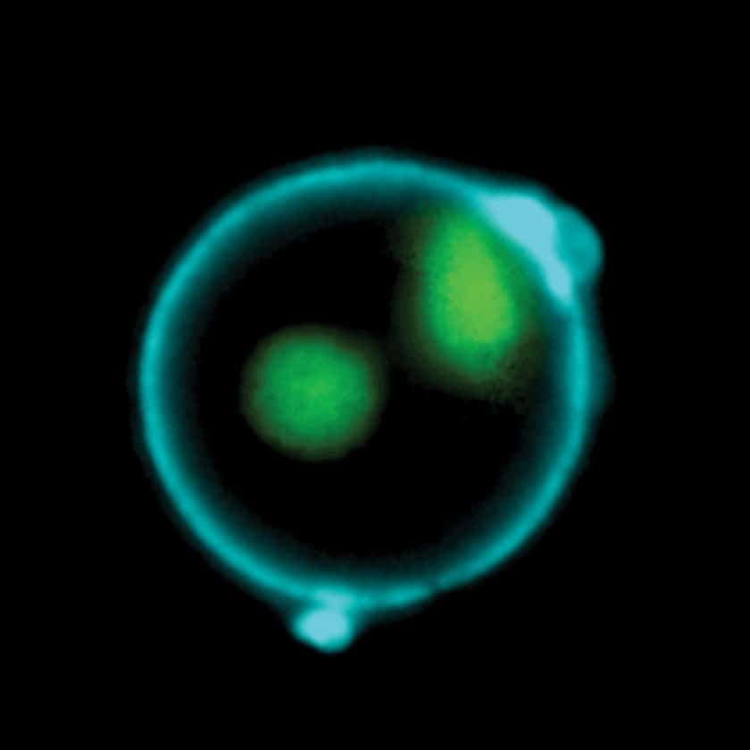
Live-cell confocal imaging (18 h) of pancreas slices treated with or without PPI ImmTAAI after addition of 200,000 IGRP-specific T cells per slice shows CF®647-labeled ImmTAAI colocalization with ENTPD3. Adapted from Becker et. al. Reproduced under CC BY 4.0.
Learn more about Biotium’s next-generation CF® Dye probes featuring exceptional brightness, photostability, and signal-to-noise, available in over 40 colors from blue to near-IR. CF® Dyes are also available in convenient Mix-n-Stain™ Labeling Kits for quick and efficient antibody labeling.
Full Citation
Matthew W. Becker et al. Beta cell–targeted PD-1 agonist inhibits cell-mediated autoimmunity in pancreas tissue slices. Sci. Adv. 12, eaec9029(2026). DOI:10.1126/sciadv.aec9029
Bioscience kits
The guaranteed shelf life from date of receipt for bioscience kits is listed on the product information sheet. Some kits have an expiration date printed on the kit box label, this is the guaranteed shelf life date calculated from the day that the product shipped from our facility. Kits often are functional for significantly longer than the guaranteed shelf life. If you have an older kit in storage that you wish to use, we recommend performing a small scale positive control experiment to confirm that the kit still works for your application before processing a large number of samples or precious samples.
Antibodies and other conjugates
The guaranteed shelf life from date of receipt for antibodies and conjugates is listed on the datasheet sheet which can be downloaded on the product page. Antibodies and other conjugates often are functional for significantly longer than the guaranteed shelf life. If you have an older conjugate in storage that you wish to use, we recommend performing a small scale positive control experiment to confirm that the product still works for your application before processing a large number of samples or precious samples.
For lyophilized antibodies, we recommend reconstituting the antibody with glycerol and antimicrobial preservative like sodium azide for the longest shelf life (note that sodium azide is not compatible with HRP-conjugates).
Chemicals, dyes, and gel stains
Biotium guarantees the stability of chemicals, dyes, and gel stains for at least a year from the date you receive the product. However, the majority of these products are highly stable for many years, as long as they are stored as recommended. Storage conditions can be found on the product information sheet or product safety and data sheet, material safety data sheet, and on the product label. Fluorescent compounds should be protected from light for long term storage.
If you have a Biotium compound that has been in storage for longer than one year that you wish to use, we recommend performing a small scale positive control experiment to confirm that the compound still works for your application before processing a large number of samples or precious samples.
Expiration date based on date of manufacture (DOM)
If your institution requires you to document expiration date based on date of manufacture for reagents, please contact [email protected] for assistance.
Chemical products with special stability considerations:
Esters
Ester compounds include the following:
Ester dyes are stable in solid form as long as they are protected from light and moisture. Esters are not stable in aqueous solution. Concentrated stock solutions should be prepared in anhydrous DMSO (see Biotium catalog no. 90082). Stock solutions in anhydrous DMSO can be stored desiccated at -20°C for one month or longer. Esters should be diluted in aqueous solution immediately before use. Succinimidyl esters (SE) should be dissolved in a solution that is free of amine-containing compounds like Tris, glycine, or protein, which will react with the SE functional group. AM esters and diacetate compounds should be dissolved in a solution that is free of serum, because serum could contain esterases that would hydrolyze the compound.
A note on CF® Dye succinimidyl ester stability
Succinimidyl esters (SE) are generally susceptible to hydrolysis, which can result in lower labeling efficiency. Many commercially available fluorescent dyes used for life science research are heavily sulfonated dyes which makes them particularly hygroscopic, worsening the hydrolysis problem. In addition, for several commercially available SE reactive dyes, the SE group is derived from an aromatic carboxylic acid, while the SE group in all of Biotium’s CF® Dyes is prepared from an aliphatic carboxylic acid. This structural difference reduces the susceptibility of CF® Dye SE reactive groups to hydrolysis, resulting in relatively stable reactive dyes with consistently higher labeling efficiency compared to other SE derivatives of other fluorescent dyes.
Maleimides, MTS and thiosulfate dyes
Like the succinimidyl ester dyes, these dyes are also susceptible to hydrolysis, although generally to a much lower degree. Thus, for long term storage, anhydrous DMSO is recommended for making stock solutions.
Other reactive dyes
Amines, aminooxy (also known as oxylamine), hydrazide, azide, alkyne, BCN, and tyramide reactive dyes, as well as dye free acids, are generally stable in aqueous solution when stored at -20°C for 6-12 months or longer, as long as no compounds are present that may react with the dye’s functional group. See the product information sheets for specific reactive dyes more information.
Coelenterazines and D-luciferin
Coelenterazines are stable in solid form when stored as recommended; they are not stable in aqueous solution. Concentrated coelenterazine stock solutions (typically 1-100 mg/mL) should be prepared in ethanol or methanol; do not use DMSO or DMF to dissolve coelenterazines, because these solvents will oxidize the compounds. Ethanol or methanol stocks of coelenterazine can be stored at -20°C or below for six months or longer; alcohol stocks may evaporate during storage, so use tightly sealing screw cap vials and wrap the vials with Parafilm for long term storage. Propylene glycol also can be used as a solvent to minimize evaporation. If the solvent evaporates, the coelenterazine will still be present in the vial, so note the volume in the vial prior to storage so that you can adjust the solvent volume to correct for evaporation if needed. Prepare working solutions in aqueous buffers immediately before use. Coelenterazines are stable for up to five hours in aqueous solution.
Aquaphile™ coelenterazines are water soluble formulations of coelenterazines. They are stable in solid form when stored as recommended. Aquaphile™ coelenterazines should be dissolved in aqueous solution immediately before use. They are stable for up to five hours in aqueous solution.
Note that coelenterazines are predominantly yellow solids, but may contain dark red or brown flecks. This does not affect product stability or performance. If your coelenterazine is uniformly brown, then it is oxidized and needs to be replaced.
D-luciferin is stable in solid form and as a concentrated stock solution when stored as recommended; it is not stable at dilute working concentrations in aqueous solution. Prepare concentrated D-luciferin stock solutions (typically 1-100 mg/mL) in water, and store in aliquots at -20°C or below for six months or longer. Prepare working solutions immediately before use.
Dyes that carry multiple negative charges can introduce background. Usually, this is more of a concern with labeled antibodies that carry many dyes, as opposed to a small toxin like bungarotoxin. When staining tissues, the endogenous autofluorescence of the tissue itself is often the most significant source of background. Endogenous fluorescence background in tissue is usually highest in the blue wavelengths (DAPI channel) and lowest in the far-red (Cy®5 channel). Our CF®633 bungarotoxin (catalog no. 00009) is a far-red conjugate for the Cy®5 channel with a low negative charge that should have low background from either the dye or autofluorescence.
We test fluorescent bungarotoxin on rat skeletal muscle sections. While the tissue shows autofluorescence, the bungarotoxin staining of motor endplates is usually much brighter than the background for all of the dye colors we've tested. However, if you are staining human tissue (especially brain), lipofuscin autofluorescence may be bright in all channels. This usually shows up as bright, punctate dots around cell nuclei. While we would usually recommend our TrueBlack® lipofuscin quenchers for human brain tissue, they are not compatible with bungarotoxin staining. We have, however, found that EverBrite TrueBlack® Mounting Medium (cat. no. 23017) can be used to mount skeletal muscle sections stained with bungarotoxin.
Cy Dye is a registered trademark of Cytiva.
Bioscience kits
The guaranteed shelf life from date of receipt for bioscience kits is listed on the product information sheet. Some kits have an expiration date printed on the kit box label, this is the guaranteed shelf life date calculated from the day that the product shipped from our facility. Kits often are functional for significantly longer than the guaranteed shelf life. If you have an older kit in storage that you wish to use, we recommend performing a small scale positive control experiment to confirm that the kit still works for your application before processing a large number of samples or precious samples.
Antibodies and other conjugates
The guaranteed shelf life from date of receipt for antibodies and conjugates is listed on the datasheet sheet which can be downloaded on the product page. Antibodies and other conjugates often are functional for significantly longer than the guaranteed shelf life. If you have an older conjugate in storage that you wish to use, we recommend performing a small scale positive control experiment to confirm that the product still works for your application before processing a large number of samples or precious samples.
For lyophilized antibodies, we recommend reconstituting the antibody with glycerol and antimicrobial preservative like sodium azide for the longest shelf life (note that sodium azide is not compatible with HRP-conjugates).
Chemicals, dyes, and gel stains
Biotium guarantees the stability of chemicals, dyes, and gel stains for at least a year from the date you receive the product. However, the majority of these products are highly stable for many years, as long as they are stored as recommended. Storage conditions can be found on the product information sheet or product safety and data sheet, material safety data sheet, and on the product label. Fluorescent compounds should be protected from light for long term storage.
If you have a Biotium compound that has been in storage for longer than one year that you wish to use, we recommend performing a small scale positive control experiment to confirm that the compound still works for your application before processing a large number of samples or precious samples.
Expiration date based on date of manufacture (DOM)
If your institution requires you to document expiration date based on date of manufacture for reagents, please contact [email protected] for assistance.
Chemical products with special stability considerations:
Esters
Ester compounds include the following:
Ester dyes are stable in solid form as long as they are protected from light and moisture. Esters are not stable in aqueous solution. Concentrated stock solutions should be prepared in anhydrous DMSO (see Biotium catalog no. 90082). Stock solutions in anhydrous DMSO can be stored desiccated at -20°C for one month or longer. Esters should be diluted in aqueous solution immediately before use. Succinimidyl esters (SE) should be dissolved in a solution that is free of amine-containing compounds like Tris, glycine, or protein, which will react with the SE functional group. AM esters and diacetate compounds should be dissolved in a solution that is free of serum, because serum could contain esterases that would hydrolyze the compound.
A note on CF® Dye succinimidyl ester stability
Succinimidyl esters (SE) are generally susceptible to hydrolysis, which can result in lower labeling efficiency. Many commercially available fluorescent dyes used for life science research are heavily sulfonated dyes which makes them particularly hygroscopic, worsening the hydrolysis problem. In addition, for several commercially available SE reactive dyes, the SE group is derived from an aromatic carboxylic acid, while the SE group in all of Biotium’s CF® Dyes is prepared from an aliphatic carboxylic acid. This structural difference reduces the susceptibility of CF® Dye SE reactive groups to hydrolysis, resulting in relatively stable reactive dyes with consistently higher labeling efficiency compared to other SE derivatives of other fluorescent dyes.
Maleimides, MTS and thiosulfate dyes
Like the succinimidyl ester dyes, these dyes are also susceptible to hydrolysis, although generally to a much lower degree. Thus, for long term storage, anhydrous DMSO is recommended for making stock solutions.
Other reactive dyes
Amines, aminooxy (also known as oxylamine), hydrazide, azide, alkyne, BCN, and tyramide reactive dyes, as well as dye free acids, are generally stable in aqueous solution when stored at -20°C for 6-12 months or longer, as long as no compounds are present that may react with the dye’s functional group. See the product information sheets for specific reactive dyes more information.
Coelenterazines and D-luciferin
Coelenterazines are stable in solid form when stored as recommended; they are not stable in aqueous solution. Concentrated coelenterazine stock solutions (typically 1-100 mg/mL) should be prepared in ethanol or methanol; do not use DMSO or DMF to dissolve coelenterazines, because these solvents will oxidize the compounds. Ethanol or methanol stocks of coelenterazine can be stored at -20°C or below for six months or longer; alcohol stocks may evaporate during storage, so use tightly sealing screw cap vials and wrap the vials with Parafilm for long term storage. Propylene glycol also can be used as a solvent to minimize evaporation. If the solvent evaporates, the coelenterazine will still be present in the vial, so note the volume in the vial prior to storage so that you can adjust the solvent volume to correct for evaporation if needed. Prepare working solutions in aqueous buffers immediately before use. Coelenterazines are stable for up to five hours in aqueous solution.
Aquaphile™ coelenterazines are water soluble formulations of coelenterazines. They are stable in solid form when stored as recommended. Aquaphile™ coelenterazines should be dissolved in aqueous solution immediately before use. They are stable for up to five hours in aqueous solution.
Note that coelenterazines are predominantly yellow solids, but may contain dark red or brown flecks. This does not affect product stability or performance. If your coelenterazine is uniformly brown, then it is oxidized and needs to be replaced.
D-luciferin is stable in solid form and as a concentrated stock solution when stored as recommended; it is not stable at dilute working concentrations in aqueous solution. Prepare concentrated D-luciferin stock solutions (typically 1-100 mg/mL) in water, and store in aliquots at -20°C or below for six months or longer. Prepare working solutions immediately before use.
For dyes or reagents that are supplied lyophilized (as solids), it is hard to compare quantities based on appearance of the dye in the tube, because during the lyophilization process the dye can dry down in different ways, either spread out all over the tube, clumped together, or coating the sides or bottom of the tube. Centrifugation of the tube may not help in collecting the dye solid to the bottom of the tube as this generally works for solutions. However, lyophilized solids are packaged based on highly accurate absorbance measurement of the reagent solution prior to drying, so the vial will contain the correct amount of dye.
Biotium ships all antibodies (primary, secondary and conjugates) at room temperature. We guarantee their quality and performance under these conditions based upon our stability testing. Antibodies were subjected to accelerated stability testing by storing them at various temperatures (4°C, room temperature, or 37°C) for 1 week to mimic simulated shipping conditions and tested in immunostaining experiments. All antibodies showed the expected brightness and specificity, even after storage at sub-optimal temperatures for a week or longer. You can also download our Product Storage Statement here.
In line with our goal to be more environmentally friendly by reducing the use of excess packaging, and lowering shipping costs for our customers, products that have passed our stability testing are shipped at room temperature.
Once you have received the antibody vial, please follow the long-term storage instructions on the product information (PI) sheet.
