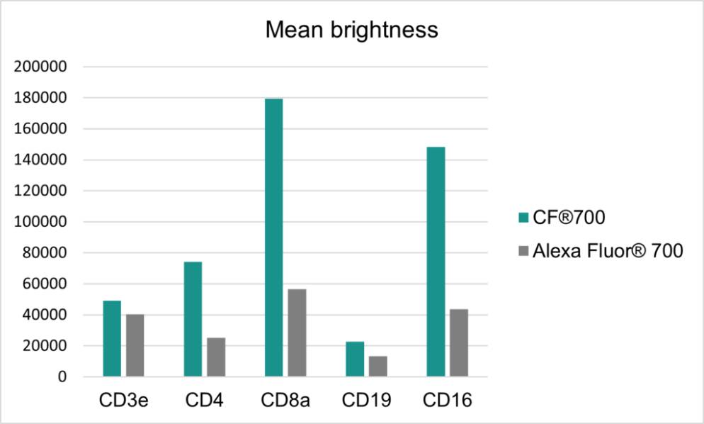
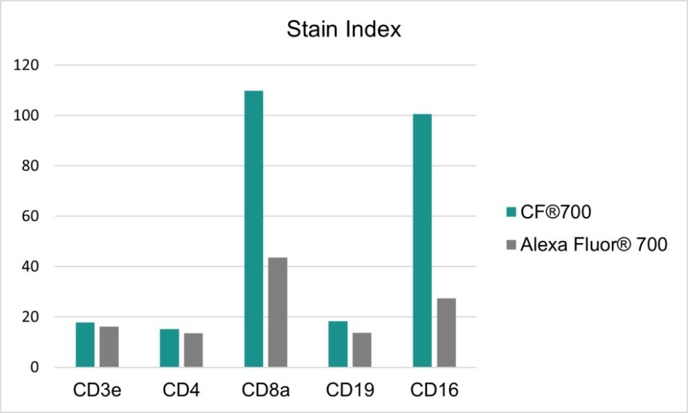
Near-IR CF®700 is Biotium’s next-generation dye with superior brightness and signal-to-noise. Unlike other near-IR dyes that suffer from aggregation and poor stability, CF®700 uses novel molecular engineering to deliver exceptional brightness. It outperforms Alexa Fluor® 700 in both microscopy and flow cytometry, offering a brighter signal and higher stain index (Figures 2–3). CF®700 is extensively validated and available in a range of formats, including Biotium Choice primary antibodies, Annexin V conjugates, Mix-n-Stain™ Antibody Labeling Kits, and more.
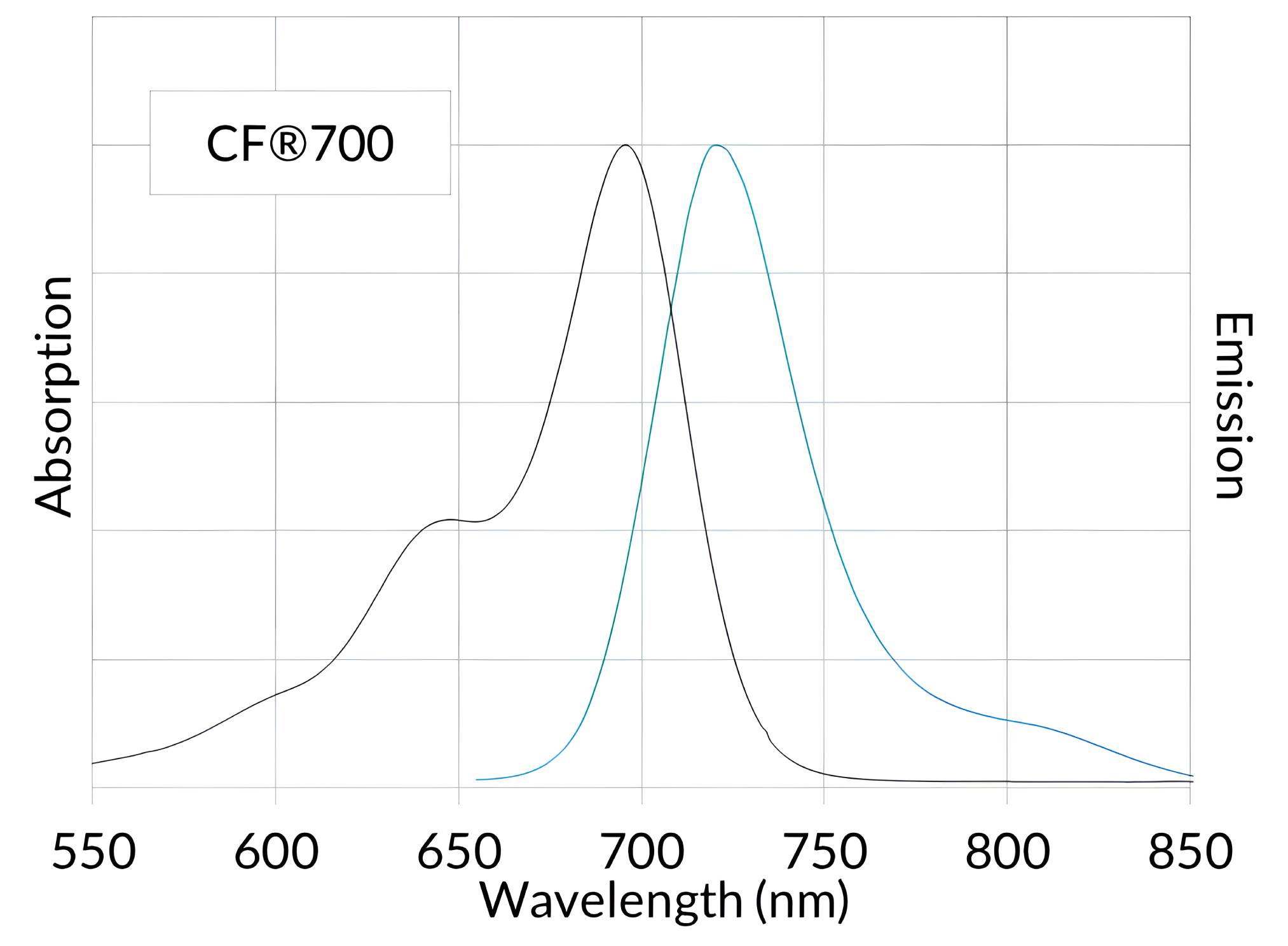
Figure 1. Normalized absorbance and emission spectra of CF®700 goat anti-mouse conjugate in PBS.
CF®700 Technical Summary
Abs/Em Maxima: 696/721 nm
Extinction coefficient: 240,000
Molecular weight: ~ 2315
Excitation laser line: 633, 635, 680 or 685 nm
Replaces: Alexa Fluor® 700, DyLight® 700-B1
CF®700 Features
Brighter near-IR alternative for Alexa Fluor® 700
Superior signal-to-noise for antibodies and other bioconjugates
Exceptional performance for microscopy and flow cytometry
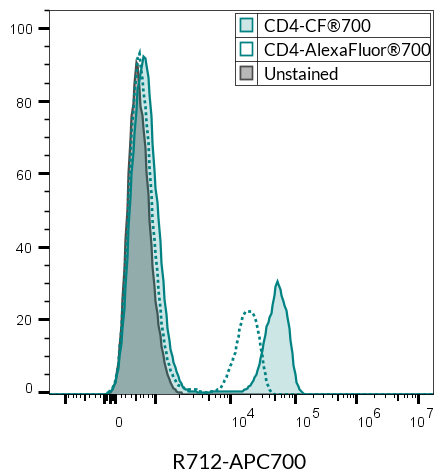
Figure 2. PBMCs stained side by side with antibodies for the same target and of the same clone, conjugated either to CF®700 or to Alexa Fluor® 700. Cells were a... See More
Figure 3. PBMCs stained side by side with antibodies for the same target and of the same clone, conjugated either to CF®700 or to Alexa Fluor® 700. Cells were a... See More
PBMCs stained with CD4(SK3)-CF®700 (filled histogram) and CD4(SK3)-Alexa Fluor®700 (BioLegend 344621, dotted line) and analyzed in the R712 (APC-700) channel of... See More
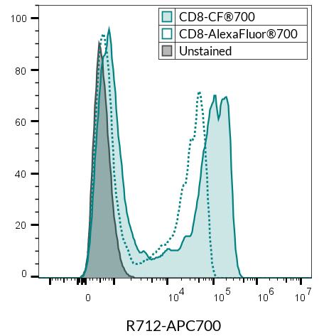
PBMCs stained with CD8(SK1)-CF®700 (filled histogram) and CD8(SK1)-Alexa Fluor®700 (BioLegend 344723, dotted line) and analyzed in the R712 (APC-700) channel of... See More
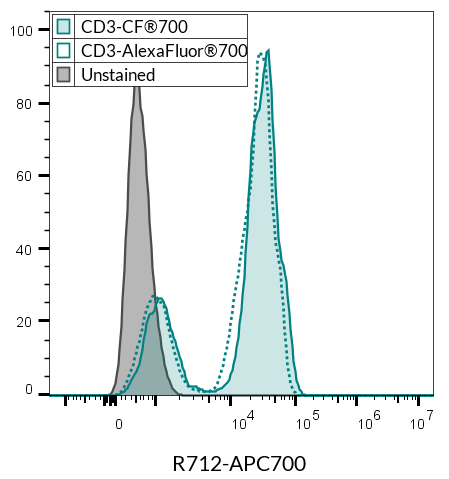
PBMCs stained with CD3(SK7)-CF®700 (filled histogram) and CD3(SK7)-Alexa Fluor®700 (BioLegend 344821, dotted line) and analyzed in the R712 (APC-700) channel of... See More
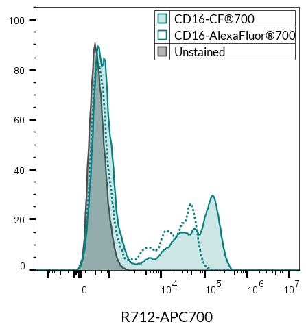
CD3- PBMCs stained with CD16(3G8)-CF®700 (filled histogram) and CD16(3G8)-Alexa Fluor®700 (BioLegend 302007, dotted line) and analyzed in the R712 (APC-700) cha... See More
Helpful Tools & Resources
Visualize and export the fluorescent dye spectrum of any CF® Dye or other dyes from Biotium.
Learn the history of fluorophores in life science research as well as the development and advantages of CF® Dyes.
Check out our handy fluorophore selection tool to help choose the best CF® Dye for your next experiment.
Download our single page reference guide for all CF® Dye fluorophores. Includes recommended channels, applications, relative brightness, and photostability.
CF is a registered trademark of Biotium, Inc.; Alexa Fluor is a registered trademark of Thermo Fisher Scientific; BioLegend is a registered trademark of Biolegend, Inc; CytoFLEX is a registered trademark of Beckman Coulter, Inc.
 New Products
 Earth-Friendly Products
 Biotium Choice Antibodies
 Special Offers