New Products
 Earth-Friendly Products
 Biotium Choice Antibodies
 Special Offers
Explore Western Blotting Solutions
Near-IR CF® Dyes for WB
TrueBlack® WB Blocking Buffer
Total Protein Prestain
Far-Red Nuclear Stain
Chromogenic Detection
Buffers & Accessories
Near-IR CF® Dye conjugates for Western Blotting
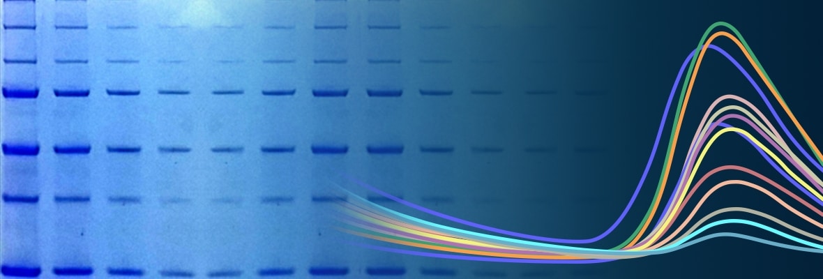
Near-infrared (near-IR) western detection is highly sensitive and offers advantages of a wider linear range and multiplexing capability compared to chemiluminescence detection. Biotium’s near-IR CF® Dyes are the brightest and most photostable available. Learn more about CF®680 and CF®770 dyes for near-IR western, In-Cell Western®, and other applications.
We offer a wide selection of primary and secondary antibodies conjugated to our exceptional near-IR CF® Dyes for western blot. We also offer HRP conjugates for chemiluminescence detection.
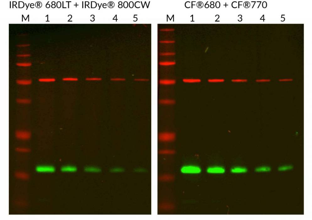
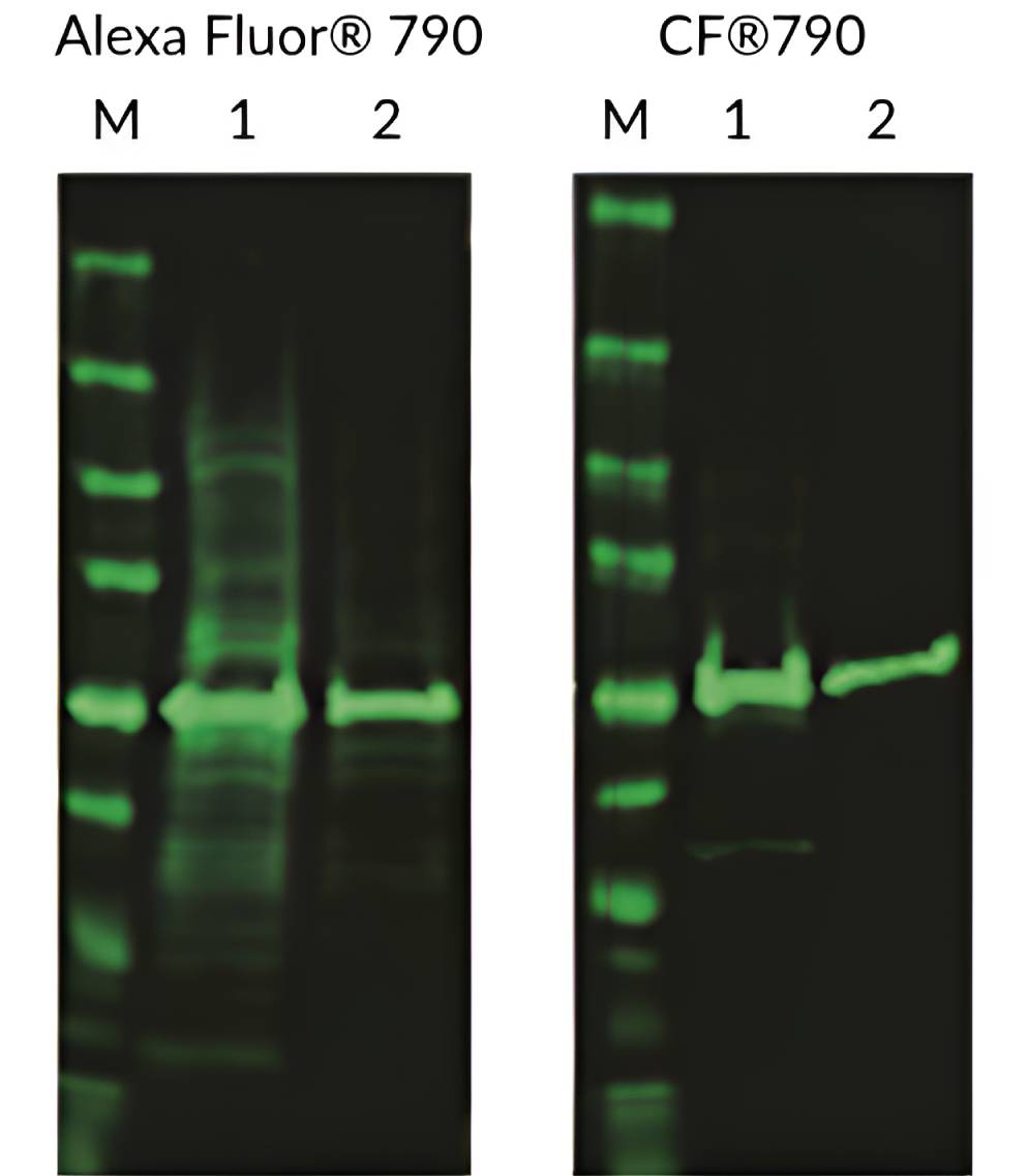
Near-IR CF® Dye Secondary Antibodies for Multiplex WB
| Conjugate | Cross-adsorption | CF®680 (681/698 nm) | CF®680R (680/701 nm) | CF®750 (755/777 nm) | CF®770 (770/797 nm) | CF®790 (784/806 nm) |
|---|---|---|---|---|---|---|
| Donkey Anti-Goat | Ck, GP, Hs, Hu, Ms, Rb, Rt, SHm | 20060 | 20196 | 20362 | 20277 | |
| Donkey Anti-Guinea Pig | Bv, Ck, Gt, Hs, Hu, Ms, Rb, Rt, Shp, SHm | 20241 | 20242 | |||
| Donkey Anti-Mouse | Bv, Ck, Gt, GP, Hs, Hu, Rb, Shp, SHm | 20194 | 20363 | |||
| Donkey Anti-Rabbit | Bv, Ck, Gt, GP, Hs, Hu, Ms, Rt, Shp, SHm | 20418 | 20195 | 20298 | 20484 | 20344 |
| Donkey Anti-Sheep | Ck, GP, Hs, Hu, Ms, Rb, Rt, SHm | 20062 | ||||
| Goat Anti-Guinea Pig | Bv, Ck, Gt, Hs, Hu, Ms, Rb, Rt, SHm, Shp | 20497 | 20496 | 20499 | 20498 | |
| Goat Anti-Mouse | Bv, Hs, Hu, Rb, Sw | 20065 | 20192 | 20463 | 20077 | 20342 |
| Goat Anti-Mouse IgG1 | Bv, Hu, Rb | 20253 | 20254 | |||
| Goat Anti-Mouse IgG2a | Bv, Hu, Rb | 20263 | 20842 | 20264 | ||
| Goat Anti-Mouse IgG2b | Bv, Hu, Rb | 20273 | 20430 | 20274 | ||
| Goat Anti-Rabbit | Hu, Ms, Rt | 20067 | 20193 | 20078 | 20343 | |
| Goat Anti-Rat | Bv, Hs, Hu, Rb | 20069 | 20383 | |||
| Rabbit Anti-Mouse | Hu | 20061 |
Bv: bovine, Ck: chicken, GP: guinea pig, Gt: goat, Hs: horse, Hu: human, Ms: mouse, Rb: rabbit, Rt: rat, SHm: Syrian hamster, Shp: sheep, Sw: swine
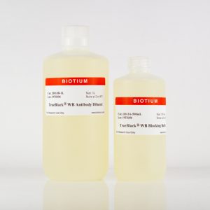
TrueBlack® WB Blocking Buffer Kit
The TrueBlack® WB Blocking Buffer Kit is a ready-to-use buffer system for fluorescence-based western blotting (WB). The buffers yield optimal specificity and sensitivity by blocking non-specific interactions of dye-labeled antibodies with proteins and the blotting membrane.
TrueBlack® WB Blocking Buffer Kit Features
- Blocks as well or better than Odyssey® Blocking Buffer, at a lower price (Figure 1)
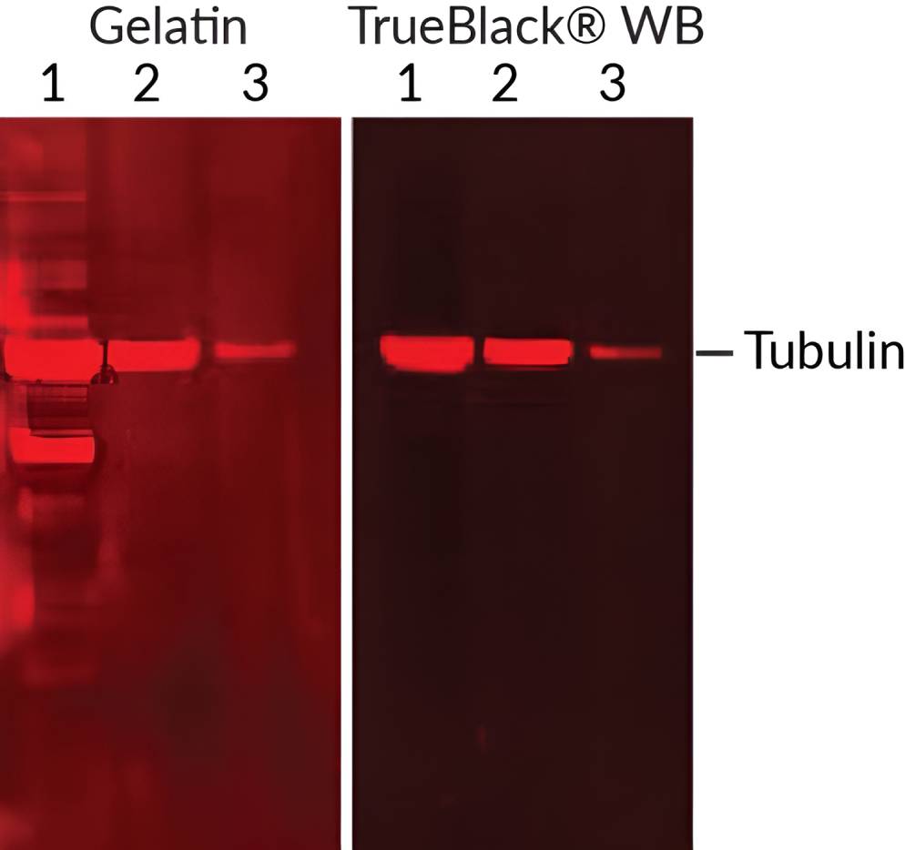
- Reduces non-specific protein bands and background over the entire membrane
- Suppresses background from charged dyes better than BSA, gelatin, or casein
- PVDF- and nitrocellulose membranes-compatible
- Free of mammalian proteins
- For visible and near-IR fluorescent westerns
Superior Western Blocking for Fluorescent Dyes
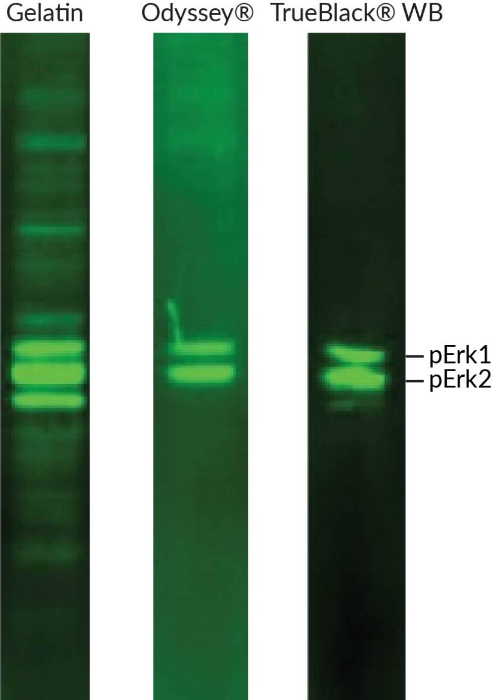
Non-specific signal in WB can arise from multiple sources, including antibody cross-reactivity, non-specific antibody adsorption, and membrane autofluorescence. Another potential cause of background is the fluorescent dyes themselves on the specificity of labeled antibodies. Highly charged dyes have improved solubility and brightness of conjugates compared to uncharged dyes. However, the extra charge carried by labeled antibodies can result in non-specific binding to proteins and membranes. TrueBlack® WB Blocking Buffer Kits block background from multiple sources, including charged dye conjugates (Figure 2). The TrueBlack® WB Blocking Buffer is especially advantageous for phosphoprotein detection, significantly improving specificity compared to conventional blocking buffers (Figure 1).
Switch from Odyssey® Blocking Buffer and Save
TrueBlack® WB Blocking Buffer performs as well or better for fluorescent WB compared to LI-COR’s Odyssey® Blocking Buffer (Figure 1), and is priced lower on a per membrane basis.
TrueBlack® WB Blocking Buffer Kit vs. Odyssey® Blocking Buffer
| Product | TrueBlack® WB Blocking Buffer Kit | Odyssey® Blocking Buffer |
|---|---|---|
| Trial Size | For 10 membranes | 125 mL for 4 membranes |
| Full Size | For 50 membranes | 500 mL for 16 membranes |
View Product Page

LI-COR and Odyssey are registered trademarks of LI-COR Inc.
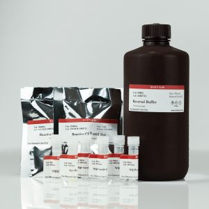
VersaBlot™ Near-IR Total Protein Normalization Kits
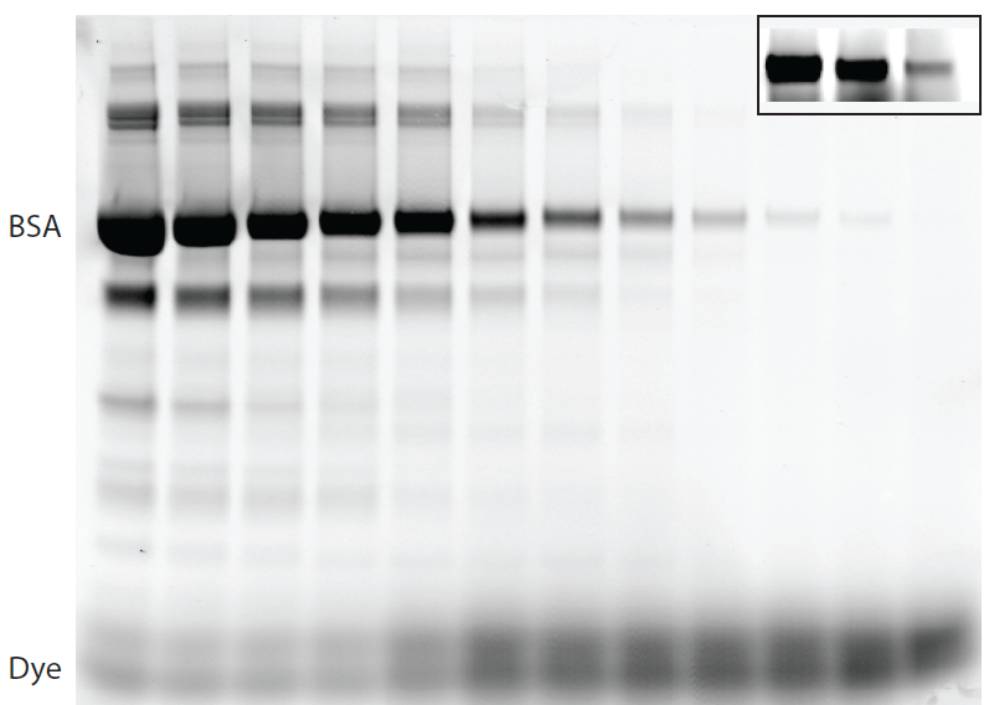
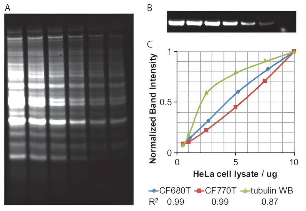
VersaBlot™ Total Protein Normalization Kits allow simple, sensitive, and highly linear protein quantitation on SDS-PAGE gels and western blot membranes. The kits label purified proteins or cell lysates with near-infrared CF® Dyes before running samples on SDS-PAGE. Proteins can be visualized on a gel or membrane using a fluorescent gel scanner. The prestain can be reversed, facilitating downstream multi-color western blot analysis.
VersaBlot™ Total Protein Prestain Features
- Reversible prestain for downstream multi-color western blot analysis
- Superior linearity for western normalization compared to housekeeping proteins
- Highly sensitive protein quantitation on PAGE gels or western membranes
- Fast and simple labeling of proteins or lysates, no purification required
- Detect as little as 1 ng protein and 10% difference in protein content
VersaBlot™ Total Protein Normalization Kits
| Product | Catalog number | Size | Abs/Em | Imaging Systems / Detection channels |
|---|---|---|---|---|
| VersaBlot™ CF®680T Total Protein Normalization Kit | 33025-T | 100 labelings | 681 / 698 nm | Amersham Typhoon™ Trio Cy®5 channel Amersham Typhoon™ 5 IR short channel LI-COR® Odyssey® 700 channel |
| 33025 | 500 labelings | |||
| VersaBlot™ CF®770T Total Protein Normalization Kit | 33026-T | 100 labelings | 764 / 787 nm | Amersham Typhoon™ 5 IR long channel LI-COR® Odyssey® 800 channel |
| 33026 | 500 labelings |
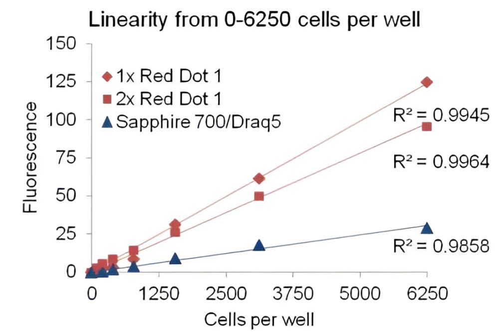
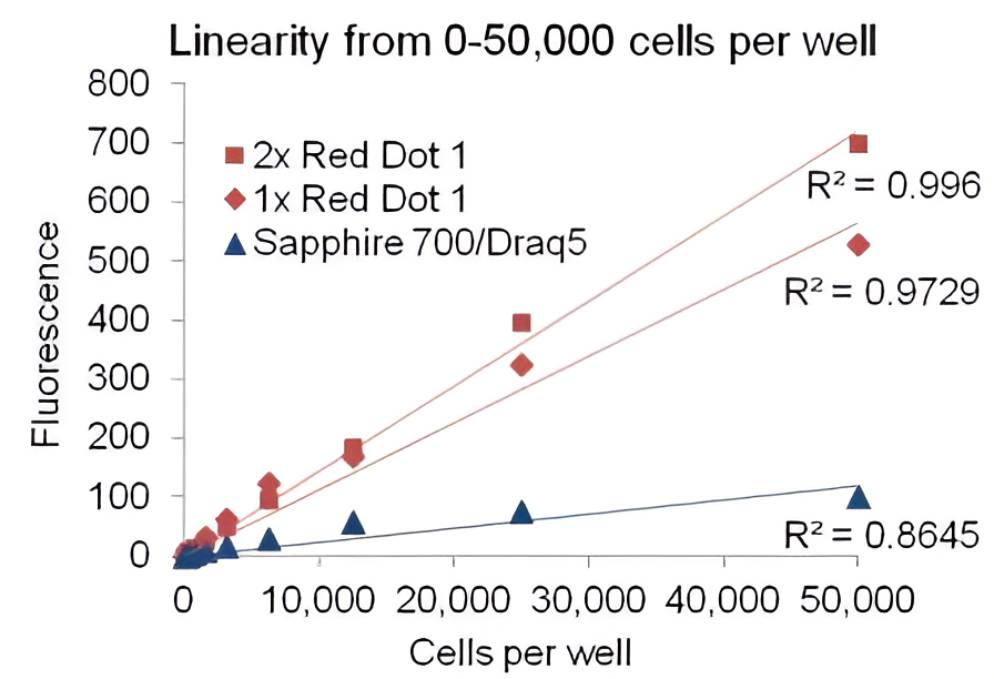
RedDot™1 Far-Red Nuclear Stain for In-Cell Western®
RedDot™1 is a far-red cell membrane-permeant nuclear dye similar to Draq5™. Compared to DRAQ5™/Sapphire700™, it delivers higher signal intensity, a broader linear range, and does not require a wash step. RedDot™1 is useful for normalizing cell numbers in In-Cell Western® assays using a near-infrared scanner. In fixed cells, RedDot™ 1 staining generates a highly linear fluorescence signal that is proportional to the number of cells.
- Ideal for normalization of In-Cell Western® in fixed cells, similar to Draq5™/Sapphire®700
- Highly thermostable and photostable, for convenient handling and demanding imaging applications
- Can also be used for DNA content analysis by flow cytometry like Vybrant™ DyeCycle™ Ruby
- λEx/λEm = 662/694 nm (with DNA), for detection in the Cy®5 channel
View Product Page
Chromogenic Detection
Chromogenic detection by horseradish peroxidase (HRP) or alkaline phosphatase is a widely used and cost-effective method for Western blot analysis. Colorimetric detection works by the production of an insoluble colored byproduct from a chemical reaction between a substrate and reporter enzyme. Biotium offers several secondary and anti-tag antibodies conjugated to either HRP or alkaline phosphatase for chromogenic detection. See the tables below for available conjugates. You can also view our entire Secondary Antibody Product Listings for other HRP and alkaline phosphatase conjugates, including anti-human IgG, IgA, and IgM antibodies. Biotium also offers several colorimetric substrates for alkaline phosphatase and a DAB substrate kit for HRP, listed in the table below.
HRP and Alkaline Phosphatase Secondary Antibodies
| Conjugate | Donkey Anti-Mouse (Min X Rat) | Donkey Anti-Rabbit | Goat Anti-Chicken | Goat Anti-Mouse | Goat Anti-Rabbit | Goat Anti-Rat | Chicken Anti-Goat | Goat Anti-Llama | Goat Anti-Mouse | Goat Anti-Rabbit |
|---|---|---|---|---|---|---|---|---|---|---|
| Cross-Adsorption | Bv, Ck, Gt, GP, Hs, Hu, Rb, Rt, Shp, SHm | Bv, Ck, Gt, GP, Hs, Hu, Ms, Rt, Shp, SHm | Bv, Gt, GP, Hs, Hu, Ms, Rb, Rt, SHm, Shp | Bv, Hs, Hu, Rb, Sw | Hu, Ms, Rt | Bv, Hs, Hu, Rb | None | None | None | None |
| HRP | 20404 | 20405 | 20474 | 20401 | 20403 | 20406 | 20839 | 20475 | 20400 | 20402 |
| Alkaline Phosphatase | 20466 | 20467 | 20464 | 20465 |
Bv: bovine, Ck: chicken, GP: guinea pig, Gt: goat, Hs: horse, Hu: human, Ms: mouse, Rb: rabbit, Rt: rat, SHm: Syrian hamster, Shp: sheep, Sw: swine
HRP and Alkaline Phosphatase Anti-Tag Antibodies
Chromogenic Substrates for HRP and Alkaline Phosphatase
| Product | Catalog No. | Size | Substrate for | Precipitate Color |
|---|---|---|---|---|
| BCIP, NA | 1001 | 100 mg | Alkaline Phosphatase | Dark Blue |
| 1001-1 | 500 mg | |||
| 1001-2 | 5 g | |||
| BCIP, Toluidine | 10002 | 100 mg | Alkaline Phosphatase | Dark Blue |
| 10002-1 | 500 mg | |||
| 10002-2 | 5 g | |||
| BCIP Pink | 10006 | 100 mg | Alkaline Phosphatase | Pink |
| BCIP Pink/NBT Kit | 10007 | 1 set | Alkaline Phosphatase | Pink |
| BCIP Red | 10004 | 100 mg | Alkaline Phosphatase | Red |
| BCIP Red/NBT Kit | 10005 | 1 set | Alkaline Phosphatase | Red |
| BCIP/NBT Kit | 10003 | 1 set | Alkaline Phosphatase | Dark Blue |
| Chromogenic Phosphatase Substrate Sampler | 10022 | 1 kit | Alkaline Phosphatase | Multiple |
| DAB Substrate Kit | 30015 | 1 kit | HRP | Brown |
Accessory Reagents for Western Blotting
See our selection of prestained markers, buffers, blocking agents, detergents, and other reagents and accessories below.
| Product | Catalog No. | Size | Features |
|---|---|---|---|
| Peacock™ Prestained Protein Marker | 21530 | 50 uL or 500 uL | • Visible color protein marker for SDS-PAGE and western • 8 blue bands ranging from 10 kDa to 180 kDa • Red/green bands at 75 kDa/25 kDa |
| Peacock™ Plus Prestained Protein Marker | 21531 | 50 uL or 500 uL | • Visible color protein marker for SDS-PAGE and western • 10 blue bands ranging from 8 kDa to 245 kDa • Red/green bands at 75 kDa/25 kDa |
| 4X Protein Loading Buffer with Orange Tracking Dye | 40136 | 15 mL | • A convenient 4X sample loading buffer for protein gel electrophoresis • Optimized for use with denaturing SDS-PAGE • Orange tracking dye avoids unwanted background fluorescence caused by blue tracking dyes |
| Ponceau S Solution | 22001 | 1 L | • Stain proteins on PVDF or nitrocellulose membranes with visible pink dye • Fast & reversible visualization of protein transfer before western detection |
| TrueBlack® WB Blocking Buffer Kit | 23013 | For 10 membranes or 50 membranes | • Superior blocking for fluorescent WB • Works as well or better than Odyssey® Blocking Buffer, at a lower cost • Suppresses background caused by charged fluorescent dyes • Reduces antibody cross-reactivity, eliminating non-specific bands |
| 10X Fish Gelatin Blocking Agent | 22010 | 100 mL | • Provides excellent blocking for IF or western • Add to buffer of your choice (PBS or TBS) • Compatible with anti-goat and anti-sheep secondaries |
| Fish Gelatin Powder | 22011 | 2 x 50 g | • Gelatin from cold water fish skin for blocking for IF or western • Compatible with anti-goat and anti-sheep secondaries |
| Bovine Serum Albumin, 30% Solution | 22014 | 100 mL | • Commonly used blocking agent and antibody or protein stabilizer • 30% solution in water • Made from IgG-free, protease-free Fraction V BSA |
| Bovine Serum Albumin Fraction V | 22013 | 50 g | • Commonly used blocking agent and antibody or protein stabilizer • IgG-free, protease-free Fraction V BSA |
| Dry Milk Powder | 22012 | 4 x 25 g | • Nonfat dry milk • Commonly used blocking agent for western |
| Tween®-20 | 22002 | 50 mL | • Detergent commonly used for western blocking and washing |
| Mini Cell Scrapers | 22003 | Pack of 200 | • For harvesting cells or cell lysates from 96-, 48- and 24-well plates • 0.5 cm (3/16") wide and 6 cm (2 3/8") long • 20 packs of 10 scrapers per pack • Polyethylene, disposable & sterile |
| Ultrafiltration Vials (3K MWCO) | 22018 | Pack of 5 | • For removing buffers, salts, and free dyes from proteins or DNA • Simple microcentrifuge spin-column format • 3 kDa molecular weight cut-off (MWCO) |
| Ultrafiltration Vials (10K MWCO) | 22004 | Pack of 5 | • For removing buffers, salts, and free dyes from proteins or DNA • Simple microcentrifuge spin-column format • 10 kDa molecular weight cut-off (MWCO) |
| DTT | 91050 | 1 g | • Reducing agent commonly used to prepare samples for SDS-PAGE |
| TCEP | 91049 | 1 g | • Odorless reducing agent • More effective and stable than DTT |