New Products
 Earth-Friendly Products
 Biotium Choice Antibodies
 Special Offers
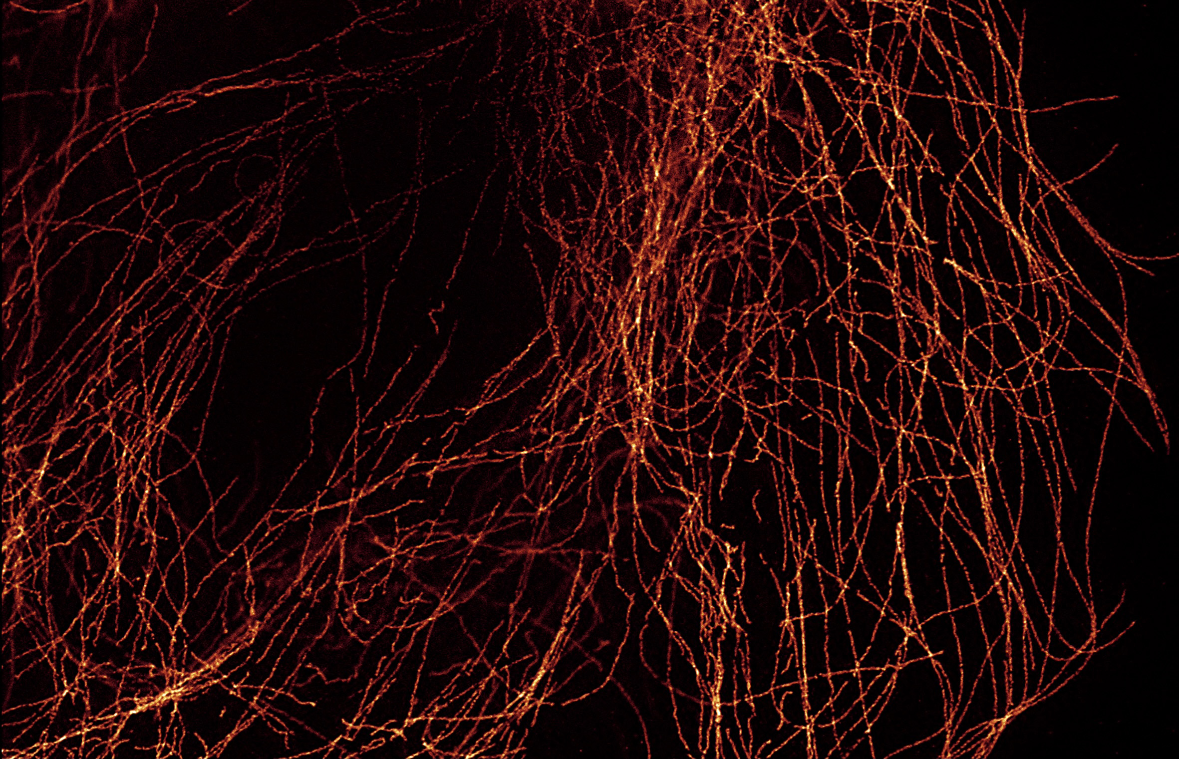
The Best Probes for Super-Resolution
Super-resolution microscopy encompasses a broad family of imaging techniques that push beyond the diffraction limit of traditional light microscopy, revealing the finer details of biological structures. These techniques rely on extremely precise control over the excitation, emission, and image acquisition of fluorescently labeled cells and tissues. Consequently, the quality and efficiency of super-resolution imaging relies heavily on the fluorescent properties of the probe. Recent advances in super-resolution microscopy have demonstrated the importance of high-performance fluorescent probes.
CF® Dye Advantages for Super-Resolution
CF® Dyes have met this demand for high-performance fluorescent probes with industry-leading brightness and photochemical switching properties. Moreover, CF® Dyes have been published in dozens of studies for a wide range of super-resolution applications.
Features
- Superior performance of labeled conjugates
- Validated in dozens of publications for STORM, STED, SIM, 2-photon, TIRF, and more
- Available in over 40 fluorophores across visible spectrum with several near-IR options
- Dye options with unique chemical switching properties ideal for STORM
CF® Dyes Validated for STED and FLIM by STELLARIS
CF® Dyes were validated for high-quality STED and FLIM imaging on Leica’s STELLARIS 8 STED FALCON platform through Biotium's research partners as well as several publications. See the tables below for validated CF® Dyes for STED, recommended depletion lasers, and fluorescent lifetime data for FLIM.


CF® Dyes Validated for STED
| CF® Dye | Abs/Em (nm) | Recommended Depletion Laser | Publication/Data Source |
|---|---|---|---|
| CF®440 | 433/512 | 592 nm | Biotium/Leica STELLARIS 8 imaging data |
| CF®488A | 490/516 | 592 nm | Angelov & Angelova, Nanoscale, 9, 9797, (2017) |
| CF®550R | 551/577 | 660 nm | Biotium Collaborator |
| CF®555 | 555/565 | 660 nm | Biotium Collaborator |
| CF®583 | 584/606 | 775 nm | Biotium Collaborator |
| CF®594 | 593/615 | 775 nm | Gonzalez Pisfil et al., Sci. Rep., 12, 14027, (2022) |
| CF®640R | 642/663 | 775 nm | Biotium/Leica STELLARIS 8 imaging data |
| CF®680R | 680/701 | 775 nm | Gonzalez Pisfil et al., Sci. Rep., 12, 14027, (2022) |
CF® Dye Lifetimes
| Dye | τ (ns) /free acid in PBS pH 7.4, ε (ns) | τ (ns) /S.Ab§ |
|---|---|---|
| CF®405S | 3.88 ± 0.05 | - |
| CF®488A | 4.11 ± 0.05 | 1.705 |
| CF®550R | - | 2.63 ± 0.04* |
| CF®568 | 3.66 ± 0.05 | 1.539 |
| CF®583 | 2.04 ± 0.02* | |
| CF®594 | - | 1.746 |
| CF®633 | 3.39* (in water) | 3* |
| CF®640R | 2.38 ± 0.05 | 1.557 |
| CF®647 | 1.07 ± 0.05 | 1.195 |
| CF®680 | 1.23 ± 0.05 | 1.277 |
| CF®680R | 1.22 ± 0.05 | 1.6 |
| CF®710 | - | 0.77 ± 0.01* |
| CF®750 | 0.58 ± 0.05 | 0.636 |
| CF®790 | 0.39 ± 0.05 | 0.54 |
§ Fluorescence lifetime measurements of CF® Dye labeled anti-mouse secondary antibodies used for immunostaining microtubules in U2OS using mouse anti-tubulin (DM1a) and mounted in ProLong™ Diamond. *Lifetime data obtained via customer communication under different experimental conditions and imaging setup. Measurements were made on a Stellaris 8 STED FALCON microscope courtesy Leica Microsystems and Mariano Gonzales Pisfil, Steffen Dietzel, Core Facility Bioimaging, Biomedical Center, Ludwig-Maximilians-University, Munich, Germany.
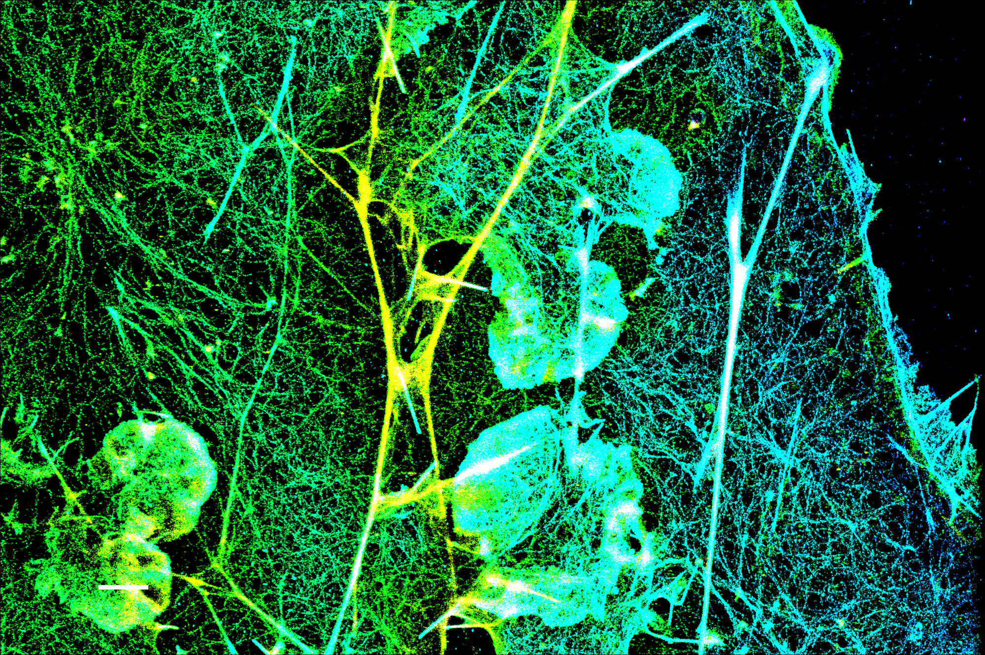
Superior Performance for STORM Imaging
Several CF® Dyes were developed and validated for optimal performance in Stochastic Optical Reconstruction Microscopy (STORM). This includes Biotium’s red-excited CF® Dyes such as CF®647, CF®660C, and CF®680 which display superior brightness and unique photoswitching properties that are ideal for STORM. However, while STORM imaging commonly relies on photoswitchable red-excited dyes, finding comparable dyes for other excitation sources to produce quality multi-color STORM images remains a challenge. In response, Biotium has developed several green-excited dyes developed for optimal performance in STORM.
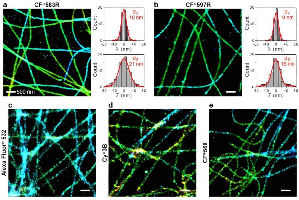
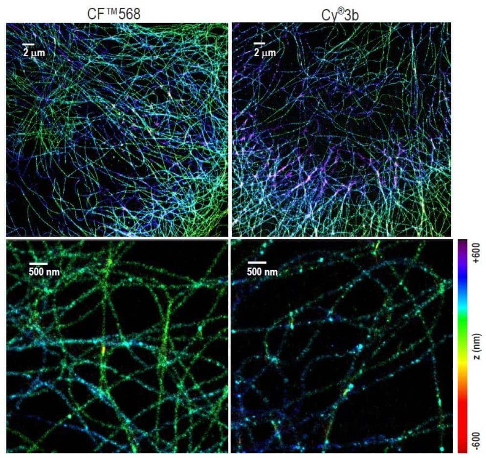
Specifically, green-excited CF®583R and CF®597R were developed in collaboration with UC Berkeley and STORM scientist Ke Xu, PhD for exceptional performance in multi-color STORM imaging. In addition, green-excited CF®568 has also demonstrated better performance than Cy®3b. See below for a full list of validated CF® Dyes for STORM and available products.
“Working closely with Biotium chemists and communicating the ideal chemical properties needed for optimal STORM dyes, our collaboration was able to yield a framework for developing more fluorescent colors that expand the color palette for multicolor STORM imaging.”
Dr. Ke Xu, Ph.D. Professor of Chemistry at UC Berkeley
CF® Dyes for STORM Show Superior STORM Performance

Literature Digest
Literature Digest: CF® Dyes for STORM Super-Resolution Microscopy
CF® Dyes Validated for Super-Resolution
| Dye | Abs/Em (nm) | STORM | STED | SIM | 2-Photon | TIRF | Other Applications | Features |
|---|---|---|---|---|---|---|---|---|
| CF®405S | 411/431 | ✓ | ✓ | • Brighter than Alexa Fluor® 405 | ||||
| CF®405M | 416/452 | ✓ | ✓ | • More photostable than Pacific Blue™ • Excellent choice for SIM imaging |
||||
| CF®440 | 433/512 | ✓ | • More photostable than spectrally similar dyes | |||||
| CF®488A | 490/516 | ✓ | ✓ | ✓ | ✓ | ✓ | DNA-PAINT | • Less non-specific binding than Alexa Fluor® 488 |
| CF®498 | 498/519 | ✓ | • Less non-specific binding than Alexa Fluor® 488 • Use with CF®568/CF®583R and CF®647/Alexa Fluor®647 for multicolor STORM |
|||||
| CF®505 | 505/519 | ✓ | • Identical to ATTO 488 | |||||
| CF®535ST | 535/569 | ✓ | • Orange dye designed specifically for STORM imaging | |||||
| CF®550R | 551/577 | ✓ | • A spectrally unique orange/red dye | |||||
| CF®555 | 554/568 | ✓1 | ✓ | ✓1 | • Brighter and more photostable than Cy®3 • Less non-specific binding than Alexa Fluor® 555 |
|||
| CF®568 | 562/584 | ✓1 | ✓ | ✓ | ✓ | • Yields much brighter conjugates than Alexa Fluor® 568 • Outperforms Cy®3b in STORM • Pairs well with CF®647 and CF®680 for multi-color STORM - see publication |
||
| CF®583 | 584/606 | ✓ | • Spectrally similar red fluorescent dye with exceptional brightness • Serves as an alternative for Cy®3.5 and Texas Red® |
|||||
| CF®583R | 585/609 | ✓1 | • One of two top-performing dyes specifically designed for STORM with green laser (also see CF®597R) - see publication |
|||||
| CF®594 | 593/615 | ✓ | ✓ | • Significantly brighter than Alexa Fluor® 594 and Texas Red® • Extremely photostable |
||||
| CF®597R | 597/619 | ✓1 | • Deep-red fluorescent dye designed specifically for STORM • Top-performing dye specifically designed for STORM with green laser (also see CF®583R) - see publication |
|||||
| CF®633 | 629/650 | ✓ | FIONA, gSHRImp, SMT | • Significantly brighter than spectrally similar far-red dyes • Far more photostable than Alexa Fluor® 647 |
||||
| CF®640R | 642/663 | ✓ | ✓ | ✓ | ✓ | FLImP | • Offers improved brightness and photostability over ATTO 647N and spectrally similar dyes |
|
| CF®647 | 652/668 | ✓1 | • Spectrally similar to Cy®5 and Alexa Fluor® 647 • Pairs well with CF®568 for multi-color STORM • The best far-red dye for demixing-based multi-color (d)STORM imaging when paired with CF®680 - see publication |
|||||
| CF®660R | 662/682 | SMLM, DNA-PAINT | • Much brighter than Alexa Fluor® 660 • The most photostable 660 nm dye • Validated for use with DNA-PAINT SMLM - see publication |
|||||
| CF®660C | 667/685 | ✓1 | MINFLUX | • Much brighter and more photostable than Alexa Fluor® 660 • Ideal for long high-intensity 3D (d)STORM image acquisitions with minimal photobleaching - see publication |
||||
| CF®680 | 681/698 | ✓1 | Dual-color 3D SMLM, MINFLUX | • The brightest among spectrally similar 680 nm dyes • Pairs well with CF®568 for multi-color STORM • The best near-IR dye for demixing-based multi-color (d)STORM imaging when paired with CF®647 - see publication |
||||
| CF®680R | 680/701 | ✓1 | ✓ | ✓ | Single-molecule spectroscopy, SMT | • The most photostable 680 nm dye • Suitable for labeling nucleic acids and small biomolecules |
||
| CF®750 | 755/779 | ✓ | • Exceptionally bright and photostable near-IR dye • Patented pegylated dye for superior performance |
1 Dye was validated in multi-color STORM experiments
FLImP: Fluorophore localization imaging with photobleaching; SIM: Structured illumination microscopy; STED: Stimulated emission depletion; STORM: Stochastical optical reconstruction microscopy; TIRF: Total internal reflection fluorescence; FIONA: Fluorescence imaging with one-nanometer accuracy; ExM: Expansion microscopy; SMT: Single-molecule tracking; SMLM: Single-molecule localization microscopy.
CF® Dye Products for Super-Resolution Microscopy
Antibody Labeling Kits
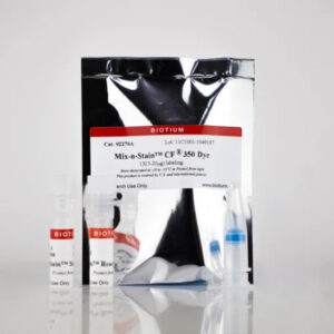
Our Mix-n-Stain™ STORM CF® Dye Antibody Labeling Kits allow you to label 50 ug of your antibody with one of Biotium’s STORM CF® Dyes to produce an antibody conjugate with a low 1-2.5 DOL (degree of labeling) optimal for super-resolution STORM (Stochastic Optical Reconstruction Microscopy). Labeling takes just 30 minutes, with minimal hands-on time and no purification. Click here to learn more about Biotium’s antibody labeling kits.
Features
- Choice of 8 CF® Dye colors ideal for STORM
- CF®583R and CF®597R are novel green-excited STORM dyes
- Labeling optimized to provide low 1-2.5 DOL
- Less than 30 seconds of hands-on time
- 30 minutes total reaction time
- No purification, 100% recovery
Nanobody® Labeling Kits
Nanobodies® (also called camelid single variable or VHH domains), are smaller ~15 kDa single-domain antibodies that enable deeper tissue penetration, faster staining times, and improved access to tightly packed or masked epitopes—making them ideal for high-resolution and super-resolution imaging.
Biotium offers labeling kits designed specifically for labeling single-domain Nanobodies® with one of Biotium’s bright and photostable CF® Dyes.
Mix-n-Stain™ Nanobody Labeling Kits are for standard labeling of VHH, while the Mix-n-Stain™ Nanobody Thiol Labeling Kits were designed for labeling VHH containing a single cysteine (1x Cys) residue. The kits allow optimal labeling of between 5-50 ug of Nanobody®, with minimal hands-on time and no purification.
Biotium also offers MiniMab™ single-domain antibodies (SdAbs) labeled with best-in-class CF® Dyes for STORM. MiniMab™ SdAbs are available for popular neuroscience targets or as secondary antibodies.
Mix-n-Stain™ Nanobody Labeling Kit Features
- Ideal for standard labeling of VHH
- Label 5-20 ug or 20-50 ug Nanobody®
- Choice of 7 CF® Dye colors or biotin
- Simple 30 minute labeling, no purification
- Compatible with common stabilizers
- Prevents over-labeling
Mix-n-Stain™ Nanobody Thiol Labeling Kits Features
- Designed for labeling VHH with a single cysteine (1x Cys) residue
- Label 5-20 ug or 20-50 ug Nanobody®
- Choice of 7 CF® Dye colors
- Simple 2 hour labeling, no purification
- No purification, 100% recovery
Single-Label Secondary Antibody Conjugates
Secondary antibodies with a low degree of labeling (DOL, or number of dye molecules per antibody molecule) have been reported to be optimal for STORM (Bittel et al. 2015). Biotium offers single-label secondary antibody conjugates with an average DOL of one for STORM applications.
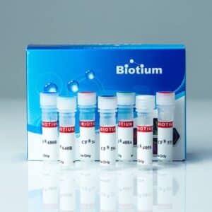
Reactive CF® Dyes
Probes for Cytoskeleton and Other Cell Structures
- Phalloidin conjugates for F-actin labeling available in 16 CF® Dyes. CF®647 and CF®680 phalloidins ideal for STORM imaging.
- Con A, WGA, and PNA lectin CF® Dye conjugates for labeling glycoproteins and cellular surfaces
- Streptavidin and biotinylated CF® Dye conjugates
- CF® Dye nucleotides and probes for apoptosis, endocytosis, and other cellular processes
Learn More

Membrane & Organelle Stains for Super-Resolution
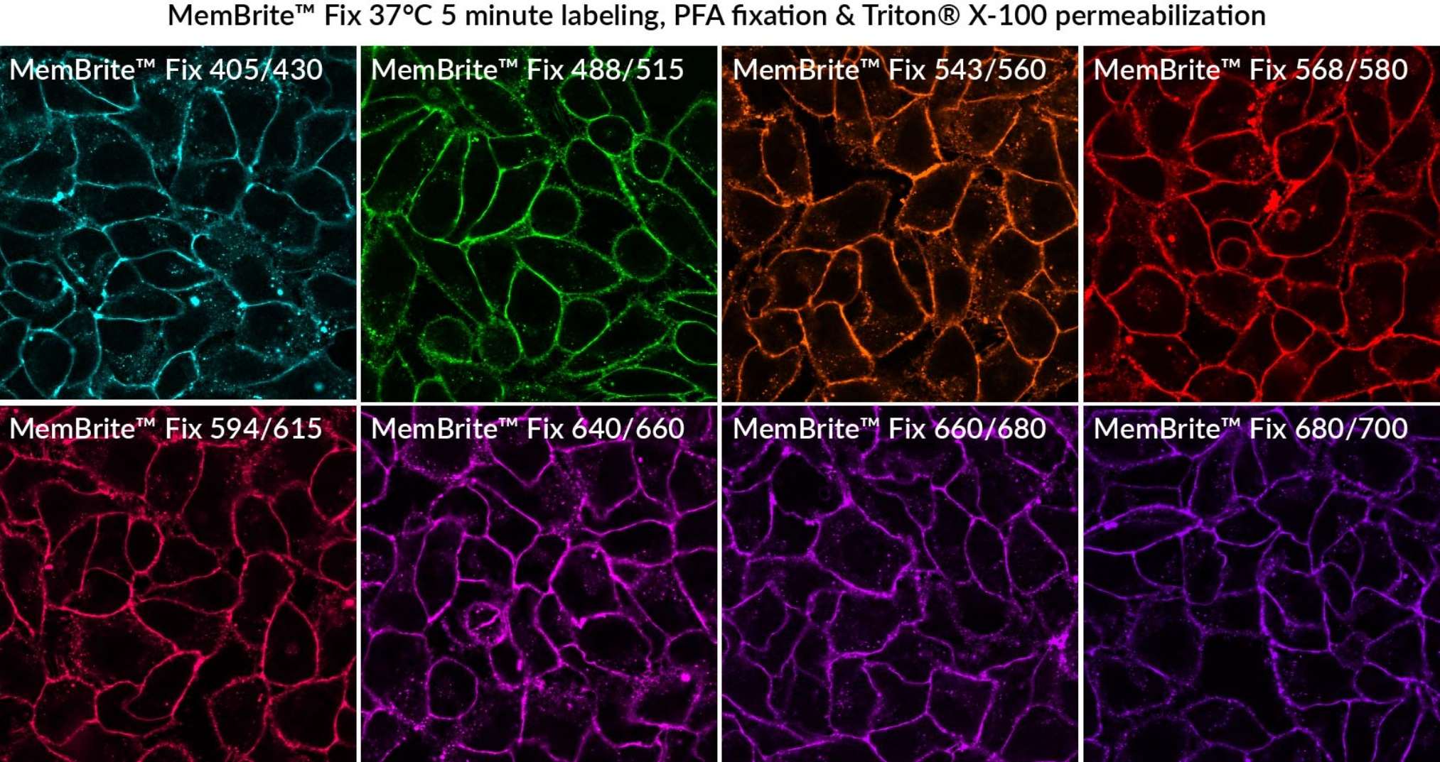
MemBrite® Fix for Specialized Imaging Applications
MemBrite® Fix Membrane Stains are novel reactive fluorescent dye stains that react irreversibly with cell surface proteins, for staining that can withstand formaldehyde or alcohol fixation, and detergent permeabilization. Several MemBrite® Fix dyes have been validated in super-resolution imaging applications or 2-photon microscopy. MemBrite® Fix-ST dyes are specialized dyes recommended for super-resolution imaging by STORM.
- Rapid, uniform cell surface labeling that can be fixed & permeabilized
- Choose from 12 bright & photostable dye colors from blue to near-IR
- MemBrite® Fix-ST designed specifically for STORM imaging
- Several MemBrite® Fix colors validated for super-resolution and other specialized imaging applications
| Catalog number | Dye | Specialized applications |
|---|---|---|
| 30092 | MemBrite® Fix 405/430 | SIM, TIRF |
| 30093 | MemBrite® Fix 488/515 | STED, TIRF, 2-photon microscopy |
| 30094 | MemBrite® Fix 543/560 | N/A |
| 30095 | MemBrite® Fix 568/580 | SIM, STORM, TIRF |
| 30096 | MemBrite® Fix 594/615 | 2-photon microscopy |
| 30097 | MemBrite® Fix 640/660 | FLImP, SIM, TIRF |
| 30098 | MemBrite® Fix 660/680 | N/A |
| 30099 | MemBrite® Fix 680/700 | STORM1, Single-molecule imaging, STED, 2-photon microscopy |
| 30101 | MemBrite® Fix-ST 650/665 | STORM |
| 30102 | MemBrite® Fix-ST 667/685 | STORM |
| 30103 | MemBrite® Fix-ST 681/698 | Single-molecule imaging, STORM |
| 30104 | MemBrite® Fix-ST 755/777 | STORM |
1. MemBrite® Fix-ST 681/698 dye is reported to have better performance in STORM imaging than MemBrite® Fix 680/700 dye.
View Product Page
Lysosome & Lipid Stains Validated for Super-Resolution
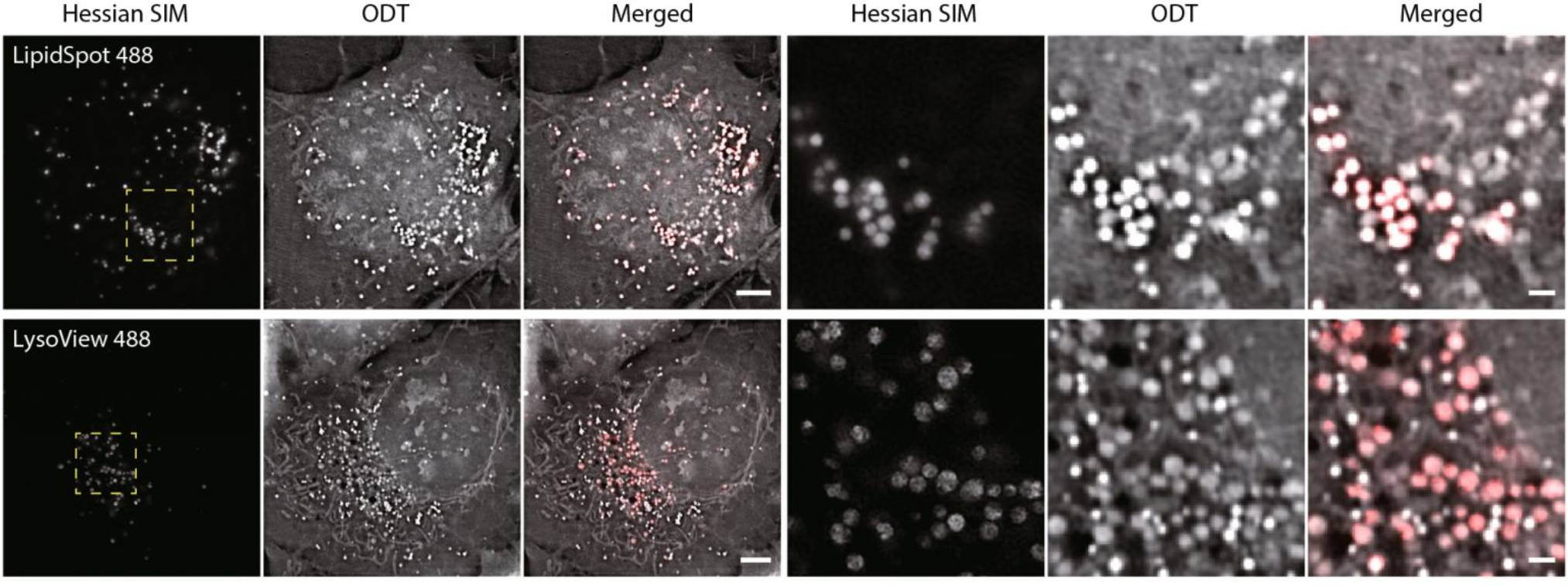
LysoView™ Dyes are fluorescent stains for imaging lysosome localization and morphology in live cells. The dyes accumulate in the low pH environment of the lysosomes, resulting in highly specific lysosomal staining without the need for a wash step. LysoView™ 540 and LysoView™ 633 exhibit pH-sensitive fluorescence. LysoView™ 488 has been validated in super-resolution imaging by SIM, while LysoView™ 650 fluorophore is compatible with SIM and STED.
LipidSpot™ Lipid Droplet Stains are fluorogenic neutral lipid stains that rapidly stain lipid droplets. The dyes can be used to stain lipid droplets in both live and fixed cells, with no wash step required. Cells also can be fixed and permeabilized after staining. LipidSpot™ stains show minimal background staining of cellular membranes or other organelles, unlike dyes like Nile Red. Available with green or deep-red/far-red fluorescence.
View Product Page
LysoView™ 488 and LipidSpot™ 488 are Published for SIM
Highlighted Citation: In a publication in Light: Science & Applications, Dong et al. developed a dual-mode high-speed super-resolution microscope for live cells by combining optical diffraction tomography (ODT) with structured illumination microscopy (SIM). This novel technique, deemed super-resolution fluorescence assisted diffraction computational tomography (SR-FACT), was used to acquire ~100 nm resolution images merged with 3D imaging of live cells in real time. The authors validated the technique by visualizing organelles within COS-7 cells labeled by expressed GFP fusion proteins or synthetic fluorescent probes. SIM visualization of LysoView™ 488 and LipidSpot™ 488 were shown to colocalize in the ODT channel with lysosomes and lipid droplets respectively.
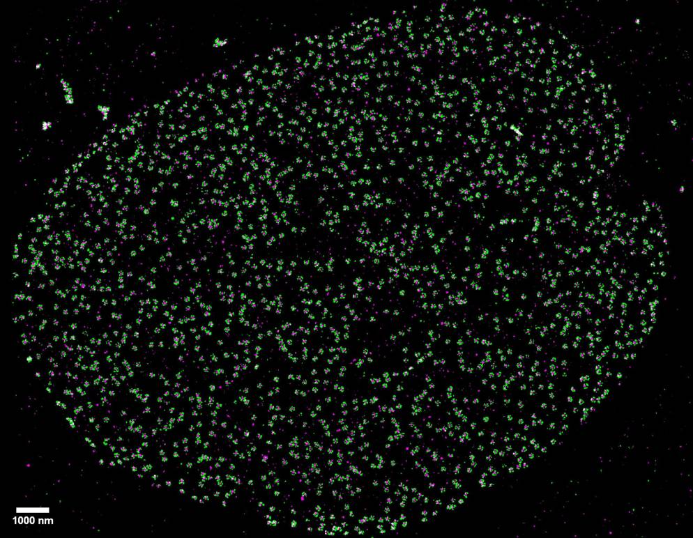
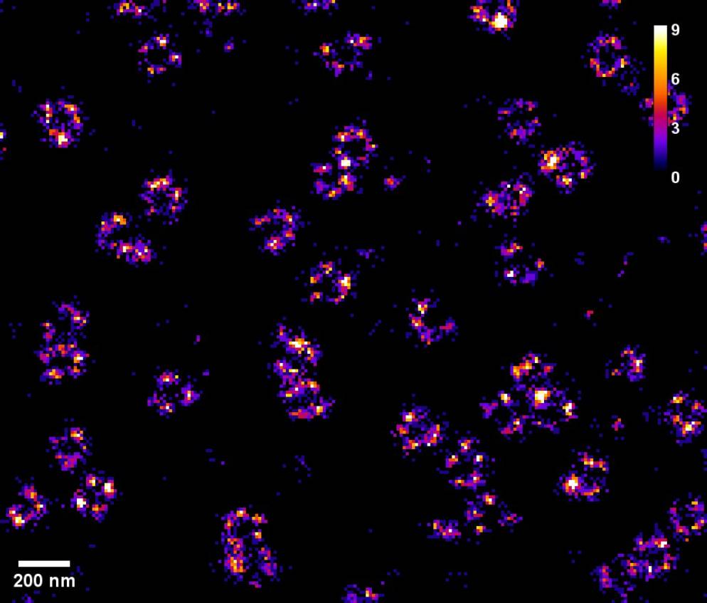
EV Stains Featuring STORM-Optimized CF® Dyes
Characterizing exosomes and EVs by imaging is challenging due to their small size and the resolution limits of conventional light microscopy. Super-resolution techniques like direct stochastic optical reconstruction microscopy (dSTORM) overcome these limits, providing single-molecule resolution of subcellular structures such as EVs. This enables deeper insights into EV biogenesis, behavior, and morphology.
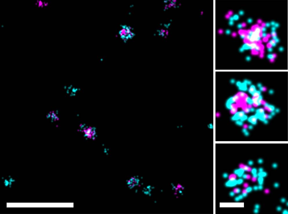
ExoBrite™ STORM Antibodies and ExoBrite™ STORM CTB EV Stains harness CF® Dyes optimized for STORM imaging, delivering ultra-bright, high-resolution EV imaging. The ExoBrite™ STORM Antibodies are formulated for detecting EV markers with high signal-to-noise, while ExoBrite™ STORM CTB EV Stains enable sharp, specific membrane labeling via cholera toxin subunit B. These advanced reagents provide exceptional clarity for STORM imaging.
Note: ExoBrite™ CTB stains have been found to label EVs derived from several tested cell lines, but may not stain EVs from every source. Please visit the product page to view a full list of validated EV sources.
Advantages of ExoBrite™ STORM Antibodies
- Validated antibodies for detecting EV markers CD9, CD63, and CD81
- Available with excellent STORM dyes
- CF®498, CF®568, CF®583R, and CF®647Plus
- Proprietary buffer for enhanced signal-to-noise
- Available in 4 colors for FITC, Cy®3, Texas Red®, and Cy®5 channels
Advantages of ExoBrite™ STORM CTB EV Stains
- Optimally formulated CTB conjugates for imaging EVs by STORM
- Available with excellent STORM dyes CF®505, CF®583R, CF®647, and CF®680
- Allows antibody co-staining and localization studies with EV biomarkers
ExoBrite™ STORM Antibodies
| Antibody | Conjugates | Detection Channels | Catalog Number |
|---|---|---|---|
| ExoBrite™ STORM CD9 Antibody | CF®498 CF®568 CF®583R CF®647Plus | FITC Cy®3 Texas Red® Cy®5 | P003-498ST-500 P003-568ST-500 P003-583RST-500 P003-647PST-500 |
| ExoBrite™ STORM CD63 Antibody | CF®498 CF®568 CF®583R CF®647Plus | FITC Cy®3 Texas Red® Cy®5 | P004-498ST-500 P004-568ST-500 P004-583RST-500 P004-647PST-500 |
| ExoBrite™ STORM CD81 Antibody | CF®498 CF®568 CF®583R CF®647Plus | FITC Cy®3 Texas Red® Cy®5 | P005-498ST-500 P005-568ST-500 P005-583RST-500 P005-647PST-500 |
| ExoBrite™ STORM CD9 (Mouse) Antibody | CF®568 CF®647Plus | Cy®3 Cy®5 | P018-568ST-500 P018-647PST-500 |
| ExoBrite™ STORM CD63 (Mouse) Antibody | CF®568 CF®647Plus | Cy®3 Cy®5 | P022-568ST-500 P022-647PST-500 |
| ExoBrite™ STORM CD81 (Mouse/Rat) Antibody | CF®568 CF®647Plus | Cy®3 Cy®5 | P019-568ST-500 P019-647PST-500 |
ExoBrite™ STORM CTB EV Staining Kits
| Product | Ex/Em (nm) | Laser Line(s) (nm) | Detection Channel | Size | Catalog Number |
|---|---|---|---|---|---|
| ExoBrite™ STORM CF®505 CTB EV Staining Kit | 505/519 | 488 | FITC | 100 Labelings | 30115-T |
| 500 Labelings | 30115 | ||||
| ExoBrite™ STORM CF®583R CTB EV Staining Kit | 583/609 | 555 or 561 | Rhodamine or Texas Red® | 100 Labelings | 30116-T |
| 500 Labelings | 30116 | ||||
| ExoBrite™ STORM CF®647 CTB EV Staining Kit | 652/668 | 633-640 | Cy®5 | 100 Labelings | 30117-T |
| 500 Labelings | 30117 | ||||
| ExoBrite™ STORM CF®680 CTB EV Staining Kit | 681/698 | 633-640 | Cy®5.5 | 100 Labelings | 30118-T |
| 500 Labelings | 30118 |
Cellular Stains Validated for Super-Resolution
FLImP: Fluorophore localization imaging with photobleaching; SIM: Structured illumination microscopy; STED: Stimulated emission depletion; STORM: Stochastical optical reconstruction microscopy; TIRF: Total internal reflection fluorescence; ExM: Expansion microscopy.
NANOBODY is a registered trademark of Ablynx.