New Products
 Earth-Friendly Products
 Biotium Choice Antibodies
 Special Offers
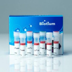
Simplify Your Panel Design with CF® Dyes
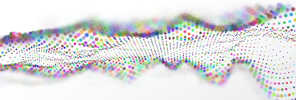
Our industry-leading CF® Dyes boast superior brightness over alternative fluorophores with exceptional signal-to-noise for flow cytometry. In addition, CF® Dyes offer the widest selection of spectrally unique dyes to accommodate highly complex panels.
Learn more about CF® Dyes and learn how CF® Dyes offer advantages for enhanced multiplexing in spectral flow cytometry.
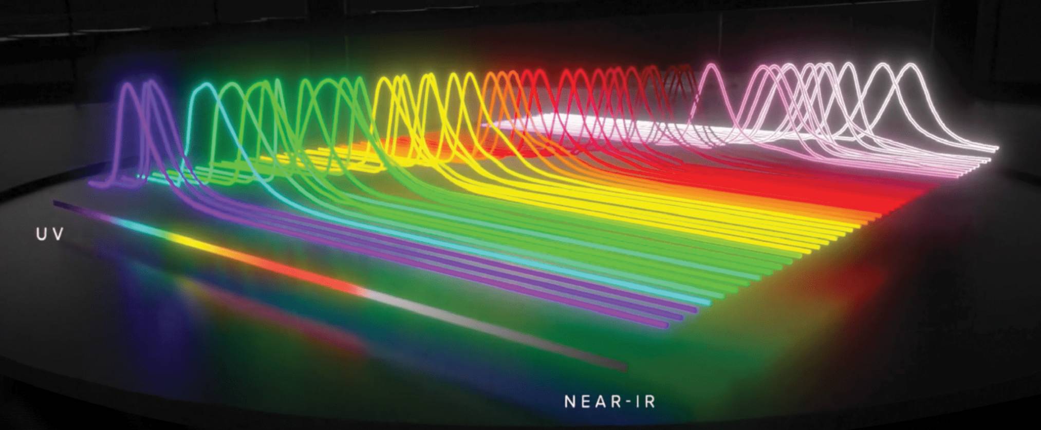
- Available in over 40 colors, offering the greatest coverage across visible and near-IR wavelengths
- Widest selection of spectrally unique dyes to accommodate highly complex panels
- CF® Dye antibody conjugates have superior brightness over alternative fluorophores with exceptional signal-to-noise
Download our CF® Dyes for Flow Cytometry Poster!

Poster
CF® Dyes for Flow Cytometry Poster
Antibodies & Labeling Kits
Sharper Signals, Flexible Panels with Biotium Choice
Biotium Choice fluorescent conjugated primary antibodies are carefully curated and validated in-house to offer exceptional signal-to-noise. Labeled with our advanced CF® Dyes and Astral Leap™ tandem dyes, they are our top-recommended antibodies for flow cytometry and other applications.
Biotium Choice Antibody Features
- Robust, validated clones against common targets
- Developed and optimized for flow cytometry and other applications
- Conjugated to bright, photostable CF® Dyes for superior signal and clarity
- Also available with Astral Leap™ tandem dyes for expanding multiplexing
- New antibody clones and dye conjugates continuously in development
Identify Biotium Choice Antibodies
Look for this symbol next to the product name on product pages and search results.
Also view our selection of Biotium Choice Antibodies for popular immunology targets below.

Biotium Choice Antibodies for Immunology
Explore Our Full Primary Antibody Catalog
- Growing collection of more than 2000 monoclonal antibodies
- Choose from antibodies conjugated to one of 6 bright CF® Dyes or biotin
- Isotype control antibodies available conjugated to the same dyes and labels
- Available BSA-free, ready to use with Mix-n-Stain™ labeling
- Affordable 100 uL sizes available
Browse our assortment of primary antibodies, or search with Antibody Finder to locate specific antibodies based on categories and product attributes.
We also have streptavidin and anti-biotin secondary antibodies available in 21 bright CF® Dyes, R-PE, APC, and PerCP. See the full list of anti-tag secondary antibodies.
Primary Antibodies – sizes and formats
| Format | Concentration | Size |
|---|---|---|
| CF® Dye conjugates (6 colors) | 0.1 mg/mL | 100 uL or 500 uL |
| Biotin conjugates | 0.1 mg/mL | 100 uL or 500 uL |
| Purified, with BSA | 0.2 mg/mL | 100 uL or 500 uL |
| Purified, BSA-free (Mix-n-Stain™ Ready) | 1 mg/mL | 50 uL |
Antibody Labeling Kits
Labeling your own antibodies? Our Mix-n-Stain™ Antibody Labeling Kits dramatically simplify the process of preparing fluorescently-labeled antibodies, offering bright and stable conjugates in only 15 minutes. Each Mix-n-Stain™ kit comes with everything you need to perform the conjugation. Without a separation step, you will have an optimally labeled antibody conjugate ready to be used in any immunofluorescence staining experiment. The resulting conjugates offer comparable performance to antibodies labeled with other commercially available labeling kits. Because the labeling is covalent, the conjugates are stable for long-term storage, and ideal for multi-color imaging and multiplex flow cytometry.
See How Easy the Labeling Process Is in Our Video
- Label with your choice of 32 CF® Dyes in only 15 minutes
- Labeling Kits for FITC, Biotin, and other labels also available
- Stable covalent labeling with no purification required
- Compatible with common antibody stabilizers
- Labeling kits designed for Nanobodies® (i.e., camelid single variable or VHH domains) also available
Live-Or-Dye™ Fixable Viability Staining Kits
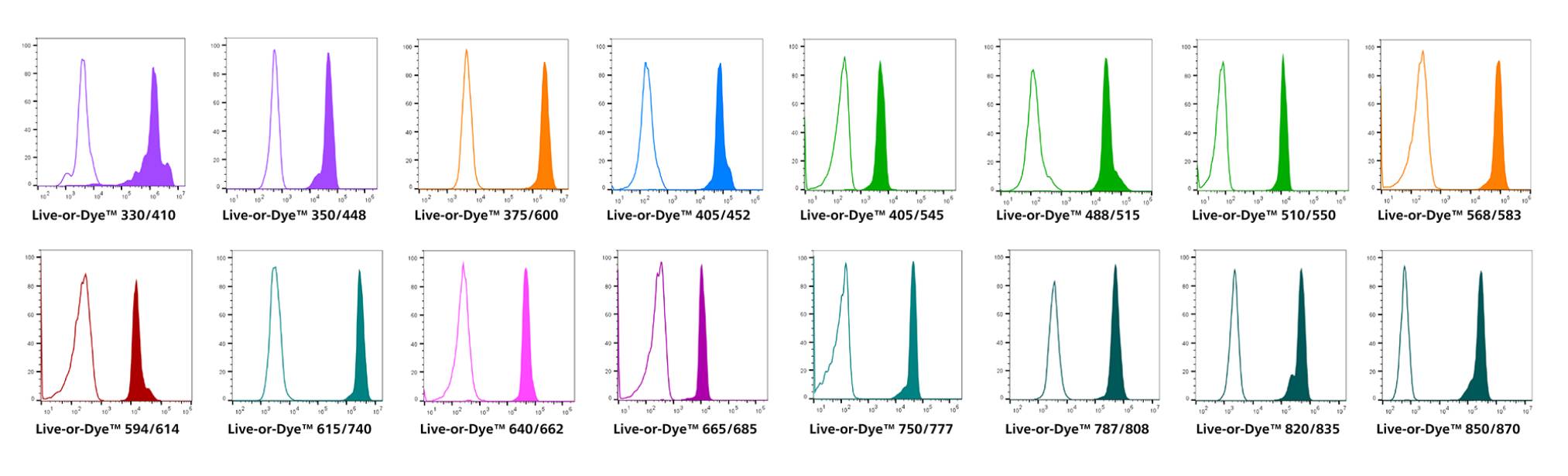
Live-or-Dye™ fixable dead cell stains are cell membrane-impermeant, amine-reactive dyes. The dyes enter dead cells that have compromised membrane integrity and covalently label free amines on intracellular proteins. Live-or-Dye™ labeling is extremely stable, allowing the cells to be fixed and permeabilized without loss of fluorescence or dye transfer between cells. The staining protocol has been optimized to maximize live/dead discrimination with minimal live cell staining, in order to prevent interference with immunostaining. Biotium currently offers a wide selection of 16 Live-or-Dye™ stains spanning the fluorescence spectrum for maximal flexibility in designing multi-color flow panels.
The Best Fixable Dead Cell Stains for Flow Cytometry
- Wide selection of dye colors for easy panel design
- Exceptionally bright dyes for excellent live/dead separation
- No loss of brightness after fixation
- For flow cytometry or fluorescence microscopy
- Dyes designed to fit gaps in spectral flow panels
- The most options for the 633 nm and 808 nm lasers

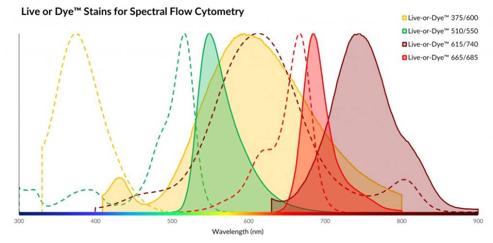
Live-or-Dye™ Stains for Spectral Flow
Spectral flow cytometry is a rapidly growing technology with enhanced multiplexing capabilities over conventional flow cytometry. Where conventional flow cytometer instruments can detect panels with more than a dozen fluorophores, spectral flow cytometers can accommodate panels with upwards of 40 dyes. Biotium offers spectrally unique Live-or-Dye™ stains designed specifically to take advantage of enhanced multiplexing by spectral flow cytometry.
Live-or-Dye™ Sampler Kits
Live-or-Dye™ stains are available in two sampler kits, each containing five different dye colors, for excitation from all of the popular flow cytometry laser lines (see Live-or-Dye™ Sampler Kits table below). The Live-or-Dye™ Sampler Kit, Standard (32016) was designed for use on the most common flow cytometer laser and filter configurations, with dyes excitable by UV, Violet, Blue, Yellow-Green, and Red lasers. The Live-or-Dye™ Sampler Kit, Spectral (32017) was designed for use in spectral scanning flow cytometry. It contains dyes excitable by UV, Violet, Blue, and Red lasers, all of which have been validated on the Cytek® Aurora spectral cytometer, and chosen for their ability to fill the gaps in many spectral flow panels.
Learn more about Live-or-Dye™ Fixable Viability Stains.
View Product Page

Live-or-Dye™ Fixable Viability Staining Kits
Cat. No. | Viability Dye | Compatible lasers (nm) | Optimal detection channels | Notes |
|---|---|---|---|---|
| 32018, 32018-T | Live-or-Dye™ 330/410 | 355, 375 | BUV395 | |
| 32002, 32002-T | Live-or-Dye™ 350/448 | 355, 375 | DAPI | |
| 32014,32014-T | Live-or-Dye™ 375/600 | 355, 375, 405 | Spectral scan, BUV615, BV605 | Developed for spectral cytometry |
| 32003, 32003-T | Live-or-Dye™ 405/452 | 405 | Pacific Blue®, BV421, BV450 | |
| 32009, 32009-T | Live-or-Dye™ 405/545 | 405 | AmCyan, BV510 | |
| 32004, 32004-T | Live-or-Dye™ 488/515 | 488 | FITC | Validated for microscopy |
| 32012, 32012-T | Live-or-Dye™ 510/550 | 488, 532 | Spectral scan | Developed for spectral cytometry |
| 32005, 32005-T | Live-or-Dye™ 568/583 | 488, 532, 561 | PE, PI | Validated for microscopy |
| 32006, 32006-T | Live-or-Dye™ 594/614 | 488, 532, 561 | PI, PE-CF®594, PE-Texas Red® | Validated for microscopy |
| 32015, 32015-T | Live-or-Dye™ 615/740 | 633-640 | Spectral scan, APC-Cy®7 | Developed for spectral cytometry |
| 32007, 32007-T | Live-or-Dye™ 640/662 | 633-640 | APC | Validated for microscopy |
| 32023, 32023-T | Live-or-Dye™ 650/720 | 633-640 | APC-700 | |
| 32013, 32013-T | Live-or-Dye™ 665/685 | 633-640 | Spectral scan, AF700 | Developed for spectral cytometry |
| 32024, 32024-T | Live-or-Dye™ 700/745 | 633-640, 785 | APC-Cy®7 | Our best dye for the APC-Cy®7 channel |
| 32008, 32008-T | Live-or-Dye™ 750/777 | 633-640, 785 | APC-Cy®7, IR840 | |
| 32011, 32011-T | Live-or-Dye™ 787/808 | 785, 808 | APC-Cy®7, IR800 | |
| 32021, 32021-T | Live-or-Dye™ 820/835 | 808 | IR840 | |
| 32022, 32022-T | Live-or-Dye™ 850/870 | 808 | IR885 |
Live-or-Dye™ Sampler Kits
Kit Name | Catalog No. | Included dyes: Ex/Em (Cat No.) | Application |
|---|---|---|---|
| Live-or-Dye™ Fixable Viability Sampler Kit, Standard | 32016 | • 350/448 (32002A) • 405/545 (32009A) • 488/515 (32004A) • 568/583 (32005A) • 640/662 (32007A) | Designed for use on most standard flow cytometry laser and filter configurations |
| Live-or-Dye™ Fixable Viability Sampler Kit, Spectral | 32017 | • 350/448 (32002A) • 375/600 (32014A) • 510/550 (32012A) • 615/740 (32015A) • 665/685 (32013A) | Designed for use in spectral flow cytometry, to fill in gaps between common fluorophores |
Dead Cell Selective Counterstains
Cyanine-based, cell membrane-impermeant, nucleic acid stains, such as Oxazole Blue (PO-PRO™-1), Oxazole Yellow (YO-PRO®-1), and Thiazole Red (TO-PRO®-3), are dead cell-selective stains suitable for flow cytometry. Unlike Live-or-Dye™ Fixable Viability Stains, these dyes are not fixable, but are fluorogenic and highly specific for nucleic acids. This results in bright and sensitive staining localized to the nucleus in dead or dying cells, and staining with these dyes can be done directly in cell culture medium without the need for washing.
View our full selection of nuclear-specific stains suitable for flow cytometry or microscopy.
Product | Equivalent to | Color (Ex/Em) | Catalog No. |
|---|---|---|---|
| Oxazole Blue, 1 mM in DMSO | PO-PRO™-1 | Blue (434/457 nm) | 40091 |
| Oxazole Blue Homodimer, 1 mM in DMSO | POPO™-1 | Blue (433/457 nm) | 40093 |
| Oxazole Yellow, 1 mM in DMSO | YO-PRO®-1 | Green (491/506 nm) | 40089 |
| Oxazole Yellow Homodimer, 1 mM in DMSO | YOYO®-1 | Green (491/508 nm) | 40090 |
| TO Iodide, 1 mM in DMSO | TO-PRO®-1 | Green (515/531 nm) | 40088 |
| Thiazole Orange Homodimer, 1 mM in DMSO | TOTO®-1 | Green (514/531 nm) | 40079 |
| Oxazole Red, 1 mM in DMSO | YO-PRO®-3 | Far-red (613/629 nm) | 40105 |
| Oxazole Red Homodimer, 1 mM in DMSO | YOYO®-3 | Far-red (612/631 nm) | 40106 |
| Thiazole Red, 1 mM in DMSO | TO-PRO®-3 | Far-red (642/657 nm) | 40087 |
| Thiazole Red Homodimer, 1 mM in DMSO | TOTO®-3 | Far-red (642/661 nm) | 40080 |
ViaFluor® Cell Proliferation Kits
Cell Proliferation Monitoring by Flow Cytometry
Cell proliferation dyes diffuse passively into live cells and are used for long-term cell labeling. They are initially non-fluorescent esters, but are converted to fluorescent dyes by intracellular esterases and will covalently react with amine groups on intracellular proteins at the same time, forming fluorescent conjugates that are retained in the cell. Immediately after staining, a single bright fluorescent population will be detected by flow cytometry. Each cell division that occurs after labeling is revealed by the appearance of a successively dimmer fluorescent peak on a flow cytometry histogram. Cell proliferation dyes can be used to track cell divisions in vivo or in vitro. The staining can withstand fixation and permeabilization for subsequent immunostaining.
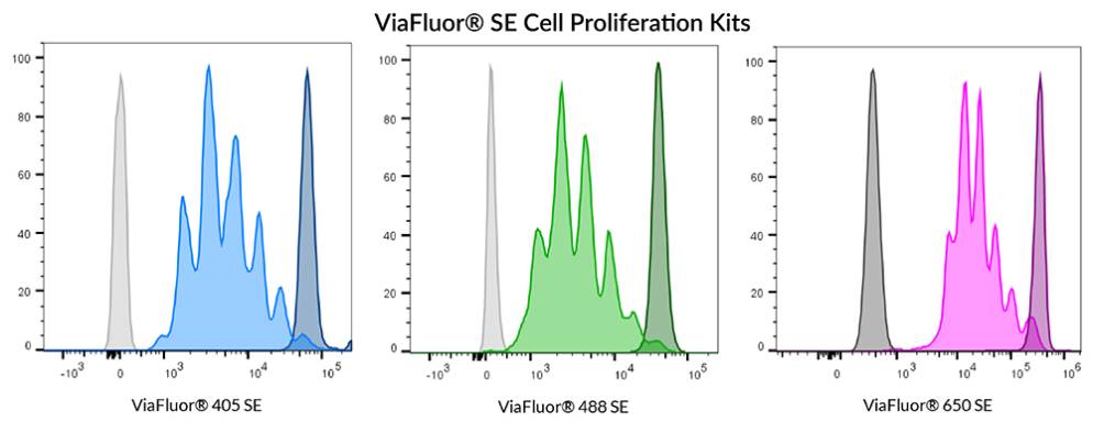
ViaFluor® SE Cell Proliferation Kits
CFSE is widely used to monitor cell proliferation by flow cytometry. Biotium offers CFSE under the tradename ViaFluor® CFSE. However, CFSE has several drawbacks, including leakage from cells, cell toxicity, and bleed-through into the PE and PE-TexasRed® channels.
ViaFluor® SE Cell Proliferation Kits were developed to provide superior cell staining, fixability, and low toxicity. ViaFluor® 405 gives great peaks with no toxicity and behaves identically to CellTrace™ Violet. ViaFluor® 488 and ViaFluor® CFSE are both detected in the FITC channel, but ViaFluor® 488 is a less toxic, less leaky, and more fixable alternative to the classic dye CFSE. It also has less bleed-through into other channels such as PE and PE-Texas-Red®. ViaFluor® 650 was designed as an improved and cost-effective alternative to CellTrace™ Far Red for monitoring cell proliferation in the APC channel.
Learn more about ViaFluor® SE Cell Proliferation Kits.
Cell Division | Catalog No. | Ex/Em (nm) | Flow detection | Features |
|---|---|---|---|---|
| ViaFluor® 405 SE Cell Proliferation Kit | 30068 | 387/446 | Pacific Blue® | • ViaFluor® 405 SE replaces CellTrace™ Violet |
| ViaFluor® 488 SE Cell Proliferation Kit | 30086 | 495/524 | FITC | • ViaFluor® 488 SE is a unique, improved green dye to replace CFSE |
| ViaFluor® 650 SE Cell Proliferation Kit | 30139 | 653/682 | APC | • ViaFluor® 650 SE is an improved and cost-effective alternative to CellTrace™ Far Red |
| ViaFluor® CFSE Cell Proliferation Kit | 30050 | 495/519 | FITC | • Classic cell division tracing dye, see our ViaFluor® 488 SE for an improved alternative |
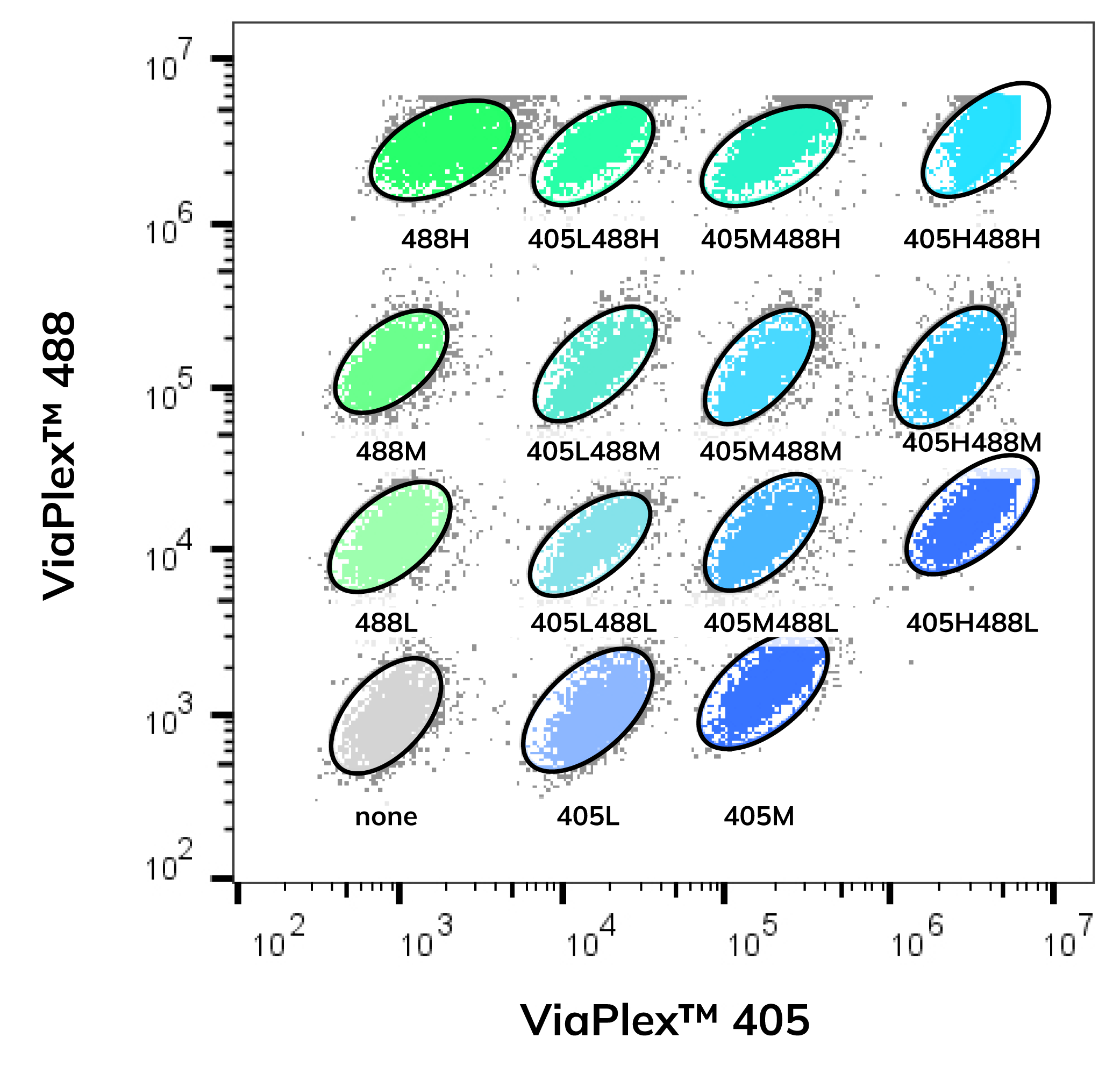
ViaPlex™ 2-Color Cell Barcoding Kits
Analyze 15 Samples in a Single Tube
The ViaPlex™ Cell Barcoding Kit enables convenient and flexible cell barcoding of up to 15 cell populations in a single tube, saving time and reagents. Cells are labeled using covalently bound fluorescent dyes with up to three concentrations per dye. The barcode labeling is compatible with surface and intracellular antibody staining workflows, and barcoding can be performed either before or after cell treatment to accommodate a wide range of experimental designs. Unlike many other barcoding methods, ViaPlex™ labeling does not require fixation or permeabilization, enabling assays on live cells. The covalent dye labeling also remains stable if cells are later fixed and permeabilized for intracellular detection.
ViaPlex™ Advantages
- Saves time and reagent cost
- Multiplex up to 15 samples in one staining reaction; optional 16th barcode via compensation
- Compatible with surface and/or intracellular staining
- Works with live cells; no fixation required
- Covalent dyes stably label cells for clean sample separation
- Uses Pacific Blue® and FITC channels
View Product Page
Cell Cycle Analysis
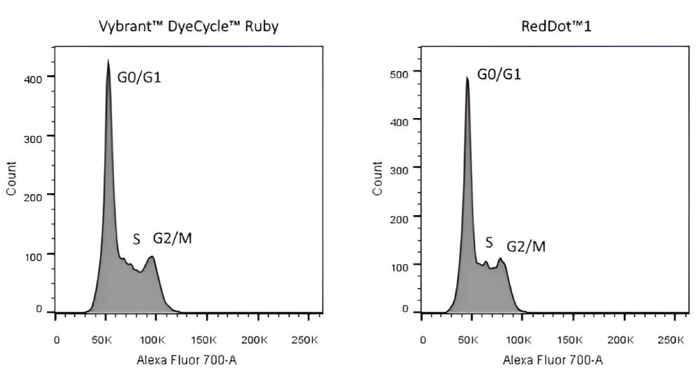
RedDot™1 Far-Red Nuclear Stain
RedDot™1 is a far-red, cell membrane-impermeant, nuclear stain. This live cell stain is similar to DRAQ®5 and can be used for cell cycle analysis by flow cytometry. Also, RedDot™1 cell cycle analysis does not require an RNase step, unlike the classic nuclear stain Propidium Iodide (PI). RedDot™1 can be used as a far-red nuclear counterstain for live cells in microscopy. The dye is highly thermostable and photostable, for convenient handling.
View Product Page
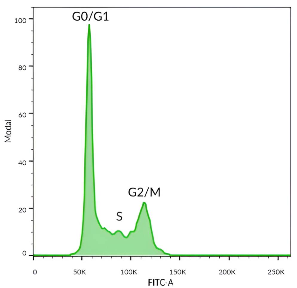
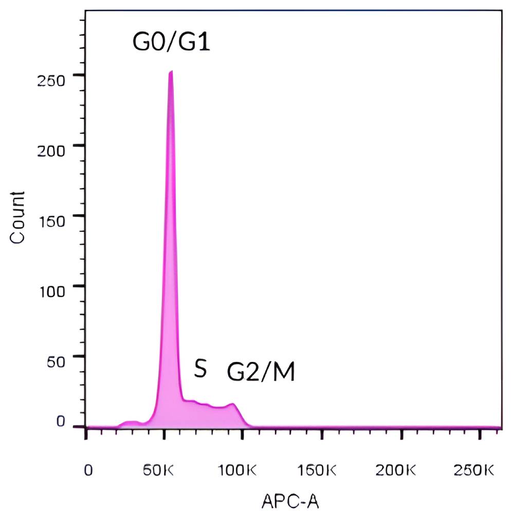
NucSpot® 470 Nuclear Stain
NucSpot® 470 is a cell membrane-impermeant green fluorescent DNA stain for nuclear counterstaining of fixed cells, or selective staining of dead cells.
- Nuclear-specific green counterstain for fixed cells
- Selective detection of dead cells by flow cytometry in the FITC channel
- Perform cell cycle profiling by flow cytometry in fixed/permeabilized cells
- No wash required
View Product Page
NucSpot® Far-Red Nuclear Stain
NucSpot® Far-Red is an improved alternative to the popular flow cytometry dead cell dye 7-AAD. It has red-shifted fluorescence emission compared to 7-AAD, for less bleed-through fluorescence in the PE-Texas Red® channel.
- Improved alternative to 7-AAD with less bleed-through fluorescence in the PE-Texas Red® channel
- Selective detection of dead cells by flow cytometry in the PE-Cy®5 or APC channel
- Perform cell cycle profiling by flow cytometry in fixed/permeabilized cells
- Substitute for 7-AAD in any standard protocol
View Product Page
Dyes for Cell Cycle Analysis | Catalog No. | Live or Fixed Cells? | RNase treatment required? | Color (Ex/Em*) | Features |
|---|---|---|---|---|---|
| NucSpot® 470 Nuclear Stain, 1000X in DMSO | 40083 | Fixed | No | Green (460/546 nm) | • Nuclear-specific green counterstain for fixed cells • Selectively stains dead cells in live cultures • Excellent match for blue LED excitation sources |
| Propidium Iodide, 100 mg | 40016 | Fixed | Yes | Red (530/622 nm) | • Widely used dead cell stain • Can be excited by 488 nm laser line for detection in the PE channel |
| Propidium Iodide, 1 mg/mL in Water | 40017 | ||||
| Propidium Iodide Buffer, 50 ug/mL | 40048 | ||||
| 7-AAD, 1 mg | 40037 | Fixed | No | Far-red (546/647 nm) | • Far-red dye for detection in the PE-Cy®5 channel • Can be excited by the 488 nm or 532 nm laser line |
| 7-AAD, 1 mg/mL solution | 40084 | ||||
| NucSpot® Far-Red, 1000X in DMSO | 40085 | Fixed | No | Far-red (597/667 nm) | • Designed as improved replacement for 7-AAD • For the PE-Cy®5 or APC channel • Less bleed into the PE-Texas Red® channel |
| RedDot™1 Far-Red Nuclear Stain, 200X in Water | 40060 | Live | No | Far-red (662/694 nm) | • For short-term live cell staining (≤4 hours) • Useful for cell number normalization for In Cell Western® • Can be excited at wavelengths between 488 and 647 nm • Detect in Cy®5 or APC channel |
Cy Dye is a registered trademark of Cytiva. In Cell Western is a registered trademarks of LI-COR Inc.
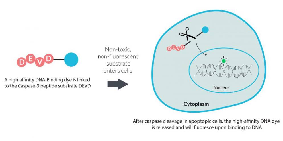
NucView® Caspase-3 Substrates
Live Cell Apoptosis Monitoring
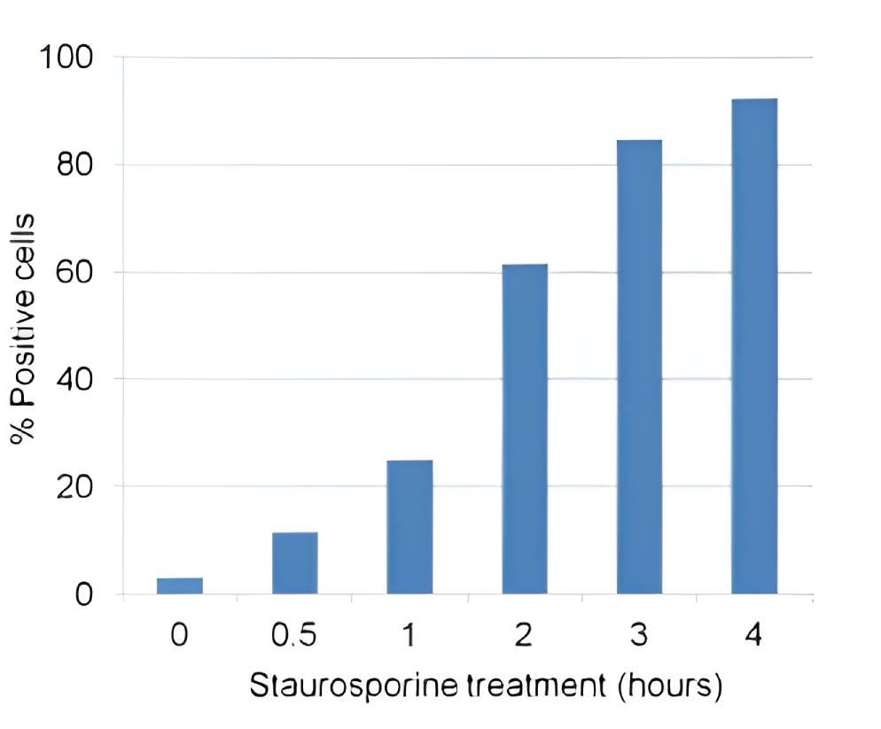
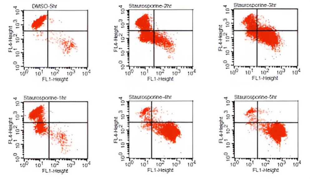
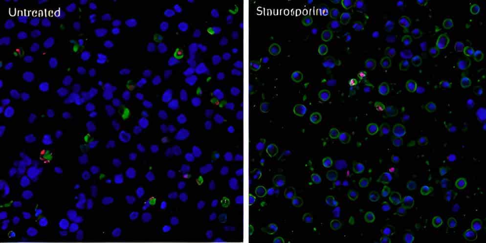
NucView® Caspase-3 Substrates are novel fluorogenic substrates to detect apoptosis in intact cells in real-time. The NucView® caspase-3/7 substrate peptide DEVD is attached to a fluorogenic DNA-binding dye. Before cleavage, the dye is non-fluorescent and unable to bind to DNA. The substrate enters the cell cytoplasm where it is cleaved by caspase-3/7 in apoptotic cells to release the fluorogenic DNA dye, which stains the nucleus. NucView® can be fixed with formaldehyde after staining, but it cannot be used to stain fixed cells or tissues.
NucView® Advantages
- Simple, homogenous assay for endpoint assay or real-time detection in live cells
- Non-toxic, allowing for multi-day experiments
- Detect caspase-3 activity and see apoptotic nuclear morphology
- For flow cytometry, fluorescence microscopy, or live cell imaging
- Blue, green, or red fluorescence
- NucView® 488 validated in 100+ cell types and 200+ publications
NucView® Caspase-3 Substrates and Kits | Catalog No. | Features |
|---|---|---|
| NucView® 405 Caspase-3 Substrate, 1 mM in DMSO | 10405 | Blue fluorescence for flow cytometry in the Pacific Blue® channel or microscopy with the 405 nm laser |
| NucView® 405 Caspase-3 Substrate, 1 mM in PBS | 10407 | NucView® 405 substrate in PBS, for DMSO-sensitive cell types |
| NucView® 488 Caspase-3 Substrate, 1 mM in DMSO | 10402 | Green fluorescent substrate validated in more than 100 cell types and 200 publications |
| NucView® 488 Caspase-3 Substrate, 1 mM in PBS | 10403 | NucView® 488 substrate in PBS, for DMSO-sensitive cell types |
| NucView® 530 Caspase-3 Substrate, 1 mM in DMSO | 10406 | Orange fluorescence for microscopy in the Cy®3 channel or flow cytometry in the R-PE channel |
| NucView® 530 Caspase-3 Substrate, 1 mM in PBS | 10408 | NucView® 530 substrate in PBS, for DMSO-sensitive cell types |
| NucView® 488 and MitoView™ 633 Apotosis Detection Kit | 30062 | NucView® 488 and far-red fluorescent MitoView™ 633 for the Cy®5 channel |
| NucView® 488 and RedDot™ 2 Apoptosis & Necrosis Kit | 30072 | NucView® 488 and far-red dead cell DNA dye RedDot™ 2 for the Cy®5 channel |
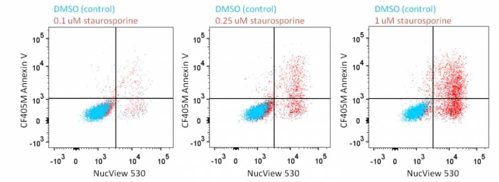
| Dual Apoptosis Assay with NucView® 488 and CF®594 Annexin V | 30067 | NucView® 488 and red fluorescent Annexin V apoptosis probe |
| Dual Apoptosis Assay with NucView® 488 and CF®640R Annexin V | 30073 | NucView® 488 and far-red fluorescent Annexin V apoptosis probe |
Cell Viability Assays
Mitochondrial Membrane Potential
Loss of mitochondrial membrane potential is a hallmark for apoptosis. MitoView™ 633 is a membrane potential-sensitive mitochondrial dye. To detect mitochondrial membrane potential and caspase-3 activity simultaneously, see the NucView®488 and MitoView™633 Apoptosis Assay.
MitoView™ 405 changes localization upon depolarization, and MitoView™ Green is a membrane potential-independent dye that can be used following depolarization, or after fixation.
In healthy cells, JC-1 dye aggregates in mitochondria as a function of membrane potential, resulting in red fluorescence with brightness proportional to the membrane potential. Conversely, in apoptotic and necrotic cells with diminished mitochondrial membrane potential, JC-1 exists in a green fluorescent monomeric form in the cytosol, allowing of cell viability to be assessed by measuring the ratio of red to green fluorescence by flow cytometry or fluorescence microplate reader. Biotium offers Aquaphile™ JC-1, novel formulation of the JC-1 dye with improved solubility and minimal dye aggregation over JC-1 chloride and iodide salts. Rhodamine 123 is a green fluorescent mitochondrial membrane potential dye. Red fluorescent TMRM and TMRE are the preferred dyes for quantitative membrane potential measurements.
Mitochondrial Dyes | Abs/Em | Detection channel | Potential-dependent? | Catalog No. | Size |
|---|---|---|---|---|---|
| MitoView™ 633 | 622/648 nm* | Cy®5, APC* | Yes | 70055-T | 50 ug |
| 70055 | 20x50 ug | ||||
| NucView® 488 & MitoView™ 633 Apoptosis Kit | 500/530 nm (NucView®488) 622/648 nm* (MitoView™ 633) | FITC/Cy®5* | Yes | 30062 | 100 assays |
| MitoView™ 405 | 398/440 nm | DAPI | Partial† | 70070-T | 50 ug |
| 70070 | 20x50 ug | ||||
| MitoView™ Green | 490/523 nm | FITC, GFP | No | 70054-T | 50 ug |
| 70054 | 20x50 ug | ||||
| Aquaphile™ JC-1 | 510/527 nm (cytoplasm) 585/590 nm (mitochondria) | FITC/Cy®3 | Yes | 70076 | 50 uL |
| JC-1 Mitochondrial Membrane Potential Detection Kit | 30001 | 100 assays | |||
| JC-1 Chloride Salt | 70011 | 5 mg | |||
| JC-1 Iodide Salt | 70014 | 5 mg | |||
| Rhodamine 123 | 505/534 nm | FITC | Yes | 70010 | 50 mg |
| TMRE | 548/573 nm | Cy®3 | Yes | 70016 | 25 mg |
| TMRE, 2 mM in DMSO | 70005 | 0.5 mL | |||
| TMRM | 548/573 nm | Cy®3 | Yes | 70017 | 25 mg |
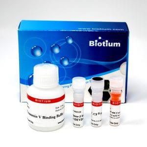
Apoptosis/Necrosis Kits & Annexin V
Annexin V is a protein with a high affinity for phosphatidylserine (PS). During apoptosis, PS is translocated from the inner to the outer leaflet of the plasma membrane, where it can be stained by one of our Annexin V conjugates.
We offer a wide range of staining kits with diverse dye combinations, along with dead cell nucleic acid stains. See the table below for more details on the various combination kits.
Apoptosis and Necrosis Staining Kits | Catalog No. | Features |
|---|---|---|
| CF®488A Annexin V and PI Apoptosis Kit | 30061 | • CF®488A stains apoptotic cells green for the FITC channel • PI stains necrotic cells red for the Cy®3 or PE channels • For microscopy or flow cytometry |
| CF®488A Annexin V and 7-AAD Apoptosis Kit | 30060 | • CF®488A stains apoptotic cells green for the FITC channel • 7-AAD stains necrotic cells red for the PE-Cy®5 or FL3 channels • For flow cytometry |
| Apoptosis & Necrosis Quantitation Kit Plus | 30065 | • CF®488A stains apoptotic cells green for the FITC channel • EthD-III stains dead cells red for the Cy®3 or PE channels • EthD-III has higher affinity and quantum yield than PI or 7-AAD • For microscopy or flow cytometry |
| Apoptotic, Necrotic & Healthy Cells Quantitation Kit Plus | 30066 | • CF®488A stains apoptotic cells green for the FITC channel • EthD-III stains dead cells red for the Cy® channel • Hoechst stains all cell nuclei blue for the DAPI channel • For microscopy |
| NucView®488 and RedDot™2 Apoptosis and Necrosis Kit | 30072 | • NucView®488 stains apoptotic nuclei green for the FITC channel • RedDot™2 stains necrotic cells far-red for the Cy®5 or PE-Cy®5, APC, or FL3 channels • For microscopy or flow cytometry |
| Annexin V Conjugates | Multiple | • Annexin V with our bright and photostable CF® dyes and a selection of other labels |
| Annexin V Conjugates, Azide-Free | Multiple | • Annexin V with our bright and photostable CF® dyes and a selection of other labels • Lyophilized and preservative-free for real-time cell imaging |
| Annexin V Near-IR CF® Dye Conjugates | Multiple | • Annexin V conjugated to our superior Near-IR CF® dyes • Lyophilized and preservative-free for in vivo use |
| Annexin V (His Tag) | 29088 | • Recombinant Annexin V (His Tag) expressed from E.coli |
| Annexin Binding Buffer | 99902 | • Concentrated Annexin Binding Buffer for cell staining |
| NucSpot® 470 Nuclear Stain | 40083 | • Green fluorescent dead cell stain • Also useful as a nuclear-specific counterstain for fixed cells |
| RedDot™2 Far-Red Nuclear Stain | 40061 | • Far-red nuclear stain for dead cells or fixed cells • Detect in the Cy®5 channel |
| Ethidium homodimer III | 40051 | • Red fluorescent dead cell stain • Higher affinity and quantum yield than PI |
| Propidium Iodide | 40017 | • Widely used red fluorescent dead cell stain |
| 7-AAD | 40037 | • Fluorescent dead cell stain for the PE-Cy®5/FL3 channel |
| NucSpot® Far-Red | 40085 | • Designed as an improved replacement for 7-AAD • Less bleed into the PE-Texas Red® channel compared to 7-AAD |
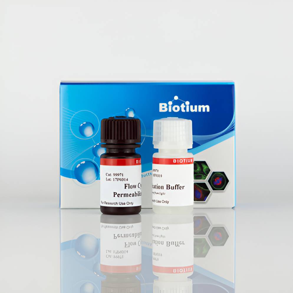
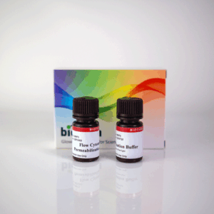
Buffers, Fixatives, & Other Accessories
We offer the following accessory products for your flow cytometry experiments:
- Flow Cytometry Fixation/Permeabilization Kit
- Fixation Buffer
- Permeabilization Buffer
- Permeabilization and Blocking Buffer (5X)
- Mini Cell Scrapers
View all of our flow cytometry accessory products.