Neurotoxins & Fluorescent Toxin-Based Receptor Probes
α-Bungarotoxin (BTX) & Bungarotoxin Conjugates
α-Bungarotoxin is a potent inhibitor for the nicotinic acetylcholine receptor with sub-nanomolar affinity. Fluorescent conjugates of α-bungarotoxin can be used for labeling motor endplates in tissue sections. We offer pure α-bungarotoxin as well as conjugates with a selection of our CF® dyes and other labels. CF® dyes offer advantages in brightness and photostability compared to Alexa Fluor® dyes and other next generation fluorescent dyes. Learn more about CF® Dyes, or download the CF® Dye Selection Guide.

Tetrodotoxin (TTX)
Tetrodotoxin (TTX) reversibly blocks excitable sodium channels and has been a widely used tool for studies of excitable membranes of nerve and muscle cells. Available lyophilized in citrate buffer, or citrate-free.
TTX, With or Without Citrate
| Catalog number | Size | Product |
|---|---|---|
| 00060 | 1 mg | Tetrodotoxin, Citrate-Free |
| 00061 | 1 mg | Tetrodotoxin, with Citrate |
Cholera Toxin Subunit B
Cholera toxin is the symptom-causing toxin produced by the bacteria Vibrio cholerae during cholera infection. The toxin is composed of two subunits, A and B. Subunit A is the toxic enzymatic subunit present in one copy per toxin. Cholera toxin subunit B (CT-B) is the receptor binding subunit that is found as a pentamer in each toxin and is relatively non-toxic, making it useful for cell biological studies.
CT-B has been used as a neuronal tracer and has also been shown to bind to GM1 gangliosides that are found in lipid rafts on the surface of mammalian cells. Therefore, fluorescently labeled conjugates of CT-B have been used as lipid raft markers and endocytic tracers for live imaging or on fixed cells. Cholera Toxin Subunit B is available with a wide selection of our bright and photostable CF® dyes.
α-Bungarotoxin & Conjugates
| Product | Ex/Em | Catalog number | Size |
|---|---|---|---|
| α-Bungarotoxin | N/A | 00010-1 | 1 mg |
| CF®405S α-Bungarotoxin | 404/431 nm | 00002 | 100 ug or 0.5 mg |
| CF®488A α-Bungarotoxin | 490/515 nm | 00005 | |
| CF®543 α-Bungarotoxin | 541/560 nm | 00026 | |
| CF®555 α-Bungarotoxin | 555/565 nm | 00018 | |
| CF®568 α-Bungarotoxin | 562/583 nm | 00006 | |
| CF®594 α-Bungarotoxin | 596/614 nm | 00007 | |
| CF®633 α-Bungarotoxin | 630/650 nm | 00009 | |
| CF®640R α-Bungarotoxin | 642/662 nm | 00004 | |
| CF®680R α-Bungarotoxin | 680/701 nm | 00003 | |
| Biotin-XX-α-Bungarotoxin | N/A | 00017 | 0.5 mg |
| FITC α-Bungarotoxin | 494/518 nm | 00011 00013 | 10×50 ug or 0.5 mg |
| Tetramethylrhodamine α-Bungarotoxin | 553/577 nm | 00012 00017 |
|
| Sulforhodamine-101 (Texas Red®) α-Bungarotoxin | 593/613 nm | 00015 00016 |
CF® Dye Cholera Toxin Conjugates
| Conjugation | Ex/Em | Size | Catalog No. | Dye Features |
|---|---|---|---|---|
| CF®405M | 408/452 nm | 100 ug | 00068 | CF®405M Features |
| CF®488A | 490/515 nm | 100 ug | 00070 | CF®488A Features |
| CF®532 | 527/558 nm | 100 ug | 00074 | CF®532 Features |
| CF®543 | 541/560 nm | 100 ug | 00075 | CF®543 Features |
| CF®568 | 562/583 nm | 100 ug | 00071 | CF®568 Features |
| CF®594 | 593/614 nm | 100 ug | 00072 | CF®594 Features |
| CF®633 | 630/650 nm | 100 ug | 00077 | CF®633 Features |
| CF®640R | 642/662 nm | 100 ug | 00073 | CF®640R Features |
| CF®647 | 650/665 nm | 100 ug | 00069 | CF®647 Features |
| CF®660R | 663/682 nm | 100 ug | 00078 | CF®660R Features |
| CF®680R | 680/701 nm | 100 ug | 00079 | CF®680R Features |
| CF®740 | 742/767 nm | 100 ug | 29127 |
Nerve Terminal Dyes
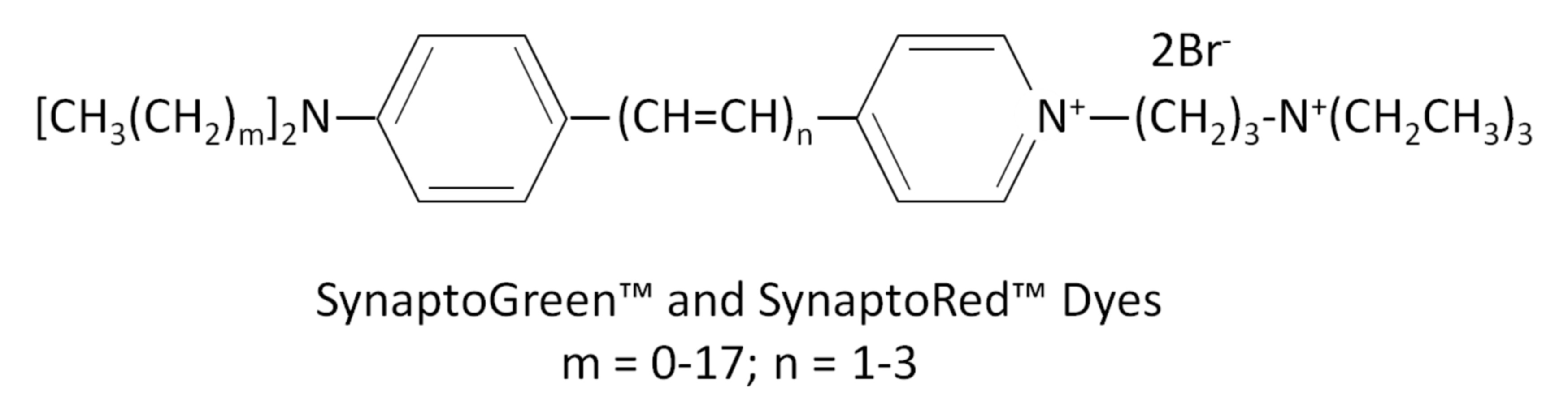
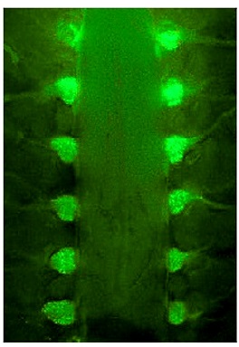
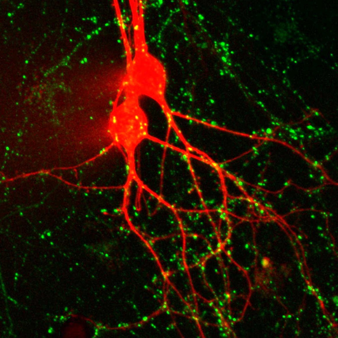
SynaptoRed™ & SynaptoGreen™
SynaptoGreen™ and SynaptoRed™ nerve terminal probes (originally called FM® dyes) are membrane dyes used to trace endocytic vesicles. They are a series of fluorescent cationic styryl dyes developed to follow synaptic activity at neuromuscular junctions or synapses. The dyes label synaptic vesicles in neuronal tissues and cultured neurons in an activity-depending fashion. They also can be used to label endocytic vesicles in other cell types.
Nerve terminal dyes have highly hydrophilic, cationically charged head group at one end with lipophilic tails at the other end. They are virtually non-fluorescent in aqueous solution, but become intensely fluorescent in membranes. Following nerve stimulation, the dye molecules are internalized in newly formed endocytic vesicles. During exocytosis, the dyes are released from the vesicles along with neurotransmitters, causing a decrease in fluorescence signal. As a result, the change in fluorescent intensity reflects the amount of endocytosis/exocytosis or synaptic activity. The rate of fluorescence increase during endocytosis (on-rate), and the rate of fluorescence decrease during exocytosis (off-rate) vary from dye to dye. AM dyes and HM dyes are fixable nerve terminal dyes. After staining with these dyes, cells can be fixed and permeabilized for subsequent immunostaining. See the table below for a list of nerve terminal dyes and their properties.

Background Reducers & Nerve Terminal Staining Kits
A common problem encountered with nerve terminal dyes is background fluorescence due to residual membrane staining, even after extensive washing. To reduce this background fluorescence, we offer three quencher or dye-clearing agents. ADVASEP-7, a sulfonated β-cyclodextrin, forms a water soluble inclusion complex with SynaptoGreen™ C4 that can be removed more effectively by washing. Biotium’s unique quencher, SCAS, reduces background fluorescence as soon as it is added to the preparation without the need for washing. Sulforhodamine 101 quenches SynaptoGreen™ background staining via fluorescent resonance energy transfer (FRET). We offer these reagents as individual products and in kits with dyes and the quencher/dye-clearing agents.
Properties of Nerve Terminal Dyes
| Nerve Terminal Dye | m* | n* | Fixable? | Size | Catalog number | Features |
|---|---|---|---|---|---|---|
| SynaptGreen™ Dyes (Ex/Em ~480/660 nm in membranes) | ||||||
| SynaptoGreen™ C1 | 0 | 1 | No | 5 mg, 5 x 1 mg | 70042, 70043 | • Green nerve terminal probe • Shortest tail for slowest on-rate & fastest off-rate |
| SynaptoGreen™ C2 (equivalent to FM®2-10) | 1 | 1 | No | 70044, 70045 | • Equivalent to FM®2-10 | |
| SynaptoGreen™ C3 | 2 | 1 | No | 70023, 70026 | • Green nerve terminal probe | |
| SynaptoGreen™ C4 (equivalent to FM®1-43) | 3 | 1 | No | 70020, 70022 | • Equivalent to FM®1-43 | |
| SynaptoGreen™ C5 (equivalent to FM®1-84) | 4 | 1 | No | 70046, 70047 | • Equivalent to FM®1-84 | |
| SynaptoGreen™ C18 (equivalent to FM®3-25) | 17 | 1 | No | 70048, 70049 | • Equivalent to FM®3-25 | |
| AM1-43 | 3 | 1 | Yes | 1 mg | 70024 | • Fixable version of SynaptoGreen C4 • Equivalent to FM®1-43FX |
| AM1-44 | 4 | 1 | Yes | 70038 | • Improved fixability over AM1-43 | |
| AM2-10 | 1 | 1 | Yes | 70036 | • Fixable analog of SynaptoGreen™ C2 | |
| AM3-25 | 17 | 1 | Yes | 70051 | • Fixable far-red nerve terminal probe | |
| HM1-43 | 3 | 1 | Yes | 70053 | • Fixable red nerve terminal probe | |
| SynaptoRed Dyes™ (Ex/Em ~510/750 nm in membranes) | ||||||
| SynaptoRed™ C1 | 0 | 3 | No | 5 mg, 5 x 1 mg | 70040, 70041 | • One carbon shorter than SynaptoRed™ C2 |
| SynaptoRed™ C2 (equivalent to FM®4-64) | 1 | 3 | No | 70021, 70027 | • Equivalent to FM®4-64 | |
| SynaptoRed™ C2M** (equivalent to FM®5-95) | 1 | 3 | No | 70019, 70028 | • More water soluble than SynaptoRed™ C2 • Equivalent to FM®5-95 |
|
| AM4-64 | 1 | 3 | Yes | 1 mg | 70025 | • Fixable version of SynaptoRed™ C2 |
| AM4-65 | 3 | 3 | Yes | 70039 | • Fixable version of SynaptoRed™ C2 | |
| AM4-66 | 4 | 3 | Yes | 70050 | • Fixable and spectrally identical to SynaptoRed™ C2 | |
**The positively-charged end of SynaptoRed C2M is a trimethylammonium group.
FM is a registered trademark of Thermo Fisher Scientific.
Nerve Terminal Staining Kits
| Nerve Terminal Staining Kit | Nerve Terminal Dye | Background Reducer | Catalog number |
|---|---|---|---|
| Nerve Terminal Staining Kit I | SynaptoGreen™ C4 (5 x 1 mg ) | ADVASEP-7 (250 mg) | 70030 |
| Nerve Terminal Staining Kit II (A) | AM1-43 (1 mg) | ADVASEP-7 (100 mg) | 70031 |
| Nerve Terminal Staining Kit II (B) | AM1-43 (1 mg) | SCAS (100 mg) | 70031-1 |
| Nerve Terminal Staining Kit III | SynaptoGreen™ C4 (5 x 1 mg) | Sulforhodamine 101 (100 mg) | 70032 |
| Nerve Terminal Staining Kit V | SynaptoRed™ C2 (5 x 1 mg) | ADVASEP-7 (250 mg) | 70034 |
Also see our selection of fast- and slow-responding potentiometric membrane potential dyes.
Anterograde & Retrograde Axonal Tracers
Retrograde Tracers
Hydroxystilbamidine (also called Fluoro-Gold™) has been used extensively as a retrograde tracer for neurons and also a histochemical stain. Fluoro-Gold™ is used for retrograde tracing and dendrite filling.
Cholera toxin subunit B binds GM1 ganglioside in lipid rafts, and is used as a retrograde neuronal tracer. Available with a wide selection of bright and photostable CF® dyes. See Cholera Toxin Conjugates.
Retrograde & Anterograde Tracers
WGA is a glycoprotein-binding lectin that has been used for retrograde and anterograde neuronal tracing. We offer WGA CF® dye conjugates with fluorescence from UV to near-IR, plus HRP. See WGA Conjugates.
Labeled dextran amine can be used for both retrograde and anterograde tracing. CF® dye dextrans are anionic with an aldehyde-fixable free amine group, and are available with a wide selection of colors and a range of molecular weights. See Dextran Conjugates.
Biotin ethylenediamine is equivalent to Neurobiotin™, a useful anterograde and transneuronal tracer.
Anterograde & Retrograde Tracers
| Product | Ex/Em | Catalog number | |
|---|---|---|---|
| Hydroxystilbamidine (Fluoro-Gold™) | 361/536 nm | 80014 | |
| Hydroxystilbamidine (Fluoro-Gold™), 4% in H2O | 361/536 nm | 80023 | |
| Biotin ethylenediamine, hydrobromide (Neurobiotin™) | N/A | 90057 | |
| Biotin ethylenediamine, hydrochloride | N/A | 90075 | |
| Cholera Toxin Conjugates | Choice of 10 colors | ||
| Wheat Germ Agglutinin Conjugates | Choice of 13 colors, plus HRP | ||
| Dextran Conjugates | Choice of 11 colors & 5 molecular weights | ||
Cytosolic Tracers for Cell Morphology & Gap Junctions
Biotin derivatives
Formaldehyde-fixable biocytin and biocytin hydrazide are widely used microinjectable polar tracers. Biocytin has been used as an anterograde tracer and gap junction probe. Biotin derivatives can be detected with labeled streptavidin or anti-biotin antibodies. Biotin ethylenediamine is equivalent to Neurobiotin™, a useful anterograde and transneuronal tracer. We also offer fluorescent CF® dye biotin and biocytin conjugates.
Lucifer Yellow and Related Dyes
Lucifer Yellow is a classic cell-impermeant cytosolic and gap junction dye. We also offer Lucifer Yellow Cadaverine and Lucifer Yellow CH with aldehyde-fixable groups.
Membrane-Permeant Cytosolic Stains
Calcein-AM is a membrane-permeant, non-fluorescent compound that can be loaded into cultured cells by incubation. Once inside the cytoplasm, it is hydrolyzed inside viable cells to release the green fluorescent, membrane impermeant dye calcein, which fills the entire cell. Calcein-AM can be used to assess cell viability, and for short term cytoplasmic labeling.
ViaFluor® SE Cell Proliferation Dyes are membrane-permeant compounds that are hydrolyzed in the cytoplasm to release amine-reactive fluorescent dyes. The staining fills the entire cell, is stable for several days to weeks, and can withstand fixation and permeabilization. Available with blue and green fluorescence.
CF® Dye Hydrazides
Hydrazides are non-toxic, highly water soluble membrane-impermeant tracers that can be used to fill cells by microinjection. See our large selection of bright, photostable CF® dye hydrazides.
Cytosolic Tracers & Fluid Phase Markers
| Product | Features |
|---|---|
| CF® Hydrazides | • Microinjectable, fixable fluorescent tracers • Wide selection of bright & photostable CF® dyes |
| Calcein | • Water soluble green fluorescent tracer for microinjection |
| Calcein AM | • Membrane-permeable compound is hydrolyzed in live cells to release water soluble green fluorescent dye • Uniform intracellular labeling • Dead cells don’t retain dye, for true endpoint viability assay |
| ViaFluor® SE Dyes | • Membrane-permeable compound is hydrolyzed in live cells to release amine-reactive dye • Covalent, fixable intracellular labeling • Choice of blue or green fluorescence |
| Lucifer Yellow Derivatives | • Widely used green fluorescent tracers for neuronal morphology and gap junction studies • Fixable Lucifer Yellow CH and Lucifer Yellow Cadaverine • Lucifer Yellow Cadaverin Biotin-X for secondary detection with streptavidin |
| Hydroxystilbamidine (equivalent to Fluoro-Gold™) | • Widely used UV-excitable green fluorescent retrograde neuronal tracer • Available in solid and 4% solution in water |
| Biotin Derivatives | • Biotin-based tracers for secondary detection with streptavidin |
| CF® Dye Biotin | • Biotin conjugates of our bright & photostable CF® dyes |
| CF® Dye Biocytin | • Aldehyde-fixable biocytin conjugated with bright & photostable CF® dyes. |
Membrane Potential Dyes
Slow-Response Membrane Potential Dyes
Translational (or slow-response) voltage-sensing membrane potential dyes undergo a change in their membrane distribution as a result of changes in membrane potential.
The fluorescence of DiBAC4(3) is enhanced with membrane depolarization. The rate of fluorescence response of the dye is slower than styryl dyes like the ANEPPS dyes (see below), but the fluorescence change is significantly larger.
DiOC2(3) has been used for measuring membrane potential in bacteria. The green fluorescent dye forms red fluorescent aggregates with increasing membrane potential, allowing ratiometric potential measurements.
DiOC5(3) and DiOC6(3) are two of the most widely used carbocyanine dyes for membrane potential measurements.
Tetramethylrhodamine ethyl ester (TMRE) and Tetramethylrhodamine methyl ester (TMRM) can be used for quantitative measurements of membrane potential and mitochondrial membrane potential.
DiO/DPA Membrane Potential Kit
The membrane localization of the fluorescence quencher dipicrylamine (DPA) is a function of the polarity and magnitude of membrane potential. The DiO/DPA system detects cytoplasmic membrane potential changes using the principle of fluorescence resonance energy transfer (FRET). The green fluorescent membrane dye DiO is a “stationary” FRET donor while DPA acts as a mobile FRET acceptor, resulting in a membrane potential-dependent quenching of fluorescence by FRET. The DiO/DPA system has been reported to produce a fluorescence signal change of >56% in HEK-293 cells and >25% in neuronal cultures and brain slices per 100 mV membrane potential change.
Fast-Response Membrane Potential Dyes
Fast-response membrane voltage-sensitive dyes are styryl dyes that undergo changes in fluorescence intensity in response to changes in membrane potential, on the order of 2-10% change in fluorescence per 100 mV. The dyes also undergo spectral shift with changes in membrane potential, allowing ratiometric measurements. Fast response dyes have been used to measure electrical activity in neural and cardiac cells.
Di-4-ANNEPS has been used for studies of human stem cell-derived cardiomyocytes. Di-8-ANNEPPS is more hydrophobic and better retained in the outer leaflet of the plasma membrane than Di-4-ANNEPS, and therefore is more suitable for long-term membrane potential studies. It is also more photostable and less phototoxic than Di-4-ANNEPS.
Di-2-ANEPEQ (also known as JPW 1114) is a highly water soluble fast-responding dye that is usually introduced into cells by microinjection. Di-8-ANEPPQ and Di-12-ANEPPQ are successively more hydrophobic, and have been used for potential-sensitive retrograde labeling of neurons.
RH237, RH414, RH421, and RH795 are fast-responding potentiometric probes generally used for functional imaging of neurons. RH421 exhibits >20% fluorescence change per 100 mV on neuroblastoma cells. These dyes can differ in their physiological effects, for example RH414 causes arterial constriction during cortex staining, while the spectrally similar dye RH795 does not.
Fast-Responding Membrane Potential Dyes
| Product | Ex/Em | Catalog number |
|---|---|---|
| Di-4-ANEPPS | 496/705 nm1 | 61010 |
| Di-8-ANEPPS | 498/713 nm1 | 61012 |
| Di-2-ANEPEQ (JPW 1114) | See Note 2 | 61013 |
| Di-8-ANEPPQ | 61014 | |
| Di-12-ANEPPQ | 61015 | |
| RH237 | 528/782 nm | 61018 |
| RH414 | 532/706 nm | 61016 |
| RH421 | 515/704 nm | 61017 |
| RH795 | 530/712 nm | 61019 |
| DiO/DPA Membrane Potential Kit | 484/501 nm | 30037 |
2Spectrally similar to the ANEPPS dyes.
Amyloid Stains & Neurodegeneration Dyes
Amyloid Stains
Congo Red is commonly used to detect amyloid protein aggregates associated with Alzheimer’s disease, Bovine Spongiform Encephalopathy, and related diseases. The staining can be detected by either colorimetric or fluorescence imaging (Ex/Em 497/614 nm).
DCDAPH is a far-red fluorescent probe (Ex/Em 597/665 nm) with high affinity (Kd=27 nM) to Aβ1-42 aggregates. It has been used for fluorescent staining of brain sections, as well as in vivo small animal near-IR imaging.
Thioflavin T is a cell-permeable benzothiazole dye that exhibits enhanced fluorescence (Ex/Em 450/482 nm) upon binding to amyloid fibrils. Thioflavin T has also been used in histology and for protein characterization.
Amyloid & Neurodegeneration Stains
| Product | Ex/Em | Catalog number |
|---|---|---|
| Congo Red, High Purity | 497/614 nm | 80028 |
| DCDAPH | 597/665 nm | 80030 |
| Thioflavin T, High Purity | 450/482 nm | 80033 |
| PathoGreen™ Histofluorescent Stain, 1000X in Water | 497/520 nm | 80027 |
PathoGreen™ Histofluorescent Stain for Neurodegeneration
PathoGreen™ is an anionic green fluorescent dye functionally similar to Fluoro-Jade® dyes. These dyes stain degenerating neurons and their processes in brain sections and cell culture. The mechanism of staining by this class of dyes has not been determined, but the negatively charged dyes may bind to positively charged polyamines generated in dying neurons.
We also offer a wide selection of cell viability and apoptosis assays.
More Probes for Neuronal Research
Ion indicators
- Fluorescent indictators calcium & other ions
- Chelators, calibrations buffers, & ionophores
Cellular stains
- Live cell stains & fixable stains
- For nuclei, membranes, microtubules, & other organelles
Primary antibodies for neuroscience
- Neuron, glia, microglia, neural crest, & neuroendocrine targets
- Glioma, neuroectodermal & neuroendocrine tumor markers