New Products
 Earth-Friendly Products
 Biotium Choice Antibodies
 Special Offers
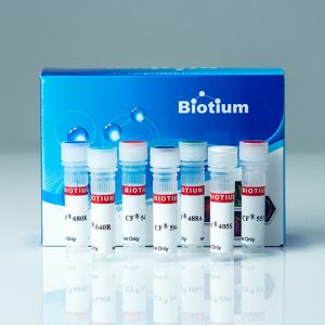
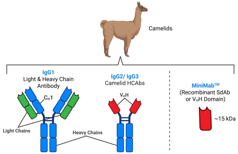
High-Performance Single-Domain Antibodies
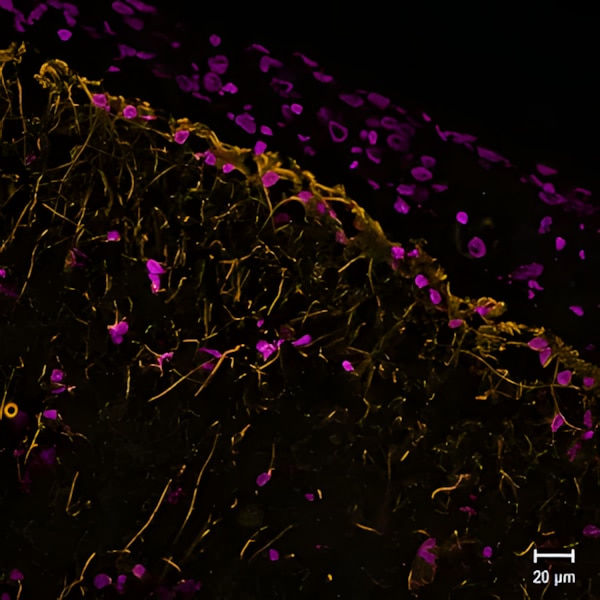
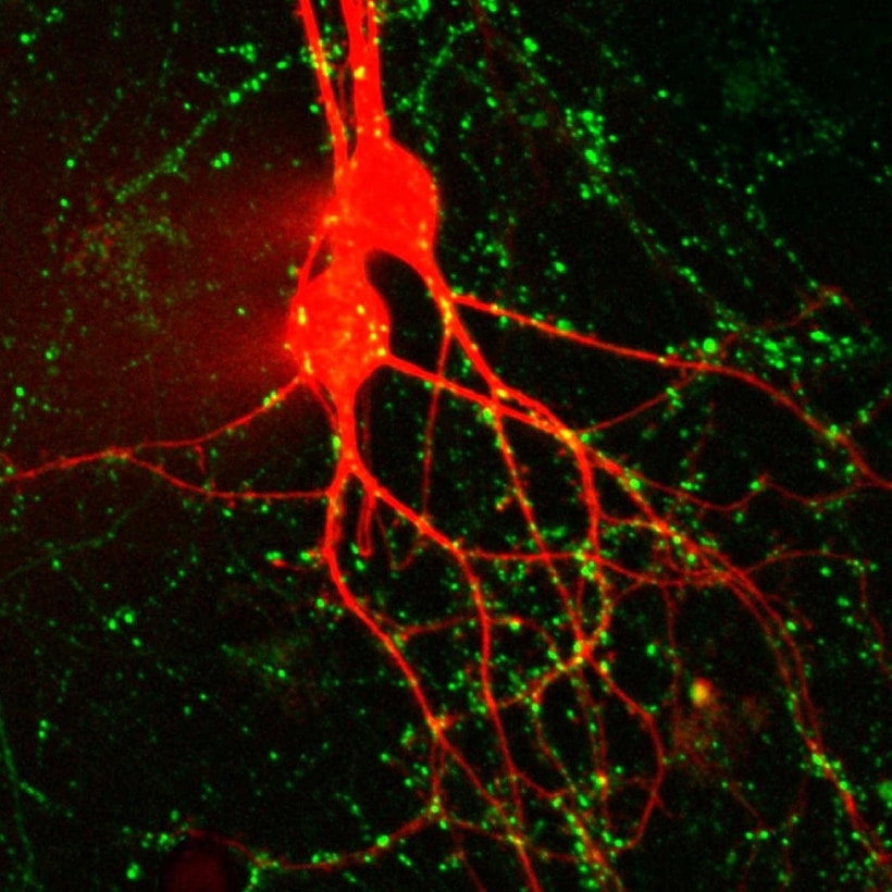
Reveal More with MiniMab™ SdAbs for Neuroscience
MiniMab™ single-domain antibodies (SdAbs) from Biotium are a next-generation labeling tool that leverage the power of SdAbs for superior performance in immunofluorescence and other applications. Their smaller ~15 kDa size relative to conventional antibodies enables deeper tissue penetration, faster staining times, and improved access to tightly packed or masked epitopes, making them ideal for high-resolution and super-resolution imaging. MiniMabs™ also offer minimal epitope-dye displacement, preserving spatial accuracy for techniques like STORM and other super-resolution methods. MiniMab™ SdAbs are available for a selection of neuroscience targets, including GFAP, VGLUT1, and SYT1. See our full selection of available targets below.
Best-in-Class CF® Dyes for IF & Super-Resolution Applications
MiniMab™ SdAbs are available are conjugated to Biotium’s industry-leading CF® Dyes, known for their exceptional brightness, photostability, and wide spectral coverage, including near-infrared options like CF®740. These dye conjugates allow for longer imaging sessions with minimal signal loss, producing consistently sharp and vivid results. With MiniMab™ CF® Dye conjugates, researchers gain a powerful and precise toolset for advancing imaging applications, from routine fluorescence microscopy to cutting-edge nanoscale visualization.
Features of MiniMab™ Single-Domain Antibodies
- Advantages over IgG antibodies: Deeper tissue penetration, higher solubility and stability, and faster staining
- Minimal epitope-dye displacement, perfect for super-resolution imaging
- Specifically developed and optimized for immunofluorescence
- Labeled with bright, photostable CF® Dyes, including near-infrared CF®740
- Available with Biotium’s best-in-class CF® Dyes for STORM
Call for Collaborators
Biotium is seeking collaborators for the evaluation of MiniMabs™ in neuroscience & advanced imaging.
Please contact us at [email protected] if you are interested in testing MiniMab™ single-domain antibodies (SdAbs) for applications focused on neuroscience research and/or advanced imaging. Single-chain variable fragments (scFv) may also be available upon request.

MiniMab™ SdAb Catalog
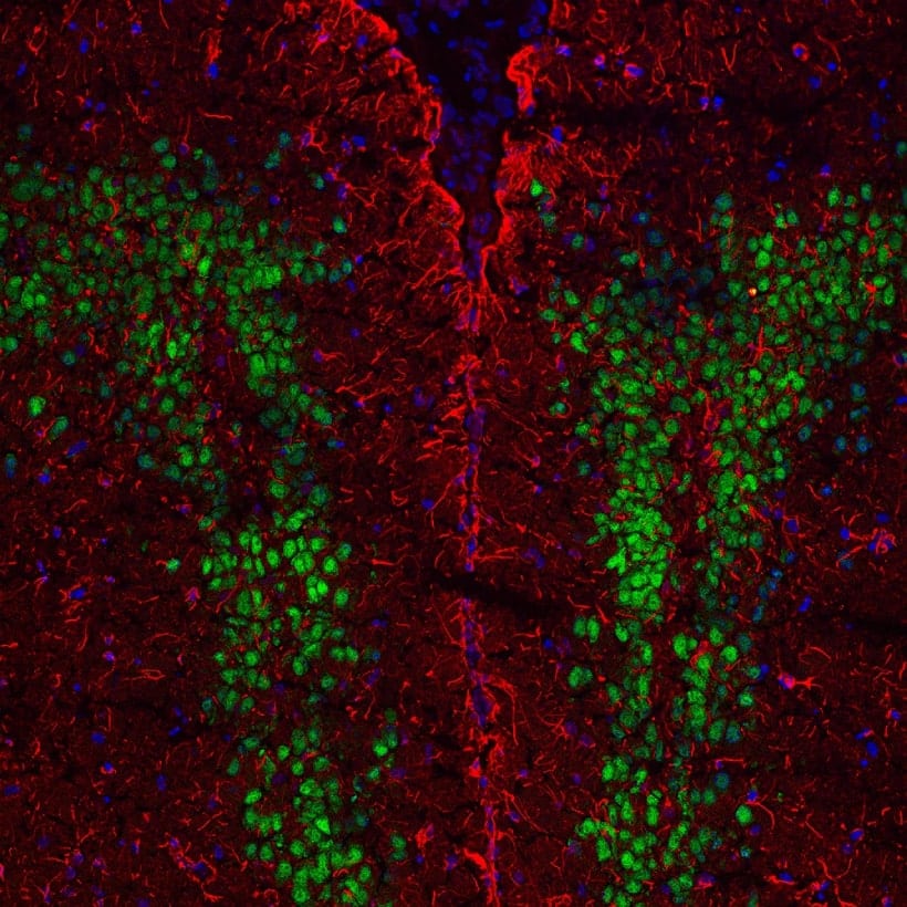
Biotium Choice Neuroscience IgG Antibodies
View Our Full Selection of Neuroscience Antibodies
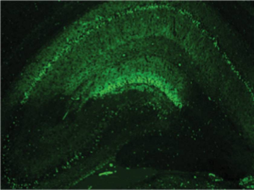
Biotium offers a broad selection of other monoclonal antibodies for neuroscience research. This includes antibody targets for brain tissue, glia, monocytes, neuroendocrine cells, neurons, and neutrophils. The antibodies are available purified, as biotin conjugates, or conjugated to Biotium’s bright and photostable CF® Dyes.
Browse Antibodies for Neuroscience by Target
Brain

Glia
Microglia
Monocytes/Macrophages
Neuroendocrine Cells
Neurons
Neutrophils
NK Cells
Skeletal Muscle
Neurotoxins & Fluorescent Toxin-Based Receptor Probes
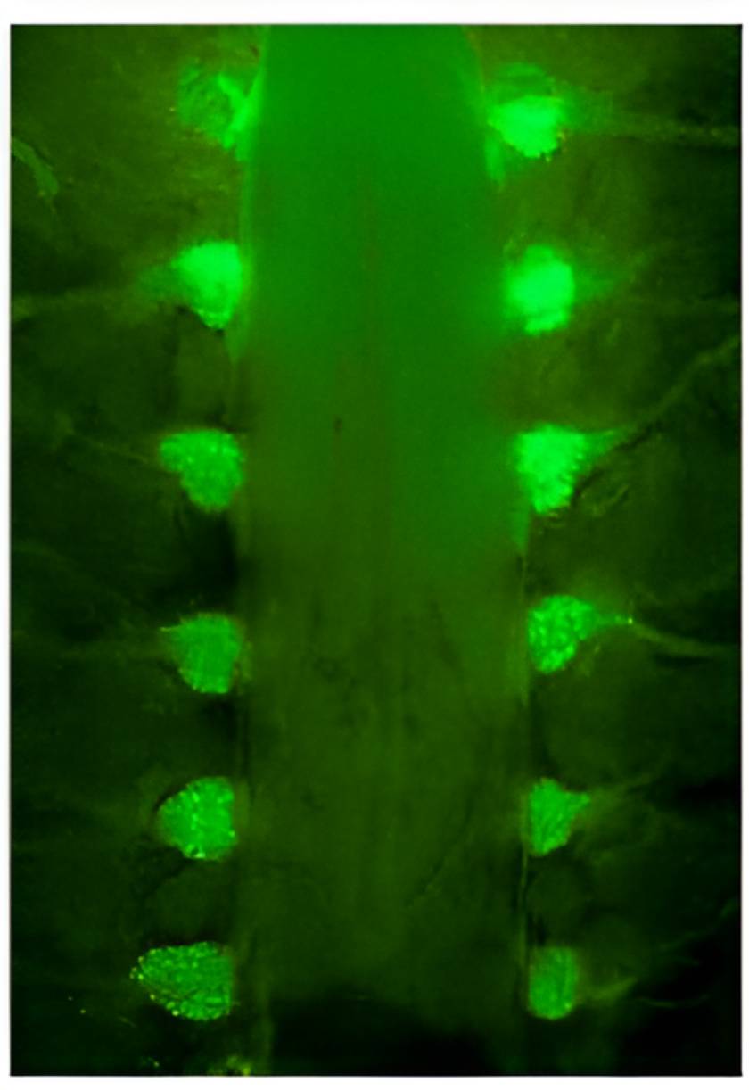
α-Bungarotoxin (BTX) & Bungarotoxin Conjugates
α-Bungarotoxin is a potent inhibitor of nicotinic acetylcholine receptors with sub-nanomolar affinity. Fluorescent conjugates of this protein can label motor endplates in tissue sections. We offer pure α-bungarotoxin and conjugates with CF® Dyes, which provide superior brightness and photostability compared to other commercially available dyes. Learn more about CF® Dyes.
α-Bungarotoxin & Conjugates
| Product | Ex/Em | Catalog number | Size |
|---|---|---|---|
| α-Bungarotoxin | N/A | 00010-1 | 1 mg |
| CF®405S α-Bungarotoxin | 404/431 nm | 00002 | 100 ug or 0.5 mg |
| CF®488A α-Bungarotoxin | 490/515 nm | 00005 | |
| CF®543 α-Bungarotoxin | 541/560 nm | 00026 | |
| CF®555 α-Bungarotoxin | 555/565 nm | 00018 | |
| CF®568 α-Bungarotoxin | 562/583 nm | 00006 | |
| CF®594 α-Bungarotoxin | 596/614 nm | 00007 | |
| CF®633 α-Bungarotoxin | 630/650 nm | 00009 | |
| CF®640R α-Bungarotoxin | 642/662 nm | 00004 | |
| CF®680R α-Bungarotoxin | 680/701 nm | 00003 | |
| Biotin-XX-α-Bungarotoxin | N/A | 00017 | 0.5 mg |
| FITC α-Bungarotoxin | 494/518 nm | 00011 00013 | 10x50 ug or 0.5 mg |
| Tetramethylrhodamine α-Bungarotoxin | 553/577 nm | 00012 00017 |
|
| Sulforhodamine-101 (Texas Red®) α-Bungarotoxin | 593/613 nm | 00015 00016 |
Tetrodotoxin (TTX)
Tetrodotoxin (TTX) reversibly blocks excitable sodium channels and has been a widely used tool for studies of excitable membranes of nerve and muscle cells. Available lyophilized in citrate buffer, or citrate-free.
Cholera Toxin Subunit B
Cholera toxin is the symptom-causing toxin produced by the bacteria Vibrio cholerae during cholera infection. The toxin is composed of two subunits, A and B. Subunit A is the toxic enzymatic subunit present in one copy per toxin. Cholera toxin subunit B (CT-B) is the receptor binding subunit that is found as a pentamer in each toxin and is relatively non-toxic, making it useful for cell biological studies.
CT-B has been used as a neuronal tracer and has also been shown to bind to GM1 gangliosides that are found in lipid rafts on the surface of mammalian cells. Therefore, fluorescently labeled conjugates of CT-B have been used as lipid raft markers and endocytic tracers for live imaging or on fixed cells. Cholera Toxin Subunit B is available with a wide selection of our bright and photostable CF® Dyes.
| Conjugation | Ex/Em | Size | Catalog No. | Dye Features |
|---|---|---|---|---|
| CF®405M | 408/452 nm | 100 ug | 00068 | CF®405M Features |
| CF®488A | 490/515 nm | 100 ug | 00070 | CF®488A Features |
| CF®532 | 527/558 nm | 100 ug | 00074 | CF®532 Features |
| CF®543 | 541/560 nm | 100 ug | 00075 | CF®543 Features |
| CF®568 | 562/583 nm | 100 ug | 00071 | CF®568 Features |
| CF®594 | 593/614 nm | 100 ug | 00072 | CF®594 Features |
| CF®633 | 630/650 nm | 100 ug | 00077 | CF®633 Features |
| CF®640R | 642/662 nm | 100 ug | 00073 | CF®640R Features |
| CF®647 | 650/665 nm | 100 ug | 00069 | CF®647 Features |
| CF®660R | 663/682 nm | 100 ug | 00078 | CF®660R Features |
| CF®680R | 680/701 nm | 100 ug | 00079 | CF®680R Features |
| CF®740 | 742/767 nm | 100 ug | 29127 | CF®740 Features |
| CF®790 | 783/808 nm | 100 ug | 00065 | CF®790 Features |
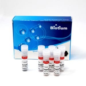
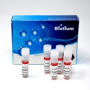
Nerve Terminal Dyes
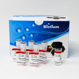
SynaptoRed™ & SynaptoGreen™
SynaptoGreen™ and SynaptoRed™ (formerly FM® dyes) are fluorescent styryl membrane dyes used to trace endocytic vesicles and monitor synaptic activity at neuromuscular junctions or synapses. They label synaptic vesicles in neurons in an activity-dependent manner and can also label endocytic vesicles in other cells.
Non-fluorescent in solution, they become highly fluorescent in membranes. Upon nerve stimulation, the dyes are internalized during endocytosis and released during exocytosis, allowing fluorescence changes to reflect synaptic activity. AM and HM fixable dyes enable post-staining fixation and immunostaining. See the table below for dye properties.


Background Reducers & Nerve Terminal Staining Kits
Nerve terminal dyes can cause persistent background fluorescence from residual membrane staining. Biotium offers three agents to reduce this effect: ADVASEP-7, which forms a water soluble inclusion complex with SynaptoGreen™ C4 that can be removed by washing; SCAS, a fast-acting quencher that reduces background without washing; and sulforhodamine 101, which quenches SynaptoGreen™ via FRET. Available individually or in convenient kits.
Properties of Nerve Terminal Dyes
| Nerve Terminal Dye | m* | n* | Fixable? | Size | Catalog number | Features |
|---|---|---|---|---|---|---|
| SynaptGreen™ Dyes (Ex/Em ~480/660 nm in membranes) | ||||||
| SynaptoGreen™ C1 | 0 | 1 | No | 5 mg, 5 x 1 mg | 70042, 70043 | • Green nerve terminal probe • Shortest tail for slowest on-rate & fastest off-rate |
| SynaptoGreen™ C2 (equivalent to FM®2-10) | 1 | 1 | No | 70044, 70045 | • Equivalent to FM®2-10 | |
| SynaptoGreen™ C3 | 2 | 1 | No | 70023, 70026 | • Green nerve terminal probe | |
| SynaptoGreen™ C4 (equivalent to FM®1-43) | 3 | 1 | No | 70020, 70022 | • Equivalent to FM®1-43 | |
| SynaptoGreen™ C5 (equivalent to FM®1-84) | 4 | 1 | No | 70046, 70047 | • Equivalent to FM®1-84 | |
| SynaptoGreen™ C18 (equivalent to FM®3-25) | 17 | 1 | No | 70048, 70049 | • Equivalent to FM®3-25 | |
| AM1-43 | 3 | 1 | Yes | 1 mg | 70024 | • Fixable version of SynaptoGreen C4 • Equivalent to FM®1-43FX |
| AM1-44 | 4 | 1 | Yes | 70038 | • Improved fixability over AM1-43 | |
| AM2-10 | 1 | 1 | Yes | 70036 | • Fixable analog of SynaptoGreen™ C2 | |
| AM3-25 | 17 | 1 | Yes | 70051 | • Fixable far-red nerve terminal probe | |
| HM1-43 | 3 | 1 | Yes | 70053 | • Fixable red nerve terminal probe | |
| SynaptoRed Dyes™ (Ex/Em ~510/750 nm in membranes) | ||||||
| SynaptoRed™ C1 | 0 | 3 | No | 5 mg, 5 x 1 mg | 70040, 70041 | • One carbon shorter than SynaptoRed™ C2 |
| SynaptoRed™ C2 (equivalent to FM®4-64) | 1 | 3 | No | 70021, 70027 | • Equivalent to FM®4-64 | |
| SynaptoRed™ C2M** (equivalent to FM®5-95) | 1 | 3 | No | 70019, 70028 | • More water soluble than SynaptoRed™ C2 • Equivalent to FM®5-95 |
|
| AM4-64 | 1 | 3 | Yes | 1 mg | 70025 | • Fixable version of SynaptoRed™ C2 |
| AM4-65 | 3 | 3 | Yes | 70039 | • Fixable version of SynaptoRed™ C2 | |
| AM4-66 | 4 | 3 | Yes | 70050 | • Fixable and spectrally identical to SynaptoRed™ C2 | |
**The positively-charged end of SynaptoRed C2M is a trimethylammonium group.
FM is a registered trademark of Thermo Fisher Scientific.
Nerve Terminal Staining Kits
| Nerve Terminal Staining Kit | Nerve Terminal Dye | Background Reducer | Catalog number |
|---|---|---|---|
| Nerve Terminal Staining Kit I | SynaptoGreen™ C4 (5 x 1 mg ) | ADVASEP-7 (250 mg) | 70030 |
| Nerve Terminal Staining Kit II (A) | AM1-43 (1 mg) | ADVASEP-7 (100 mg) | 70031 |
| Nerve Terminal Staining Kit II (B) | AM1-43 (1 mg) | SCAS (100 mg) | 70031-1 |
| Nerve Terminal Staining Kit III | SynaptoGreen™ C4 (5 x 1 mg) | Sulforhodamine 101 (100 mg) | 70032 |
| Nerve Terminal Staining Kit V | SynaptoRed™ C2 (5 x 1 mg) | ADVASEP-7 (250 mg) | 70034 |
Also see our selection of fast- and slow-responding potentiometric membrane potential dyes.
Anterograde & Retrograde Axonal Tracers
Retrograde Tracers
Hydroxystilbamidine (also called Fluoro-Gold™) has been used extensively as a retrograde tracer for neurons and also a histochemical stain. Fluoro-Gold™ is used for retrograde tracing and dendrite filling.
Cholera toxin subunit B binds GM1 ganglioside in lipid rafts, and is used as a retrograde neuronal tracer. Available with a wide selection of bright and photostable CF® Dyes. See Cholera Toxin Conjugates.
Retrograde & Anterograde Tracers
WGA is a glycoprotein-binding lectin that has been used for retrograde and anterograde neuronal tracing. We offer WGA CF® Dye conjugates with fluorescence from UV to near-IR, plus HRP. See WGA Conjugates.
Labeled dextran amine can be used for both retrograde and anterograde tracing. CF® Dye dextrans are anionic with an aldehyde-fixable free amine group, and are available with a wide selection of colors and a range of molecular weights. See Dextran Conjugates.
Biotin ethylenediamine is equivalent to Neurobiotin™, a useful anterograde and transneuronal tracer.
Anterograde & Retrograde Tracers
| Product | Ex/Em | Catalog number | |
|---|---|---|---|
| Hydroxystilbamidine (Fluoro-Gold™) | 361/536 nm | 80014 | |
| Hydroxystilbamidine (Fluoro-Gold™), 4% in H2O | 361/536 nm | 80023 | |
| Biotin ethylenediamine, hydrobromide (Neurobiotin™) | N/A | 90057 | |
| Biotin ethylenediamine, hydrochloride | N/A | 90075 | |
| Cholera Toxin Conjugates | Choice of 10 colors | ||
| Wheat Germ Agglutinin Conjugates | Choice of 13 colors, plus HRP | ||
| Dextran Conjugates | Choice of 11 colors & 5 molecular weights | ||
Cytosolic Tracers for Cell Morphology & Gap Junctions
Biotin Derivatives
Formaldehyde-fixable biocytin and biocytin hydrazide are widely used microinjectable polar tracers. Biocytin has been used as an anterograde tracer and gap junction probe. Biotin derivatives can be detected with labeled streptavidin or anti-biotin antibodies. Biotin ethylenediamine is equivalent to Neurobiotin™, a useful anterograde and transneuronal tracer. We also offer fluorescent CF® Dye biotin and biocytin conjugates.
Lucifer Yellow and Related Dyes
Lucifer Yellow is a classic cell-impermeant cytosolic and gap junction dye. We also offer Lucifer Yellow Cadaverine and Lucifer Yellow CH with aldehyde-fixable groups.
CF® Dye Hydrazides
Hydrazides are non-toxic, highly water-soluble membrane-impermeant tracers that can be used to fill cells by microinjection. See our large selection of bright, photostable CF® Dye hydrazides.
Membrane-Permeant Cytosolic Stains
Calcein-AM is a membrane-permeant, non-fluorescent compound that can be loaded into cultured cells by incubation. Once inside the cytoplasm, it is hydrolyzed inside viable cells to release the green fluorescent, membrane impermeant dye calcein, which fills the entire cell. Calcein-AM can be used to assess cell viability, and for short term cytoplasmic labeling.
ViaFluor® SE Cell Proliferation Dyes are membrane-permeant compounds that are hydrolyzed in the cytoplasm to release amine-reactive fluorescent dyes. The staining fills the entire cell, is stable for several days to weeks, and can withstand fixation and permeabilization. Available with blue and green fluorescence.
Cytosolic Tracers & Fluid Phase Markers
| Product | Features |
|---|---|
| CF® Hydrazides | • Microinjectable, fixable fluorescent tracers • Wide selection of bright & photostable CF® dyes |
| Calcein | • Water soluble green fluorescent tracer for microinjection |
| Calcein AM | • Membrane-permeable compound is hydrolyzed in live cells to release water soluble green fluorescent dye • Uniform intracellular labeling • Dead cells don't retain dye, for true endpoint viability assay |
| ViaFluor® SE Dyes | • Membrane-permeable compound is hydrolyzed in live cells to release amine-reactive dye • Covalent, fixable intracellular labeling • Choice of blue or green fluorescence |
| Lucifer Yellow Derivatives | • Widely used green fluorescent tracers for neuronal morphology and gap junction studies • Fixable Lucifer Yellow CH and Lucifer Yellow Cadaverine • Lucifer Yellow Cadaverin Biotin-X for secondary detection with streptavidin |
| Hydroxystilbamidine (equivalent to Fluoro-Gold™) | • Widely used UV-excitable green fluorescent retrograde neuronal tracer • Available in solid and 4% solution in water |
| Biotin Derivatives | • Biotin-based tracers for secondary detection with streptavidin |
| CF® Dye Biotin | • Biotin conjugates of our bright & photostable CF® dyes |
| CF® Dye Biocytin | • Aldehyde-fixable biocytin conjugated with bright & photostable CF® dyes. |
Membrane Potential Dyes
Slow-Response Membrane Potential Dyes
Translational (slow-response) voltage-sensitive dyes change membrane distribution with shifts in membrane potential. DiBAC4(3) shows enhanced fluorescence upon depolarization and, while slower than styryl dyes like ANEPPS, offers a larger signal change. DiOC2(3) is used in bacteria; it forms red fluorescent aggregates as membrane potential increases, enabling ratiometric measurements. DiOC5(3) and DiOC6(3) are widely used carbocyanine dyes for membrane potential. TMRE and TMRM allow quantitative assessment of overall and mitochondrial membrane potential.
DiO/DPA Membrane Potential Kit
The membrane localization of the fluorescence quencher dipicrylamine (DPA) is a function of the polarity and magnitude of membrane potential. The DiO/DPA system detects cytoplasmic membrane potential changes using the principle of fluorescence resonance energy transfer (FRET). The green fluorescent membrane dye DiO is a “stationary” FRET donor while DPA acts as a mobile FRET acceptor, resulting in a membrane potential-dependent quenching of fluorescence by FRET. The DiO/DPA system has been reported to produce a fluorescence signal change of >56% in HEK-293 cells and >25% in neuronal cultures and brain slices per 100 mV membrane potential change.
Fast-Response Membrane Potential Dyes
Fast-response voltage-sensitive styryl dyes change fluorescence intensity (~2–10% per 100 mV) and can show spectral shifts for ratiometric measurements. They are used to monitor electrical activity in neural and cardiac cells. Di-4-ANNEPS and Di-8-ANNEPS are used in cardiomyocyte studies; Di-8-ANNEPS is more photostable, membrane-retentive, photostable and less phototoxic than Di-4-ANNEPS. Di-2-ANEPEQ (also known as JPW 1114) is water-soluble and typically microinjected. Di-8/12-ANEPPQ are more hydrophobic and used for retrograde labeling. RH-series dyes (e.g., RH237, RH421) are used for neuronal imaging; RH421 shows >20% signal change per 100 mV on neuroblastoma cells. Physiological effects vary, for example RH414 causes arterial constriction while RH795 does not.
Slow-Responding Membrane Potential Dyes
Fast-Responding Membrane Potential Dyes
| Product | Ex/Em | Catalog number |
|---|---|---|
| Di-4-ANEPPS | 496/705 nm1 | 61010 |
| Di-8-ANEPPS | 498/713 nm1 | 61012 |
| Di-2-ANEPEQ (JPW 1114) | See Note 2 | 61013 |
| Di-8-ANEPPQ | 61014 | |
| Di-12-ANEPPQ | 61015 | |
| RH237 | 528/782 nm | 61018 |
| RH414 | 532/706 nm | 61016 |
| RH421 | 515/704 nm | 61017 |
| RH795 | 530/712 nm | 61019 |
| DiO/DPA Membrane Potential Kit | 484/501 nm | 30037 |
2Spectrally similar to the ANEPPS dyes.
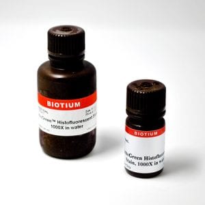
Amyloid Stains & Neurodegeneration Dyes
PathoGreen™ Stain for Neurodegeneration
PathoGreen™ is an anionic green fluorescent dye functionally similar to Fluoro-Jade® dyes. These dyes stain degenerating neurons and their processes in brain sections and cell culture. The mechanism of staining by this class of dyes has not been determined, but the negatively charged dyes may bind to positively charged polyamines generated in dying neurons.
We also offer a wide selection of cell viability and apoptosis assays.
Amyloid Stains
Congo Red is commonly used to detect amyloid protein aggregates associated with Alzheimer’s disease, Bovine Spongiform Encephalopathy, and related diseases. The staining can be detected by either colorimetric or fluorescence imaging (Ex/Em 497/614 nm).
DCDAPH is a far-red fluorescent probe (Ex/Em 597/665 nm) with high affinity (Kd=27 nM) to Aβ1-42 aggregates. It has been used for fluorescent staining of brain sections, as well as in vivo small animal near-IR imaging.
Thioflavin T is a cell-permeant benzothiazole dye that exhibits enhanced fluorescence (Ex/Em 450/482 nm) upon binding to amyloid fibrils. Thioflavin T has also been used in histology and for protein characterization.
Amyloid & Neurodegeneration Stains
| Product | Ex/Em | Catalog number |
|---|---|---|
| Congo Red, High Purity | 497/614 nm | 80028 |
| DCDAPH | 597/665 nm | 80030 |
| Thioflavin T, High Purity | 450/482 nm | 80033 |
| PathoGreen™ Histofluorescent Stain, 1000X in Water | 497/520 nm | 80027 |