New Products
 Earth-Friendly Products
 Biotium Choice Antibodies
 Special Offers
Live Cell Quantitation Assays for Microplate Reader
Absorbance-Based Cell Quantitation Assays
MTT, XTT, and resazurin are reduced by active mitochondria to yield colored products, and thus are useful for assaying cell viability and quantitating cells. MTT and XTT are reduced to colored formazin salts that can be measured by absorbance. Resazurin is reduced to form resorufin, which can be measured by either absorbance or fluorescence. These are easy-to-use homogeneous assays, but they have the disadvantage of requiring several hours for development. In addition, drugs or compounds with redox activity, like antioxidants, can interfere with the assays by directly affecting substrate reduction. MTT assay requires cell lysis before the absorbance can be measured. XTT and resazurin do not require cell lysis, allowing measurements to be made at multiple timepoints.
Fluorescence-Based Cell Quantitation Assays
Absorbance Assays
- Quantitate cells based on their metabolic activity
- Simple, easy-to-use, homogeneous assays
- Usually require several hours of development time
- Drugs or compounds with redox activity can interfere with assays
Fluorescence Assays
- Fluorescence-based assays for microplate reader
- Use Resazurin to quantitate cells based on metabolic activity
- Calcein-AM measures cellular enzymatic activity and membrane integrity
- Highly sensitive, simple, and rapid
Viability Assays for Microplate Reader | Catalog No. | Detection | Features |
|---|---|---|---|
| MTT Cell Viability Assay Kit | 30006 | Absorbance at 570 nm | • Substrates reduced by mitochondrial activity to form colored formazen salts • Widely used, simple, homogeneous assays • MTT requires cell lysis before detection, XTT does not |
| XTT Cell Viability Assay Kit | 30007 | Absorbance at 475 nm | |
| Resazurin Cell Viability Assay Kit | 30025 | Absorbance at 570 nm or Fluorescence at 571/585 nm | • Simple, homogeneous assay for mitochondrial metabolic activity • Measure absorbance or fluorescence without cell lysis • Equivalent to alamarBlue® or CellTiter-Blue® at lower cost |
| Calcein-AM Cell Viability Assay Kit | 30026 | Fluorescence at 485/530 nm | • True endpoint assay for cell viability • Measures cellular enzymatic activity and membrane integrity • Simple, rapid assay |
alamarBlue is a registered trademark of Trek Biosystems. CellTiter-Blue is a registered trademark of Promega Corporation.
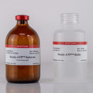
Luminescence-Based Cell Quantitation
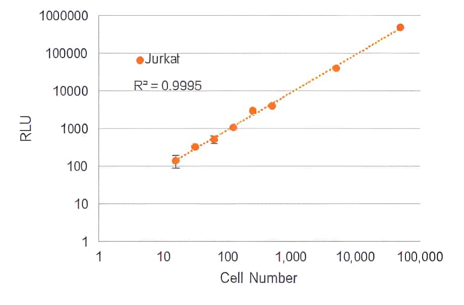
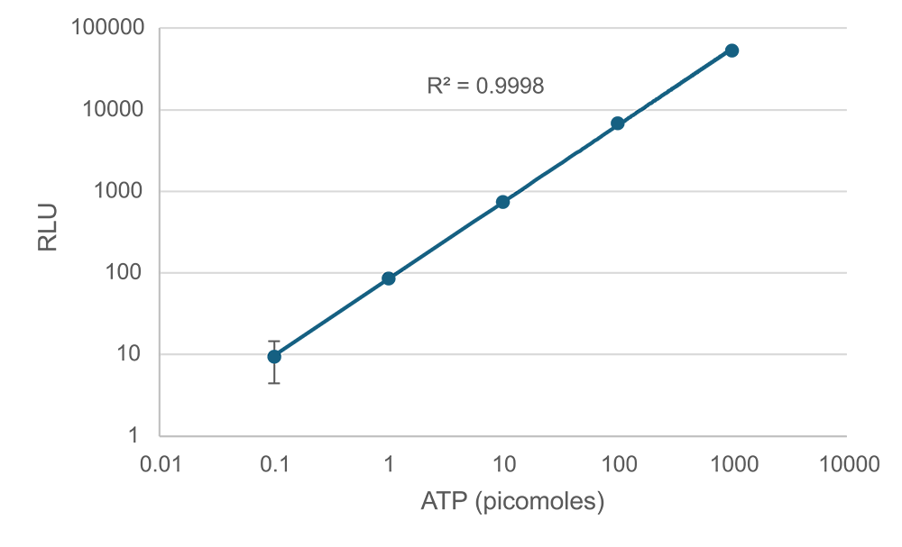
Steady-ATP™ HTS Cell Viability Assay
Steady-ATP™ HTS Viability Assay Kit allows highly sensitive quantitation of cellular ATP levels for viability assessment, with the ability to detect as low as 16 cells per well in a 384-well plate. The assay offers a robust signal half-life of over 5 hours, and a linear signal of 3-4 orders of magnitude. The procedure is designed with convenience in mind, requiring only a single addition of the homogeneous Steady-ATP™ Assay Mix directly to cells in cultured medium, with no washing or medium exchange required.
- Exceptional sensitivity, as low as 16 cells/well in a 384-well plate
- Convenient single-step assay mix
- Compatible for use with high-throughput screening platforms
- Excellent linearity, over 3-4 orders of magnitude
- Stable signal, with a half-life over 5 hours
- Comparable performance to CellTiter-Glo® at a lower cost
View Product Page

Cell Division Tracking by Flow Cytometry
ViaFluor® SE Cell Proliferation Kits
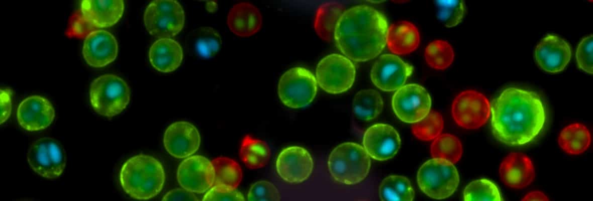
ViaFluor® SE Cell Proliferation Kits can be used to monitor cell division by flow cytometry. ViaFluor® SE dyes are membrane-permeant compounds that are initially non-fluorescent esters, but are converted to fluorescent dyes by intracellular esterases and will covalently react with amine groups on intracellular proteins at the same time, forming fluorescent conjugates that are retained in the cell. Immediately after staining, a single bright fluorescent population will be detected by flow cytometry.
With each cell division, daughter cells inherit roughly half of the fluorescent label, allowing the number of cell divisions that occur after labeling to be detected by the appearance of successively dimmer fluorescent peaks on a flow cytometry histogram compared to cells analyzed immediately after staining. Cell proliferation dyes can be used to track cell divisions in vivo or in vitro. The staining also can withstand fixation and permeabilization for subsequent immunostaining.

Cell Division | Catalog No. | Ex/Em (nm) | Flow detection | Features |
|---|---|---|---|---|
| ViaFluor® 405 SE Cell Proliferation Kit | 30068 | 387/446 | Pacific Blue® | • ViaFluor® 405 SE replaces CellTrace™ Violet |
| ViaFluor® 488 SE Cell Proliferation Kit | 30086 | 495/524 | FITC | • ViaFluor® 488 SE is a unique, improved green dye to replace CFSE |
| ViaFluor® 650 SE Cell Proliferation Kit | 30139 | 653/682 | APC | • ViaFluor® 650 SE is an improved and cost-effective alternative to CellTrace™ Far Red |
| ViaFluor® CFSE Cell Proliferation Kit | 30050 | 495/519 | FITC | • Classic cell division tracing dye, see our ViaFluor® 488 SE for an improved alternative |
Pacific Blue and Texas Red are registered trademarks of Thermo Fisher Scientific.
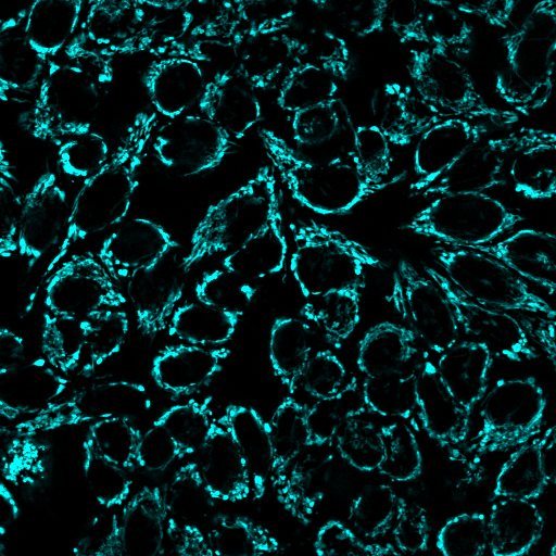
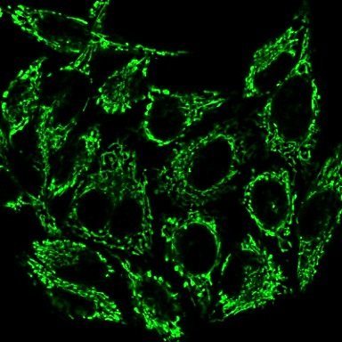
Mitochondrial Membrane Potential Dyes
MitoView™ Dyes
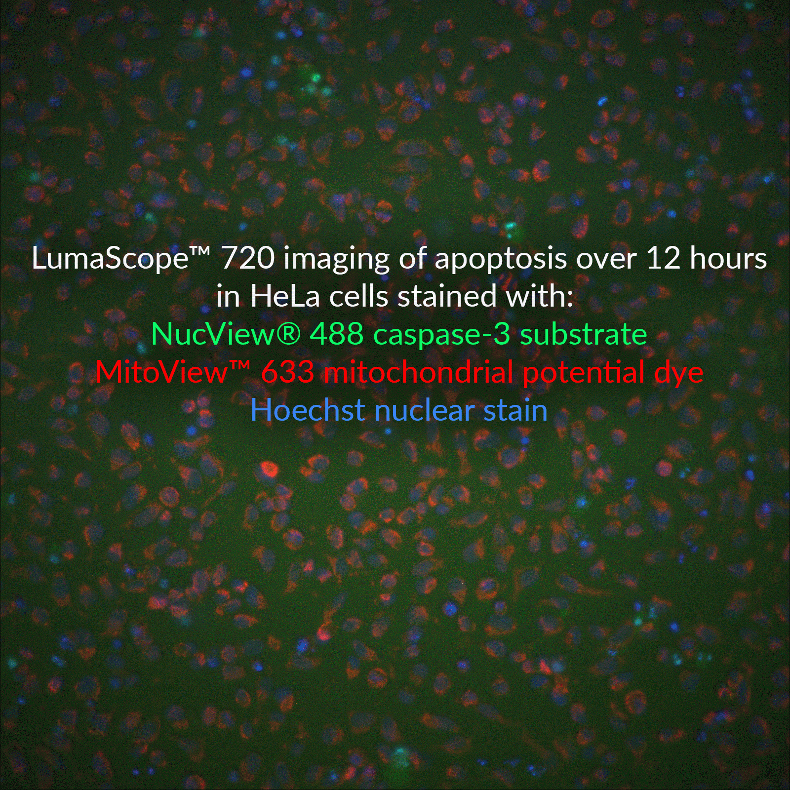
Loss of mitochondrial membrane potential is a hallmark for apoptosis, and MitoView™ 633 is a membrane potential-sensitive stain. For detection of membrane potential and caspase-3 activity, see the NucView®488 and MitoView™633 Apoptosis Assay.
MitoView™ 405 changes localization upon mitochondrial depolarization, and MitoView™ Green is a membrane potential-independent dye that can be used to image mitochondria after depolarization or fixation.
Classic Mitochondrial Membrane Potential Dyes
In healthy cells, JC-1 dye aggregates in mitochondria as a function of membrane potential, resulting in red fluorescence with brightness proportional to the membrane potential. Conversely, in apoptotic and necrotic cells with diminished mitochondrial membrane potential, JC-1 exists in a green fluorescent monomeric form in the cytosol, allowing of cell viability to be assessed by measuring the ratio of red to green fluorescence by flow cytometry or fluorescence microplate reader. Rhodamine 123 is a green fluorescent mitochondrial membrane potential dye. Red fluorescent TMRM and TMRE are the preferred dyes for quantitative membrane potential measurements.
Mitochondrial Dyes | Abs/Em | Detection channel | Potential-dependent? | Catalog No. | Size |
|---|---|---|---|---|---|
| MitoView™ 633 | 622/648 nm* | Cy®5, APC* | Yes | 70055-T | 50 ug |
| 70055 | 20x50 ug | ||||
| NucView® 488 & MitoView™ 633 Apoptosis Kit | 500/530 nm (NucView®488) 622/648 nm* (MitoView™ 633) | FITC/Cy®5* | Yes | 30062 | 100 assays |
| MitoView™ 405 | 398/440 nm | DAPI | Partial† | 70070-T | 50 ug |
| 70070 | 20x50 ug | ||||
| MitoView™ Green | 490/523 nm | FITC, GFP | No | 70054-T | 50 ug |
| 70054 | 20x50 ug | ||||
| Aquaphile™ JC-1 | 510/527 nm (cytoplasm) 585/590 nm (mitochondria) | FITC/Cy®3 | Yes | 70076 | 50 uL |
| JC-1 Mitochondrial Membrane Potential Detection Kit | 30001 | 100 assays | |||
| JC-1 Chloride Salt | 70011 | 5 mg | |||
| JC-1 Iodide Salt | 70014 | 5 mg | |||
| Rhodamine 123 | 505/534 nm | FITC | Yes | 70010 | 50 mg |
| TMRE | 548/573 nm | Cy®3 | Yes | 70016 | 25 mg |
| TMRE, 2 mM in DMSO | 70005 | 0.5 mL | |||
| TMRM | 548/573 nm | Cy®3 | Yes | 70017 | 25 mg |
Live & Dead Cell Staining
Viability/Cytotoxicity Assay Kit
View Product Page
Dead Cell Nucleic Acid Stains
In general, dead cell nucleic acid dyes cannot be fixed after staining, because the dyes do not become cross-linked to DNA. After fixation, they are free to diffuse from dead cells to live cells (which become permeabilized during fixation), resulting in poor discrimination between live and dead cell populations. For fixable dead cell stains, see our Live-or-Dye™ product line, including the nuclear-specific Live-or-Dye NucFix™ Red.
Dead Cell Nucleic Acid Stains | Catalog No. | Color (Ex/Em*) | Features |
|---|---|---|---|
| Oxazole Blue, 1 mM in DMSO | 40091 | Blue (434/457 nm) | • Blue cell-impermeant dye • Selectively stains early apoptotic cells • Equivalent to PO-PRO™-1 Iodide |
| Oxazole Blue Homodimer, 1 mM in DMSO | 40093 | Blue (433/457 nm) | • Blue cell-impermeant dye • Equivalent to POPO™-1 Iodide |
| NucSpot® 470, 1000X in DMSO | 40083 | Green (460/546 nm) | • Green cell-impermeant dye • Nuclear-specific counterstain in fixed cells • Useful for cell cycle analysis in fixed cells • Excellent match for blue LED excitation sources |
| Oxazole Yellow, 1 mM in DMSO | 40089 | Green (491/506 nm) | • Green cell-impermeant dye • Selectively stains early apoptotic cells • Equivalent to YO-PRO®-1 Iodide |
| Oxazole Yellow Homodimer, 1 mM in DMSO | 40090 | Green (491/508 nm) | • Green cell-impermeant dye • Equivalent to YOYO®-1 Iodide |
| TO Iodide, 1 mM in DMSO | 40088 | Green (515/531 nm) | • Green cell-impermeant dye • Equivalent to TO-PRO®-1 Iodide |
| Thiazole Orange Homodimer, 1 mM in DMSO | 40079 | Green (514/531 nm) | • High affinity dimeric cyanine dye • Dead cell stain and electrophoresis dye • Equivalent to TOTO®-1 Iodide |
| NucSpot® 555/570 Nuclear Stain | 41033 | Red (559/566 nm) | • Red cell-impermeant dye for the Cy®3 or PE channels • Nuclear-specific counterstain in fixed cells |
| NucSpot® 568/580 Nuclear Stain | 41036 | Red (572/583 nm) | • Red cell-impermeant dye for the Cy®3 or PE channels • Nuclear-specific counterstain in fixed cells • Suitable for multi-day live cell imaging |
| NucSpot® 594/615 Nuclear Stain | 41037 | Red (603/613 nm) | • Red cell-impermeant dye for the Texas Red® or PE-Texas Red® channels • Nuclear-specific counterstain in fixed cells • Suitable for multi-day live cell imaging |
| Propidium Iodide | 40016, 40017, 40048 | Red (530/622 nm) | • Widely used dead cell stain • Can be excited by 488 nm laser line for detection in the PE channel by flow cytometry • Useful for cell cycle analysis in fixed cells (with RNase treatment) |
| Ethidium Homodimer I | 40010, 40014 | Red (527/624 nm) | • High-affinity membrane-impermeant nucleic acid stain • >30-fold fluorescence enhancement upon binding to DNA/RNA • High-purity grade not available from other manufacturers |
| Ethidium Homodimer III | 40050, 40051 | Red (532/625 nm) | • Developed at Biotium as an alternative to Ethidium Homodimer I • 45% brighter than EthDI when bound to DNA |
| Oxazole Red, 1 mM in DMSO | 40105 | Far-red (613/629 nm) | • Far-red cell-impermeant dye for the PE-Cy®5, or APC channel • Useful dead cell stain • Equivalent to YO-PRO®-3 |
| Oxazole Red Homodimer, 1 mM in DMSO | 40106 | Far-red (612/631 nm) | • Far-red cell-impermeant dye for the PE-Cy®5, or APC channel • Useful dead cell stain • Equivalent to YOYO®-3 |
| 7-AAD | 40037, 40084 | Far-red (546/647 nm) | • Far-red dye for flow cytometry detection in the PE-Cy®5 channel • Can be excited by the 488 nm or 532 nm laser line • Useful for cell cycle analysis in fixed cells |
| NucSpot® Far-Red, 1000X in DMSO | 40085 | Far-red (597/667 nm) | • Designed as improved replacement for 7-AAD • For flow cytometry in the PE-Cy®5 or APC channel • Useful for cell cycle analysis in fixed cells • Less bleed into the PE-Texas Red® channel |
| RedDot™2 Far-Red Nuclear Stain | 40061 | Far-red (665/695 nm) | • Far-red cell-impermeant dye for the Cy®5 channel • Nuclear-specific counterstain in fixed cells • Replaces Draq7™ |
| Thiazole Red, 1 mM in DMSO | 40087 | Far-red (642/657 nm) | • Far-red cell-impermeant dye for the Cy®5 channel • Dead cell stain and electrophoresis dye • Equivalent to TO-PRO®-3 Iodide |
| Thiazole Red Homodimer, 1 mM in DMSO | 40080 | Far-red (642/661 nm) | • High affinity dimeric cyanine dye for the Cy®5 channel • Useful dead cell stain • Equivalent to TOTO®-3 Iodide |
| NucSpot® 650/665 Nuclear Stain | 41034 | Far-red (653/671 nm) | • Far-red cell-impermeant dye for the Cy®5 or APC channels • Nuclear-specific counterstain in fixed cells |
| NucSpot® 680/700 Nuclear Stain | 41035 | Near-IR (683/707 nm) | • Near-IR cell-impermeant dye for the Cy®5.5 channel • Nuclear-specific counterstain in fixed cells |
| NucSpot® 750/780 Nuclear Stain | 41038 | Near-IR (757/780 nm) | • Near-IR cell-impermeant dye for the Cy®7 or APC-Cy®7 channels • Nuclear-specific counterstain in fixed cells • Suitable for multi-day live cell imaging |
SYBR, PO-PRO, POPO, Texas Red, TOTO, TO-PRO, YO-PRO, and YOYO are trademarks or registered trademarks of Thermo Fisher Scientific. Cy Dye is a registered trademark of Cytiva. Draq7 is a trademark of Biostatus Ltd.
Fixable Live & Dead Stains
Live-or-Dye™ Fixable Dead Cell Stains
Live-or-Dye™ stains are designed for live/dead discrimination. These cell membrane-impermeant amine-reactive dyes enter dead cells and covalently label intracellular proteins.Labeling is extremely stable, so cells can be fixed and permeabilized without loss of fluorescence or dye transfer between cells. Live-or-Dye™ stains are available in a wide selection of 18 colors for flexible panel building.

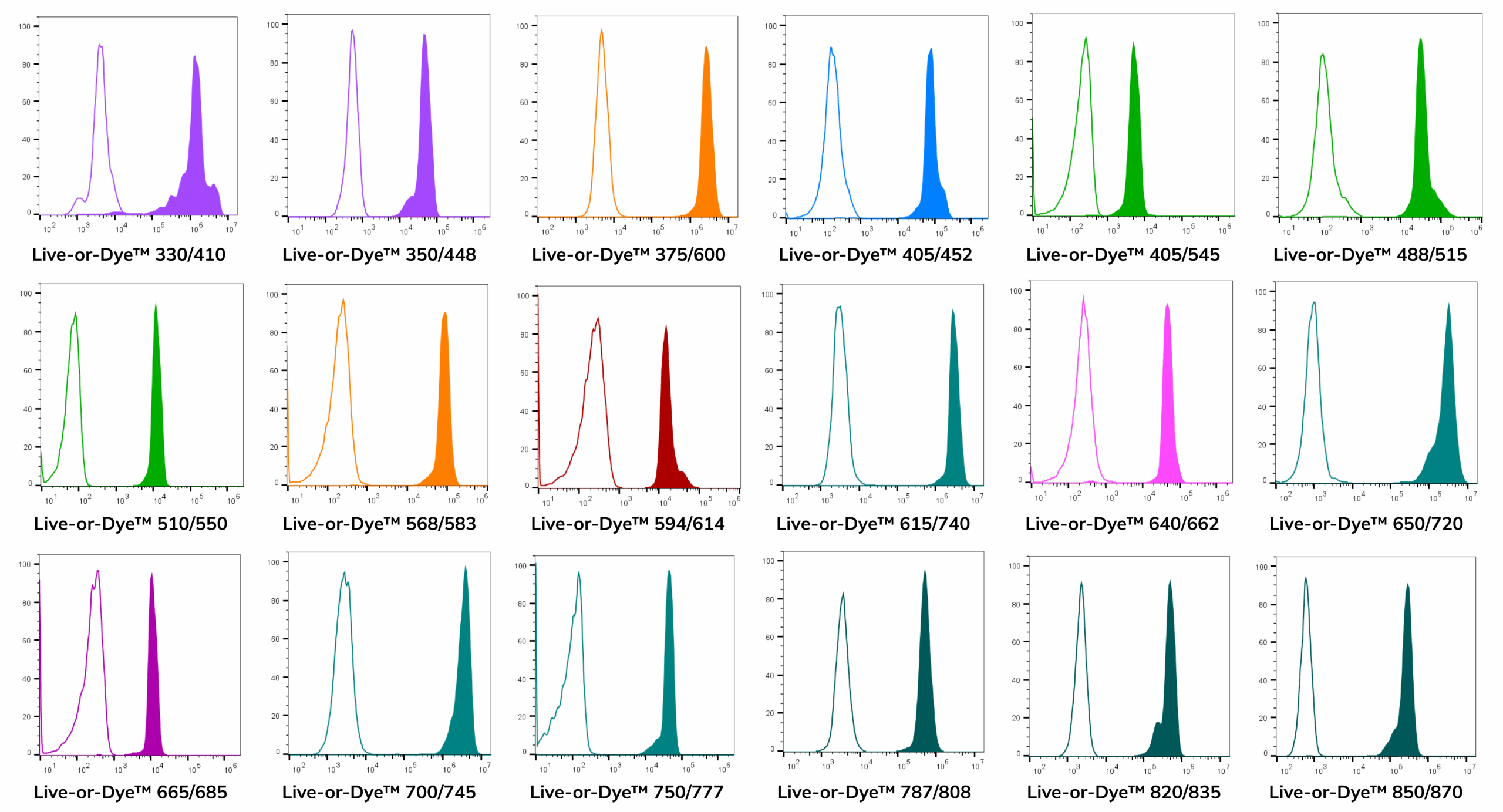
Flow Cytometry
Live/dead stains are useful probes to include when analyzing cell surface protein expression by flow cytometry, because they allow intracellular fluorescence signal from dead cells with permeable plasma membranes to be excluded from analysis. In addition, several Live-or-Dye™ stains were designed specifically for spectral flow to fill in gaps between common fluorophores: Live-or-Dye™ 510/550, 665/685, 375/600, and 615/740.
Sampler Kits Available
Fluorescence Microscopy
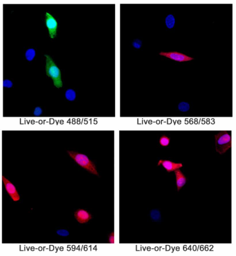
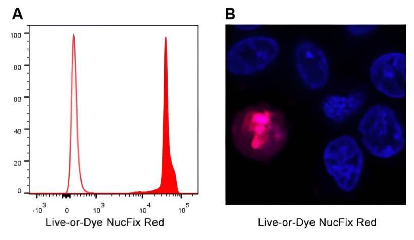
Live-or-Dye NucFix™ Red
Cat. No. | Viability Dye | Compatible lasers (nm) | Optimal detection channels | Notes |
|---|---|---|---|---|
| 32018, 32018-T | Live-or-Dye™ 330/410 | 355, 375 | BUV395 | |
| 32002, 32002-T | Live-or-Dye™ 350/448 | 355, 375 | DAPI | |
| 32014,32014-T | Live-or-Dye™ 375/600 | 355, 375, 405 | Spectral scan, BUV615, BV605 | Developed for spectral cytometry |
| 32003, 32003-T | Live-or-Dye™ 405/452 | 405 | Pacific Blue®, BV421, BV450 | |
| 32009, 32009-T | Live-or-Dye™ 405/545 | 405 | AmCyan, BV510 | |
| 32004, 32004-T | Live-or-Dye™ 488/515 | 488 | FITC | Validated for microscopy |
| 32012, 32012-T | Live-or-Dye™ 510/550 | 488, 532 | Spectral scan | Developed for spectral cytometry |
| 32005, 32005-T | Live-or-Dye™ 568/583 | 488, 532, 561 | PE, PI | Validated for microscopy |
| 32006, 32006-T | Live-or-Dye™ 594/614 | 488, 532, 561 | PI, PE-CF®594, PE-Texas Red® | Validated for microscopy |
| 32015, 32015-T | Live-or-Dye™ 615/740 | 633-640 | Spectral scan, APC-Cy®7 | Developed for spectral cytometry |
| 32007, 32007-T | Live-or-Dye™ 640/662 | 633-640 | APC | Validated for microscopy |
| 32023, 32023-T | Live-or-Dye™ 650/720 | 633-640 | APC-700 | |
| 32013, 32013-T | Live-or-Dye™ 665/685 | 633-640 | Spectral scan, AF700 | Developed for spectral cytometry |
| 32024, 32024-T | Live-or-Dye™ 700/745 | 633-640, 785 | APC-Cy®7 | Our best dye for the APC-Cy®7 channel |
| 32008, 32008-T | Live-or-Dye™ 750/777 | 633-640, 785 | APC-Cy®7, IR840 | |
| 32011, 32011-T | Live-or-Dye™ 787/808 | 785, 808 | APC-Cy®7, IR800 | |
| 32021, 32021-T | Live-or-Dye™ 820/835 | 808 | IR840 | |
| 32022, 32022-T | Live-or-Dye™ 850/870 | 808 | IR885 |
Kit Name | Catalog No. | Included dyes: Ex/Em (Cat No.) | Application |
|---|---|---|---|
| Live-or-Dye™ Fixable Viability Sampler Kit, Standard | 32016 | • 350/448 (32002A) • 405/545 (32009A) • 488/515 (32004A) • 568/583 (32005A) • 640/662 (32007A) | Designed for use on most standard flow cytometry laser and filter configurations |
| Live-or-Dye™ Fixable Viability Sampler Kit, Spectral | 32017 | • 350/448 (32002A) • 375/600 (32014A) • 510/550 (32012A) • 615/740 (32015A) • 665/685 (32013A) | Designed for use in spectral flow cytometry, to fill in gaps between common fluorophores |
Draq7 is a trademark of Biostatus, Ltd. Texas Red and Pacific Blue are registered trademarks of Thermo Fisher Scientific. Cy Dye is a registered trademark of Cytiva.
Caspase Assays & Apoptosis Inducers
NucView® Caspase Substrates for Live Cells
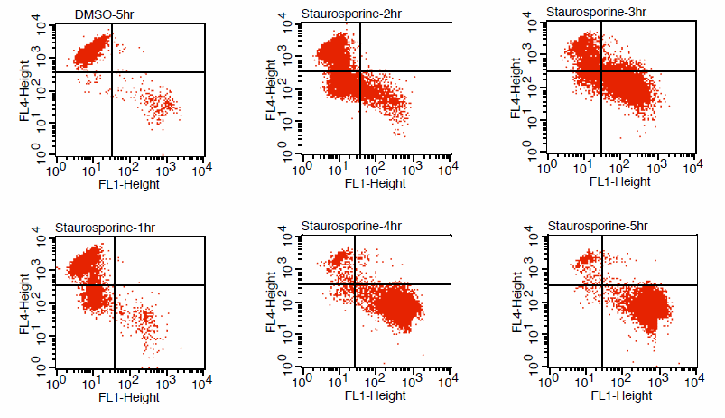
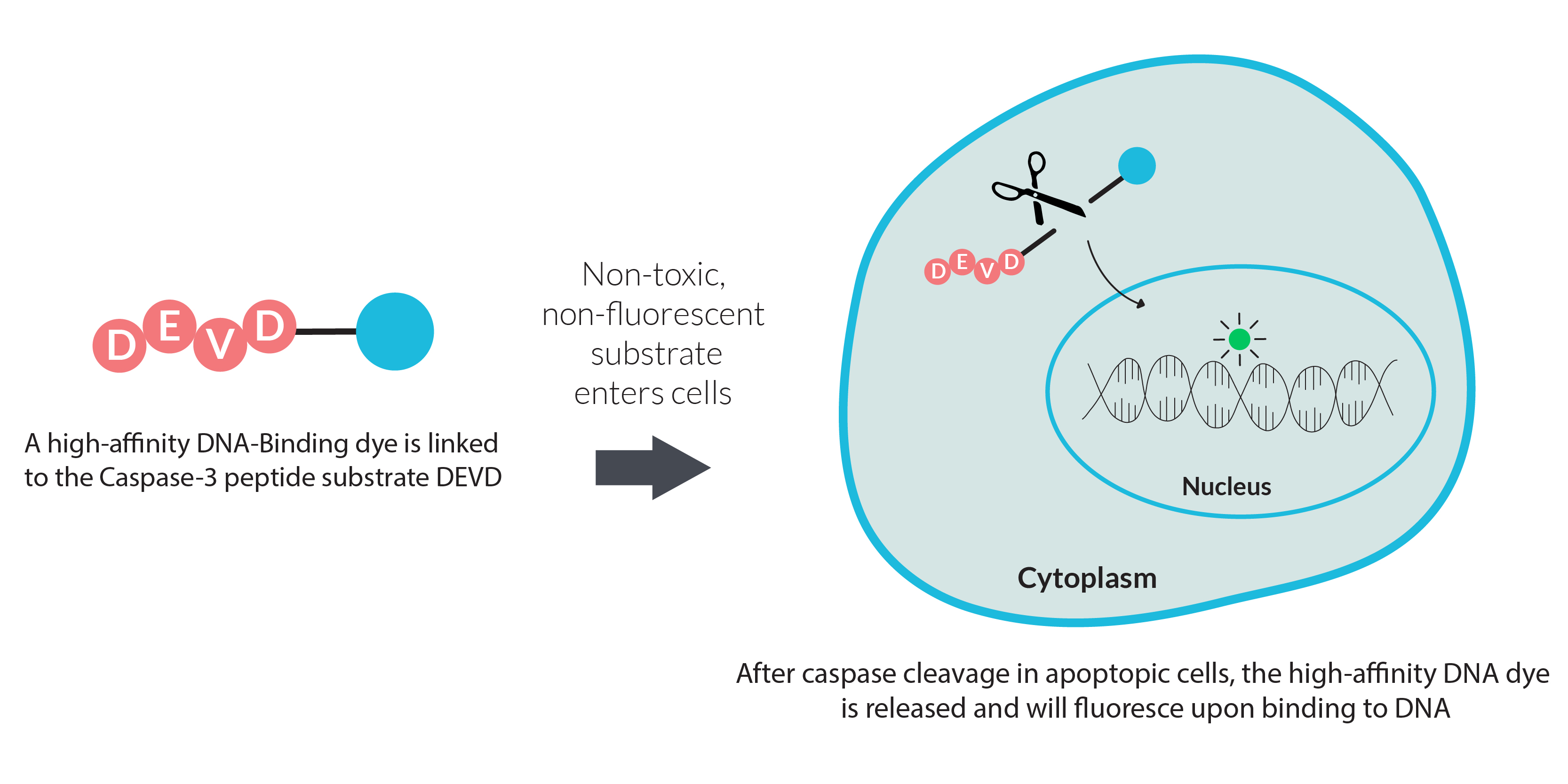
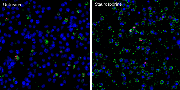
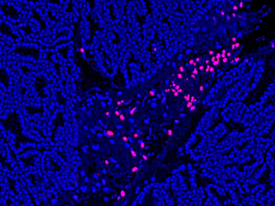
NucView® Caspase-3 Substrates are enzyme substrates attached to a fluorogenic DNA dye to detect apoptosis in intact cells in real-time. Before cleavage, the dye is non-fluorescent and unable to bind to DNA. Upon enzymatic cleavage of the substrate, the dye is released and becomes capable of binding to DNA. The caspase-3/7 substrate peptide DEVD is attached to a DNA-binding dye and when it enters the cell cytoplasm where it is cleaved by caspase-3 in apoptotic cells, the fluorogenic DNA dye is released and stains the nucleus. Unlike FLICA assays, NucView® caspase-3 substrates do not interfere with caspase activity. NucView® can be fixed with formaldehyde after staining, but it cannot be used to stain fixed cells or tissues. For staining of fixed cells or tissues, see our TUNEL Assays.
- Simple, homogenous assay for endpoint assay or real-time detection in live cells
- Non-toxic, allowing for multi-day experiments
- Detect caspase-3 activity and visualize apoptotic nuclear morphology
- For flow cytometry, fluorescence microscopy, or live cell imaging systems
- NucView® 488 validated in more than 100 cell types and 200 publications
Monitor Apoptosis in Real-Time
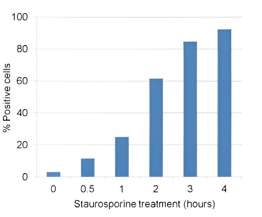

NucView® Caspase-3 Substrates and Kits | Catalog No. | Features |
|---|---|---|
| NucView® 405 Caspase-3 Substrate, 1 mM in DMSO | 10405 | Blue fluorescence for flow cytometry in the Pacific Blue® channel or microscopy with the 405 nm laser |
| NucView® 405 Caspase-3 Substrate, 1 mM in PBS | 10407 | NucView® 405 substrate in PBS, for DMSO-sensitive cell types |
| NucView® 488 Caspase-3 Substrate, 1 mM in DMSO | 10402 | Green fluorescent substrate validated in more than 100 cell types and 200 publications |
| NucView® 488 Caspase-3 Substrate, 1 mM in PBS | 10403 | NucView® 488 substrate in PBS, for DMSO-sensitive cell types |
| NucView® 530 Caspase-3 Substrate, 1 mM in DMSO | 10406 | Orange fluorescence for microscopy in the Cy®3 channel or flow cytometry in the R-PE channel |
| NucView® 530 Caspase-3 Substrate, 1 mM in PBS | 10408 | NucView® 530 substrate in PBS, for DMSO-sensitive cell types |
| NucView® 488 and MitoView™ 633 Apotosis Detection Kit | 30062 | NucView® 488 and far-red fluorescent MitoView™ 633 for the Cy®5 channel |
| NucView® 488 and RedDot™ 2 Apoptosis & Necrosis Kit | 30072 | NucView® 488 and far-red dead cell DNA dye RedDot™ 2 for the Cy®5 channel |
| Dual Apoptosis Assay with NucView® 488 and CF®594 Annexin V | 30067 | NucView® 488 and red fluorescent Annexin V apoptosis probe |
| Dual Apoptosis Assay with NucView® 488 and CF®640R Annexin V | 30073 | NucView® 488 and far-red fluorescent Annexin V apoptosis probe |
Pacific Blue is a registered trademark of Thermo Fisher Scientific. Cy Dye is a registered trademark of Cytiva.
Other Caspase Substrates and Assays
In addition to our patented NucView® technology for detecting caspase-3 activity in live cells, Biotium also offers rhodamine 110 (R110)-based assay kits for fluorescence- or absorbance-based detection of caspase-3 or caspase-8 activity in cell lysates. The HTS versions of the R110-based homogenous caspase-3 and caspase-8 assay kits are optimized for high throughput screening by fluorescence microplate reader. We also offer a coumarin (AMC)-based blue fluorogenic substrate (Ac-DEVD-AMC) for measuring caspase-3 activity in cell lysates by fluorescence microplate reader.
Apoptosis Inducers/Inhibitors
Product | Catalog No. | Size |
|---|---|---|
| Caspase-3 DEVD-R110 Fluorometric & Colorimetric Assay Kit | 30008-1 | 25 assays |
| 30008-2 | 100 assays | |
| Caspase-3 DEVD-R110 Fluorometric HTS Assay | 30009-1 | 10 assays |
| 30009-2 | 100 assays | |
| 30009-3 | 1000 assays | |
| Ac-DEVD-AMC | 10202 | 5 mg |
| Ac-DEVD-R110 | 10226 | 1 mg |
| Ac-DEVD-CHO Caspase-3 Inhibitor | 10404-1 | 1 mg |
| 10404 | 5 mg | |
| Staurosporine | 00025 | 100 ug |
| Ionomycin, Calcium Salt | 59007 | 1 mg |
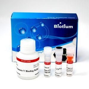
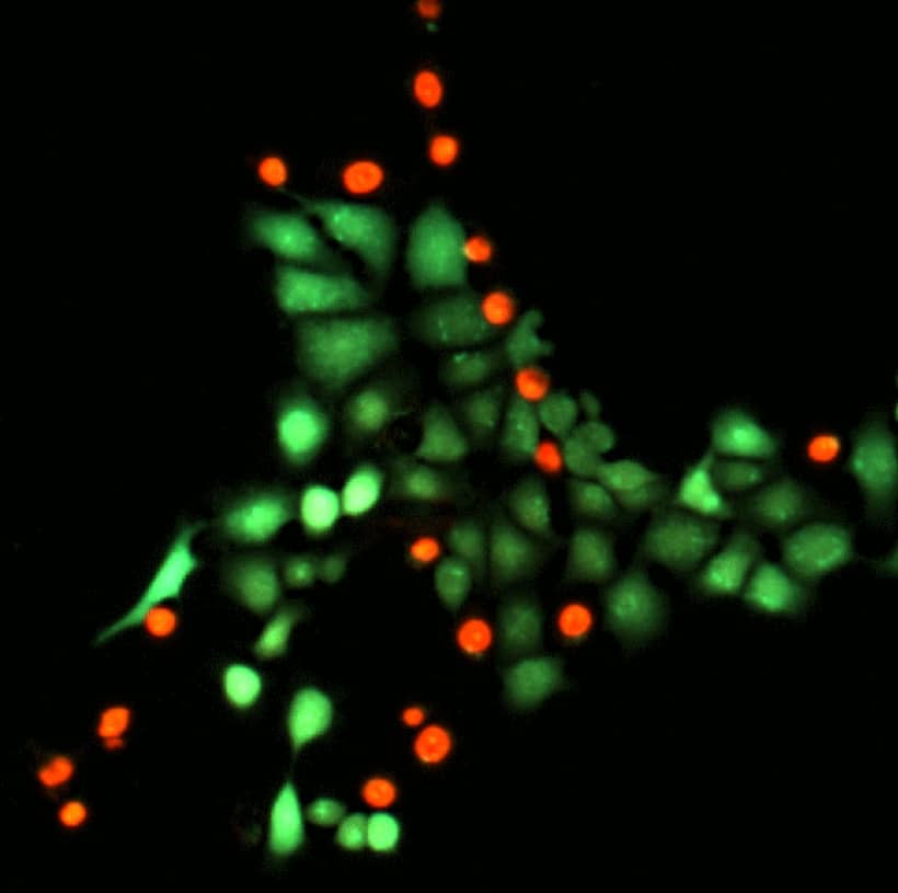
Apoptosis/Necrosis Kits & Annexin V
Dual Apoptosis & Necrosis Assay Kits
Annexin V is a protein that has high affinity for phosphatidylserine (PS). During apoptosis, PS is translocated from the inner to the outer leaflet of the plasma membrane, where it can be stained by fluorescent conjugates of Annexin V. We offer kits with CF®488A Annexin V and a variety of dead cell nucleic acid stains, including propidium iodide (PI), 7-AAD, Ethidium Homodimer III (EthDIII), and RedDot™2. These are membrane-impermeant nucleic acid dyes that stain necrotic and late apoptotic cells. The Apoptotic, Necrotic, and Healthy Cells Quantitation Kit Plus also includes blue fluorescent Hoechst DNA dye for visualizing all cells by fluorescence microscopy. Also see our apoptosis and necrosis staining kit with NucView®488 Caspase-3 Substrate and the far-red dead cell stain RedDot™2 .
Annexin V conjugates are available with a wide selection of CF® Dyes and other labels, including Near-IR CF® Dye Annexin V conjugates. We offer dead cell nucleic acid stains separately as well, including far-red fluorescent RedDot™2 far-red dye and green fluorescent NucSpot® 470 stain.
Note: Apoptosis & Necrosis Staining kits and Annexin conjugates cannot be used with fixed cells or tissues. For fixed cell stains, see our TUNEL Assays.
Apoptosis and Necrosis Staining Kits | Catalog No. | Features |
|---|---|---|
| CF®488A Annexin V and PI Apoptosis Kit | 30061 | • CF®488A stains apoptotic cells green for the FITC channel • PI stains necrotic cells red for the Cy®3 or PE channels • For microscopy or flow cytometry |
| CF®488A Annexin V and 7-AAD Apoptosis Kit | 30060 | • CF®488A stains apoptotic cells green for the FITC channel • 7-AAD stains necrotic cells red for the PE-Cy®5 or FL3 channels • For flow cytometry |
| Apoptosis & Necrosis Quantitation Kit Plus | 30065 | • CF®488A stains apoptotic cells green for the FITC channel • EthD-III stains dead cells red for the Cy®3 or PE channels • EthD-III has higher affinity and quantum yield than PI or 7-AAD • For microscopy or flow cytometry |
| Apoptotic, Necrotic & Healthy Cells Quantitation Kit Plus | 30066 | • CF®488A stains apoptotic cells green for the FITC channel • EthD-III stains dead cells red for the Cy® channel • Hoechst stains all cell nuclei blue for the DAPI channel • For microscopy |
| NucView®488 and RedDot™2 Apoptosis and Necrosis Kit | 30072 | • NucView®488 stains apoptotic nuclei green for the FITC channel • RedDot™2 stains necrotic cells far-red for the Cy®5 or PE-Cy®5, APC, or FL3 channels • For microscopy or flow cytometry |
| Annexin V Conjugates | Multiple | • Annexin V with our bright and photostable CF® dyes and a selection of other labels |
| Annexin V Conjugates, Azide-Free | Multiple | • Annexin V with our bright and photostable CF® dyes and a selection of other labels • Lyophilized and preservative-free for real-time cell imaging |
| Annexin V Near-IR CF® Dye Conjugates | Multiple | • Annexin V conjugated to our superior Near-IR CF® dyes • Lyophilized and preservative-free for in vivo use |
| Annexin V (His Tag) | 29088 | • Recombinant Annexin V (His Tag) expressed from E.coli |
| Annexin Binding Buffer | 99902 | • Concentrated Annexin Binding Buffer for cell staining |
| NucSpot® 470 Nuclear Stain | 40083 | • Green fluorescent dead cell stain • Also useful as a nuclear-specific counterstain for fixed cells |
| RedDot™2 Far-Red Nuclear Stain | 40061 | • Far-red nuclear stain for dead cells or fixed cells • Detect in the Cy®5 channel |
| Ethidium homodimer III | 40051 | • Red fluorescent dead cell stain • Higher affinity and quantum yield than PI |
| Propidium Iodide | 40017 | • Widely used red fluorescent dead cell stain |
| 7-AAD | 40037 | • Fluorescent dead cell stain for the PE-Cy®5/FL3 channel |
| NucSpot® Far-Red | 40085 | • Designed as an improved replacement for 7-AAD • Less bleed into the PE-Texas Red® channel compared to 7-AAD |
Cy Dye is a registered trademark of Cytiva.
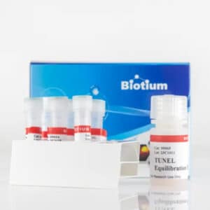
Assays & Other Stains for Fixed Cells or Tissues
CF® Dye Fluorescent TUNEL Staining Kits
TUNEL (terminal deoxynucleotidyl transferase (TdT) mediated dUTP nick-end labeling) is highly selective for the detection of apoptotic cells, but not necrotic cells or cells with DNA strand breaks resulting from irradiation or drug treatment. In this assay, TdT enzyme catalyzes the addition of labeled dUTP to the 3’ ends of cleaved DNA fragments. Fluorescent dye-conjugated dUTP can be used for direct detection of fragmented DNA in fixed cells or tissue sections. We offer TUNEL kits with dUTP conjugates of our bright and photostable CF® Dyes with green, red, or far-red fluorescence. We also offer CF® Dye dUTP Conjugates in a variety of colors.
CF® Dye TUNEL Kits | Catalog No. | Ex/Em | Detection channel |
|---|---|---|---|
| CF®488A TUNEL Assay Apoptosis Detection Kit | 30063 | 490/515 nm | FITC |
| CF®594 TUNEL Assay Apoptosis Detection Kit | 30064 | 593/614 nm | Texas Red® |
| CF®640R TUNEL Assay Apoptosis Detection Kit | 30074 | 642/662 nm | Cy®5 or APC |
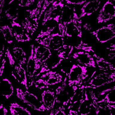
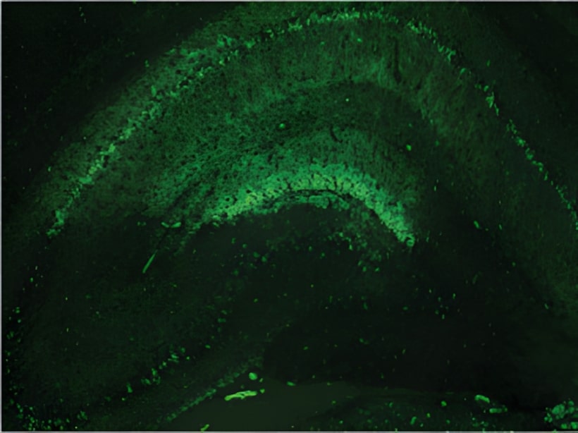
PathoGreen™ and Other Neuronal Histofluorescent Stains
PathoGreen™ Histofluorescent Stain is an anionic green fluorescent dye functionally similar to Fluoro-Jade® dyes. These dyes stain degenerating neurons and their processes in tissue sections and cultured cells. The mechanism of staining has not been determined, but it is proposed that the negatively charged dyes bind to positively charged polyamines or other molecules generated in dying neurons in response to a variety of different neurotoxic insults. We also offer stains for the detection of amyloid in live or fixed neuronal tissues, including Congo Red, DCDAPH, and Thioflavin T.
Product | Catalog No. | Size | Features |
|---|---|---|---|
| PathoGreen™, 1000X in water | 80027-5mL | 5 mL | Stains degenerating neurons in tissue sections or cultures with green fluorescence |
| 80027-50mL | 50 mL | ||
| Congo Red, High Purity Grade | 80028 | 100 mg | Colorimetric or fluorescent staining of amyloid plaques (Ex/Em 497/614 nm) |
| DCDAPH | 80030 | 5 mg | Detection of β amyloid in tissue sections and by near IR small animal imaging (Ex/Em 597/665 nm) |
| Thioflavin T, High Purity Grade | 80033 | 100 mg | Green fluorescent cell permeable fluorescent amyloid probe |
Texas Red is a registered trademark of Thermo Fisher Scientific. Cy Dye is a registered trademark of Cytiva.